https://doi.org/10.17998/jlc.18.2.103
Corresponding author : Hyung Woong Lee
Division of Hepatology, Department of Internal Medicine, Gangnam Severance Hospital, Yonsei University College of Medicine, 20 Eonju-ro 63-gil, Gangnam-gu, Seoul 06229, Korea
Tel. +82-2-2019-4360, Fax. +82-2-2019-3114 E-mail; [email protected]
http://orcid.org/0000-0002-6958-3035
Co-corresponding author : Kyung Sik Kim
Division of Hepato-biliary-pancreas Surgery, Department of Surgery, Severance Hospital, Yonsei University College of Medicine, 50-1 Yonsei-ro, Seodaemun-gu, Seoul 03722, Korea
Tel. +82-2-2228-2100, Fax. +82-2-313-8289 E-mail; [email protected] http://orcid.org/0000-0001-9498-284X
고령 환자의 간암치료 및 감시
노승윤
1∙ 이현웅
2∙ 김경식
1연세대학교 의과대학
1세브란스병원 외과학교실 간담췌외과,
2강남세브란스병원 내과학교실 소화기내과
Treatment and Surveillance of Hepatocellular Carcinoma in Elderly Patients
Seoung Yoon Rho
1, Hyun Woong Lee
2, Kyung Sik Kim
11Division of Hepato-biliary-pancreas Surgery, Department of Surgery, Severance Hospital, 2Division of Hepatology, Department of
Internal Medicine, Gangnam Severance Hospital, Yonsei University College of Medicine, Seoul, Korea
Received May. 24, 2018
Revised Aug. 20, 2018
Accepted Aug. 23, 2018
Hepatocellular carcinoma (HCC) is the third most common cancer in the digestive system based on
survey of domestic cancer incidence, and the ratio of elderly aged 65 or older is expected to rise
steadily, leading to a higher incidence of total hepatocellular carcinoma. The most important thing in
treating these older patients with HCC is to assess the benefits and risks of the treatment in advance.
In other words, the benefit of treatment should be greater than the reduction of survival period or
maladjustment due to treatment. Based on these perspectives, we examined how the detailed
treatment of hepatocellular carcinoma differs from that of general treatment in elderly patients. In
conclusion, older age was not a definite prognostic factor of survival risk-benefit comparison in the
most treatment modalities. However it should be carefully considered and approached about
possi-ble complications in treating HCC in elderly patients. (J Liver Cancer 2018;18:103-114)
Keywords: Carcinoma, Hepatocellular; Aged patients; Aging; Therapeutic; Surveillance
간 3.6% 증가하였으나, 2012-2015년 동안에는 연간 6.1%
감소하였다. 반면에, 간암 발생률은 1999-2009년 기간 동안
1.7% 감소하였으며, 2010-2015년 기간 4.2%로 감소폭이 증
가하였다. 그러나 여전히 간암은 2016년 기준 소화기암 중
에서 세 번째로 발생률이 높은 암이면서, 사망 분율을 비교
해 보았을 때에는 전체 암 사망자 중 14.1%로 첫 번째인 폐
암(23.0%)에 이어 2번째로 중증도가 높은 질환이다.
1서 론
1. 국내 간암 발생 현황
간암은 국내 암 발생률 기준(2015년) 6위로, 성별에 따르
면 남자 4위, 여자 6위를 차지하고 있다. 소화기암 중에서
위, 대장에 이어 많이 발생하는 암으로서, 모든 암의 연도별
연령표준화 발생률 추이를 보았을 때, 1999-2012년 기준 연
2. 인구의 고령화 실태와 간암 발생률의 관계
간암 발생의 최근 추세를 예측하는데 또 하나 중요한
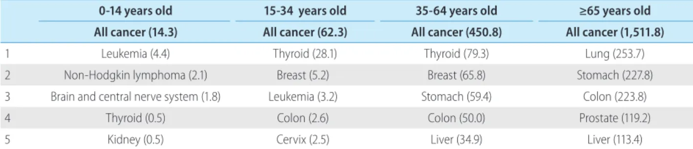
점은, 국내 인구의 고령화이다. 주요 암 연령군별 발생률을
비교해보았을 때, 남녀 전체 35-64세군뿐만 아니라, 65세
이상의 고령 환자군에서도 간암은 5위를 차지하고 있다
(Table 1). 연령 계층별 인구 구성비에 대한 2016년 통계청
보고에 의하면 향후 65세 이상의 고령 인구의 비율이 지속
적으로 상승할 것으로 예상되어 전체 간암의 발생 비율이
높아질 것으로 전망된다(Fig. 1).
2특히, 2015년 주요 암의 연령군별 발생률을 살펴볼 때, 간
암에서 남자의 경우 80-84세, 여자의 경우 85세 이상의 환
자 군에서 최대 발병률(peak incidence)을 보이고 있어, 노
령 인구의 증가에 따라 노령 간암 환자가 의미 있게 증가할
것으로 예상된다(Fig. 2).
1또한, Kim과 Park
3에 의하면 2014년
현재 간암에 의한 전체 사망 환자 중 60세 이상의 비율이
67.37%였고, 그중 70세 이상 79세 미만인 비율이 28.46%로
가장 많은 부분을 차지하였다.
본 론
1. 고령 환자의 정의
“고령”에 대한 개념을 정의하는 것이 매우 어렵다. 일반
적으로는 연령(chronological age)을 기준으로 65세 이상,
즉 은퇴를 앞두고 있는 나이가 “고령”이라고 받아들여지고
있다. 국제기구인 United Nations과 World Health
Organi-zation는 60세 이상을 “고령”으로 정의하고 있다.
4,5실제 임
상연구에서는 60세, 65세, 또는 70세 이상을 각각 “고령”으
로 다양하게 정의하고 있다.
61980년대 연구에서는 주로 65세
이상을 고령으로 정의하였으며, 1990년대 이후로는 75세
이상을 주로 고령으로 정의하였다. 특히, 고령 인구를 대상
으로 하는 연구가 활발한 일본에서는 80세 이상을 초고령
군(super-elderly population)으로 정의하고 있다.
7-10우리나
라의 경우, 2010년 통계청 총 조사인구 연령별 인구분포 결
과에 따르면, 60세 이상이 전체 인구의 16%, 65세 이상이
11.3%, 70세 이상이 7.5% 그리고 75세 이상이 4.3%를 차지
Table 1. Major cancer incidence by age group, National Cancer Information Center (2015 ; Crude rate unit : person/100,000 people)0-14 years old 15-34 years old 35-64 years old ≥65 years old All cancer (14.3) All cancer (62.3) All cancer (450.8) All cancer (1,511.8)
1 Leukemia (4.4) Thyroid (28.1) Thyroid (79.3) Lung (253.7) 2 Non-Hodgkin lymphoma (2.1) Breast (5.2) Breast (65.8) Stomach (227.8) 3 Brain and central nerve system (1.8) Leukemia (3.2) Stomach (59.4) Colon (223.8) 4 Thyroid (0.5) Colon (2.6) Colon (50.0) Prostate (119.2) 5 Kidney (0.5) Cervix (2.5) Liver (34.9) Liver (113.4)
Figure 1. Future population estimation : population ratio by age group, Korea National Statistical Office, 2016 (reference. 2).
하여 인구 분율에 따라 고령을 정의해 볼 수 있다.
2. 생물학적인 노화
노화란 생물학적인 변화와 함께 인간의 육체적, 사회적,
문화적인 활동을 포함한 여러 요인에 의하여 영향을 받는
복잡한 과정이다. 특히 생물학적인 노화는 시간의 흐름에
따라 장기에 발생하는 질병과 기능 소실의 증가를 의미하
기 때문에, 노화가 곧 질병은 아니다. 단지, 장기의 기능이
감소하고, 암과 같은 질환이 발생할 확률이 증가하는 것을
의미한다. 노화의 과정은 단순하지 않고 여러 요인이 복합
적으로 작용하는 과정으로, 주로 유전적인 영향과 환경적
인 요인에 좌우된다. 세포의 입장에서는
텔로미어(telo-mere) 단축, DNA의 변이, 산화물질에 의한 마이토콘드리
아(mitochondria) DNA 손상, 손상된 단백질의 증가 및 이
로 인한 노폐물의 축적이 결국 노화를 유도한다. 간암에서
도 분자, 세포 및 생리적인 수준에서 노화와 발암과정에 대
한 연구가 진행되고 있으며, 여러 가지 요인들이 밝혀지고
있다. 고령 인구의 증가로 이러한 기전에 대한 관심과 연구
의 증가는 암을 더욱 효과적으로 예측하고 예방하는데 반
드시 필요하다. 한가지 예로 만성 C형간염에서 산화 스트
레스(oxidative stress)와 노화가 후성적 불안정성(epigenetic
instability)의 중요한 원인으로 알려져 있다.
113. 노화에 따른 간의 변화와 고령 간암 환자의 임상적
특징
일반적으로 노화에 따라 발생하는 주요한 간담도의 변화
들 중 하나는 간 용적과 간 혈류의 감소이다. Wynne 등
12과
Zoli 등
13은 24세에서 91세 사이의 65명의 건강한 사람들의
간 용적(mL/kg body weight) 및 간 혈류(mL/min/kg body
weight)의 변화를 보았을 때, 각각 평균 41% (남 30%, 여
43%), 47% (남 43%, 여 55%)가 감소한다고 보고하였다. 뿐
만 아니라, 담즙산의 분비가 줄어들고 담즙 배설 능이 감소
한다. 이러한 변화는 간의 대사능력을 감소시키고, 간세포
의 재생능력 및 면역조절능력을 떨어뜨린다.
14이로 인하여
약제-유발 간 손상의 위험도 증가, 심각한 바이러스성 간염
발생 가능성 증가, 복강내 패혈증에 취약해지는 등의 변화
가 생긴다.
15이러한 변화를 겪는 고령 환자에서 간암의 발
생은 다음과 같은 특징을 보인다.
남성에 비하여 여성의 기대 수명 증가로 고령에서 여성의
간암 발생이 증가한다고 알려져 있는데, 간암 발생-사망률
저위험 인구지역(United States, Canada, United Kingdom
등)뿐만 아니라, 고위험 아시아 인구지역(Hongkong,
Shanghai 등)에서도 고령 환자에서 여성의 간암 발생 비율
이 남자의 발생률보다 더 높았다.
16간염 바이러스의 감염을 살펴보면, 고령 환자군에서 C형
간염에 의한 간암의 발생률이 높은데, 대한간학회에서 발
행한 『한국인 간질환백서』에 따르면 간암 발병자 중 60세
이상 연령층에서 C형간염의 비중이 급증하는 양상을 보였
다.
17,18Kim 등
19,20이 우리나라 291,314명의 인구를 대상으로
Figure 2. Major cancer incidence by age group. (A) Male, (B) female, National Cancer Information Center (2015) (reference. 1).
조사한 바에 따르면, positive anti-hepatitis C virus의 비율이
남자(989/178,808명, 0.55%)에 비하여 여자(729/112, 506명,
0.65%)에서 더 높았다.
비B형간염, 비C형간염 관련 간암의 발생률도 증가한다.
Katsuta 등
7은 간암 환자의 간절제술 후 병리학적 비교검사
상, 80세 이상의 고령 환자군에서 비B형간염, 비C형간염
환자의 비율이 유의하게 높음을 보고하였다(19/29, 66% vs.
123/433, 28%). 고령 환자에서의 간암 발생은 간경변이나
간 섬유화를 동반하지 않은, 정상간에서도 잘 발생한다고
알려져 있다. Paradis 등
21은 만성 간 손상의 원인으로 유일
하게 대사증후군만 가지고 있는 환자군이 다른 원인을 가
지고 있는 만성 간 손상의 환자군들에 비하여 간 섬유화를
보이지 않으면서 간암 발생이 많다고 보고하였다. 그리고
고령 간암 환자의 병태생리학적 특징을 살펴 보면,
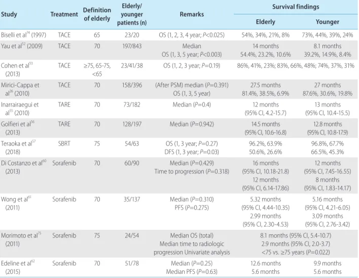
Saty-Table 2. Major finding of survival in curative aim treatment of liver cancer in elderly patientsStudy Treatment Definition of elderly Elderly/ younger patients Remarks Survival findings Elderly Younger Poon et al25 (1999) Hepatic resection 70 222/1,116 Median OS (1, 3, 5 year; P=0.940) 38 months 79%, 58%, 29% 42 months 75%, 51%, 40% Mirici-Cappa et
al26 (2010) resectionHepatic 70 614/1,104 (After PSM) median OS (1, 3, 5 year; P=0.087) (95% CI, 40.1-63.9)58 months
96.8%, 68.3%, 41.9%
42 months (95% CI, 34.1-46.8) 87.1%, 66.8%, 28.9% Santambrogio et
al38 (2017) resectionHepatic 75 53/1,115 DFS (3, 5 year; P=0.104)OS (3, 5 year; P=0.024) 65%, 46%47%, 22% 82%, 60%57%, 35%
Sato et al39 (2012) Hepatic resection ≥80, 70-79, 60-69, ≤59 423/2,261 /1,703/883
In-hospital mortality ≤59 (as reference) 70-79: OR, 3.12; 95% CI, 1.60-6.09; P=0.001 60-69: OR, 2.12; 95% CI, 1.05-4.29; P=0.04 ≥80: OR, 2.48; 95% CI, 1.04-5.92; P=0.04 Hirokawa et al45 (2013) Hepatic resection 70 100/120 OS (1, 3, 5 year; P=0.0650) DFS (1, 3, 5 year; P=0.0088) 91%, 71%, 56% 68%, 36%, 27% 93%, 79%, 64% 77%, 55%, 46% Kaibori et al46 (2017) Hepatic resection ≥75, 60-74, 40-59 2,020/7,576 /2,991 DFS (P =0.379); P=0.399 OS (P=0.016); P<0.001
60-74 vs. ≥75 years old, HR, 0.96; 95% CI, 0.88-1.05 40-59 vs. ≥75 years old, HR, 0.96; 95% CI, 0.86-1.06 60-74 vs. ≥75 years old, HR, 0.87; 95% CI, 0.78-0.97 40-59 vs. ≥75 years old, HR, 0.76; 95% CI, 0.67-0.87 Liu et al47 (2014) RFA 75 337/147 OS (1, 3, 5 year; P=0.690) 96%, 78%, 65% 95%, 81%, 65%
Takahashi et al48 (2010) RFA 75 107/354 OS (3, 5 year; P=0.824) LTPR (1, 3 year; P=0.932) 82%, 61% 6%, 14% 80%, 63% 8%, 12% Nishikawa et al31 (2013) RFA 75 130/238 OS (1, 3, 5 year; P=0.001) RFS (1, 3, 5 year; P=0.238) 90.0%, 64.1%, 44.8% 66.9%, 80.5%, 40.0% 97.6%, 83.7%, 64.0% 80.5%, 40.0%, 19.5% Fujiwara et al50 (2014)
RFA 75 353/1,048 All-cause mortality (1, 3, 5 year; P<0.001) 4.5%, 24.4%, 47.3% 0.7%, 17.7%, 37.1% Zetterman et al63 (1998) LT 60 Total 735 OS (1 year; P=0.004) GS (1 year; P=0.163) 81% 80% 90% 85% Thuluvath et al64 (2010)
LT 65 N/A GS range (5, 10 year; P=0.163) 61.5-69.9%, 41.1-56.0% 75.1-78.5%, 59.6-71.4% Taner et al65 (2012) LT 60-74, <60 635/1,304 OS (1, 3, 5 year) 88.9%, 80.4%, 73.2% 91.1%, 84.9%, 79.2% Kim et al66 (2014) LT >65, 50-64, 35-49 1,569/6,790 /1,671 OS (1, 5 year; P=0.0008) DSS (1, 5 year; P=0.8582) 85%, 60% 87%, 65% 89%, 67% 97%, 85% 97%, 86% 97%, 87% OS, overall survival; PSM, propensity score matching; CI, confidence interval; DFS, disease-free survival; OR, odds ratio; HR, hazard ratio; RFA, radiofrequency ablation; LTPR, local tumor progression rate; RFS, relapse-free survival; LT, liver transplantation; N/A, not available; GS, graft survival.
anarayana 등
22과 Plentz 등
23은 노화에 따른 세포단계에서의
Telomere의 단축이 간의 재생능을 제한 및 고갈시키게 되
고, 간경변이 진행함과 동시에 P53의 손실이 텔로미어의
기능 부전을 촉진하여 간 세포암의 발생이 진행된다고 하
였다. 또 다른 기전으로, Nishida
11은 C형간염에 의한 지속
적인 염증 반응과 노화에 따른 low-level methylation이 간세
포 내의 암 관련 유전자들의 epigenetic alteration의 축적을
가속화한다고 보고한 바 있다.
이러한 내용을 요약하자면, 고령에서 간암은 평균 수명
이 상대적으로 긴 여성에서 빈도가 상승하고, 성인에서 발
생하는 C형간염의 빈도가 높다. 또한, 지방간질환의 빈도
가 상승하여 간경변증이 동반되지 않은 간 세포암의 발생
빈도가 상승한다.
4. 고령 간암 환자에서의 치료
고령 환자를 치료하는 데 있어 가장 중요한 것은 치료로
인한 이익과 위험을 미리 평가하는 것이다. 즉, 치료로 인한
생존 기간의 감소나 육체적, 정신적 스트레스 유발로 인한
부적응을 유발하는 것보다 치료로 인한 이득이 더 많아야
한다. 이러한 관점에 기초하여, 고령의 환자에서 세부적인
간암 치료방법이 일반적인 치료방침과 어떤 차이를 보이
는 지 살펴보았는데, 간절제술, 고주파 열치료, 간이식 등
근치 목적의 치료(Table 2)와 경동맥화학색전술, 경동맥방
사선색전술, 정위신체방사선치료, 분자표적치료 등의 완
화 목적의 치료(Table 3)로 나누어 정리하였다. 추가로, 고
령 환자들은 대부분 동반질환이 있기 때문에, 재활, 장기간
Table 3. Major finding of survival in palliative aim treatment of liver cancer in elderly patients
Study Treatment Definition of elderly
Elderly/ younger patients (n)
Remarks Survival findings Elderly Younger
Biselli et al74 (1997) TACE 65 23/20 OS (1, 2, 3, 4 year; P<0.025) 54%, 34%, 21%, 8% 73%, 44%, 39%, 24%
Yau et al52 (2009) TACE 70 197/843 Median
OS (1, 3, 5 year; P<0.003) 14 months 54.4%, 23.2%, 10.6% 8.1 months 39.2%, 14.9%, 8.4% Cohen et al53 (2013) TACE ≥75, 65-75, <65 23/41/38 OS (1, 2, 3 year; P=0.19) 86%, 41%, 23%; 83%, 66%, 48%; 74%, 37%, 31% Mirici-Cappa et al26 (2010)
TACE 70 158/396 (After PSM) median (P=0.391) OS (1, 3, 5 year) 27.5 months 81.4%, 38.5%, 6.9% 27 months 87.6%, 30.6%, 19.8% Inarrairaegui et
al55 (2010) TARE 70 73/182 Median (P=0.4) (95% CI, 4.2-15.7)12 months (95% CI, 10.4-15.5)13 months
Golfieri et al56
(2013)
TARE 70 128/197 Median (P=0.942) 14.5 months (95% CI, 10.6-16.8) 12.8 months (95% CI, 10.8-17.9) Teraoka et al57 (2018) SBRT 75 54/63 OS (1, 3 year; P=0.27) DFS (1, 3 year; P=0.03) 96.2%, 63.9% 50.6%, 26.6% 96.8%, 67.7% 66.5%, 45.3% Di Costanzo et al60 (2013) Sorafenib 70 60/90 Median (P=0.429) Time to progression (P=0.318) 16 months (95% CI, 10.18-21.8) 12 months (95% CI, 6.14-17.86) 12 months (95% CI, 7.45-16.55) 8 months (95% CI, 1.83-14.17) Wong et al61 (2011) Sorafenib 70 35/137 Median (P=0.310) PFS (P=0.275) 5.32 months (95% CI, 4.44-10.35) 2.99 months (95% CI, 2.30-4.53) 5.16 months (95% CI, 4.21-6.05) 3.09 months (95% CI, 2.76-3.42) Morimoto et al75 (2011)
Sorafenib 75 24/54 Median OS (total) Median time to radiologic progression Univariate analysis
8.1 months (95% CI, 5.4-10.7) 2.9 months (95% CI, 2.0-3.7) <75 vs. ≥75 years (P=0.022) Edeline et al62 (2015) Sorafenib 70 51/78 Median (P=0.25) Median PFS (P=0.63) 12.6 months 5.6 months 9.9 months 5.6 months TACE, transarterial chemoembolization; OS, overall survival; TARE, transarterial radioembolization; PSM, propensity score matching; CI, confidence interval; SBRT, stereotactic body radiation therapy; DFS, disease-free survival; PFS, progression-free survival.
의 돌봄에 대한 체계화된 분류와 평가가 함께 고려된다면,
향후 좀 더 효과적인 치료방침을 결정할 수 있으리라 판단
된다.
5. 간절제술
현재 대한간암학회에서 발간한 간세포암종 진료 가이드
라인에 따르면, 간절제술은 간경변증이 없는 간에 국한된
단일 간세포암종 환자에서의 1차 치료법이며, 간경변증이
있는 경우에도, 잔존 간 기능이 충분하다고 예상되는 경우,
우선적으로 고려할 수 있으나, 고령 환자에서는 간 재생능
력이 감소하기 때문에 대량 간 절제는 신중히 고려되어야
한다고 명시하고 있다. 그러나 진료지침에 고령 환자 치료
에 대한 세부 내용은 없다.
24Poon 등
25은 1,116명의 70세 미만 간암 환자와 222명의
70세 이상 고령 간암 환자에서 간절제술을 시행한 비율은
각각 299명(27%), 31명(14%)이었으며(P<0.001), 두 군 간
의 전체 생존율 비교에서 유의한 차이가 없음을 보고하였
다(
P=0.940). Mirici-Cappa 등
26의 대규모 다기관 후향적 코
호트연구에서 70세 이상 간암 환자 614명과 70세 미만
1,104명을 비교하였고, 간절제술을 시행받은 환자가 각각
43명(7%), 142명(12.9%)이었다. Propensity score matching
후 두 군 간 전체 생존율을 비교하였을 때, 5년 생존율은
41.9%, 28.9% (P=0.087)로 유의한 차이가 없었다.
26다른 연
구에서도 고령 간암 환자에서 나이 자체는 간절제술의 절
대적 금기가 아니며, 간 기능이 보존되어 있는 고령 환자에
서 치료성적 또한 비고령 환자군과 유의미한 차이가 없었
다.
9,10,27-37Santambrogio 등
38은 53명의 75세 이상 환자군과 115명의
75세 미만군의 간절제술 후 성적을 비교하였을 때, 무병 생
존율의 유의미한 차이는 없었으나(P =0.104), 전체 생존율
에서 75세 이상의 고령 환자군의 성적이 더 나빴다고 보고
하였다(P=0.024). 그러나 원인을 분석해보면, 간암이나 간
경변증에 의한 사망의 비율은 고령 환자와 비고령 환자군
간에 차이가 없었기 때문에(36% vs. 34%), 심혈관계질환
또는 다른 악성 종양과 같은 동반질환에 의한 영향으로 판
단된다.
38Sato 등
39에 따르면 54,145명의 간암 환자 중 간절제술을
시행받은 5,270명의 환자를 분석하였을 때, 간절제술 시행
후 병원내 사망률이 59세 미만을 기준으로, 상대위험도가
70-79세 환자에서 3.12배(95% 신뢰구간 1.60-6.09, P=0.001),
80세 이상에서 2.48배(95% 신뢰구간 1.04-5.92, P=0.04)까
지 증가하였다. 고령 환자에서 대량 간절제술과 제한 절제
술(쐐기절제술 혹은 구역절제술[segmentectomy])을 비교
한 연구에서, Portolani 등
40은 70세 이상과 미만의 두 그룹
간 전체 생존율의 유의미한 차이는 없었으나, 제한 절제술
을 한 경우가 고령 환자군에서 수술 후 사망률을 낮추고(상
대위험도 0.67, P =0.035), 좋은 전체 생존율을 보여주었다
(상대위험도 0.38, P=0.011).
Iida 등
8은 80세 이상의 44명의 간암 환자와 351명의 80세
미만 환자군을 비교하였고, 전체 생존율 및 무병 생존율이
두 군 간 유의한 차이는 없었다. 그러나 80세 이상의 환자군
에서 생존율에 영향을 미치는 인자를 분석해보았을 때, 수
술 후 합병증이 중요한 예후인자였다(상대위험도 4.30,
95% 신뢰구간 1.49-13.25, P<0.01). 그리고 수술 후 합병증
의 주요한 원인이 영양상태이며, 수술 전후의 영양관리가
중요함을 보고하였다.
8Kim 등
41은 70세 이상 환자군(60명)
에서 수술 후 폐렴 발생이 60-69세 환자군(219명)에서 보다
유의하게 높음을 보고하였고, Kishida 등
42의 연구에 따르
면, Clavien-Dindo grade 3a 이상의 합병증 발생률이 75세
이상 고령 환자군(n=22)에서 41%로, 75세 미만 환자군
(n=82)에서 15%에 비하여 의미 있게 높았다(
P=0.006). 이
에 대하여, Ide 등
43과 Nanashima 등
44은 고령의 간암 환자
에서 수술 후 합병증을 예측하는 방법으로, 나이, 심장/폐
질환의 유무, 당뇨 유무, performance status index,
Ameri-can society of anesthesiologists score 등으로 구성된
preop-erative risk score와 comprehensive risk score가 유용하다 하
였다.
Hirokawa 등
45에 따르면 70세 이상의 100명의 환자와 120명
의 70세 미만 환자의 비교에서, 전체 생존율에는 유의미한
차이가 없으나(P =0.0650), 무병 생존율에선 70세 미만 환
자군이 더 좋았다(P=0.0088). 또한, 70세 이상 환자군에서
여성의 비율(P =0.023)과 C형간염의 비율(P =0.009), 수술
후 폐합병증 비율(P=0.0484)이 더 높았으며, 낮은
interfer-on 치료에 대한 반응률(
P =0.0203)을 보였다. 고령화 사회
로 접어든지 상당한 시간이 지난 일본사회에서 대규모 코
호트연구가 진행된 바 있는데, Kaibori 등
46은 일본 전역
68,806명의 간암 환자를 대상으로 간절제술을 받은 환자
12,587명(75세 이상 2,020명, 60-74세 7,576명, 40-59세
29,91명)의 생존율을 분석하였다. 세 그룹 간의 전체 생존
율은 75세 이상 환자군이 다른 군들에 비하여 통계학적으
로 유의미하게 낮았으나(60-74 vs. ≥75, hazard ratio [HR],
0.87, P=0.016; 40-59 vs. ≥75, HR 0.76, P<0.001), 무병 생존
율에는 유의미한 차이가 없었다.
46요약하면, 고령의 간암
환자라도 심혈관계질환과 같은 동반질환이 없다면, 간 절
제술과 같은 적극적인 치료가 도움이 되지만, 간 재생능력
을 고려하여 가능한 제한된 절제술을 고려하며, 수술 후 합
병증 예방을 위하여 수술 전후 영양공급 및 감염 예방에 주
의해야 한다.
6. 고주파 열치료(radiofrequency ablation, RFA)
고주파 열치료(RFA)는 간절제술이 불가능한 환자에서
좋은 대안으로 알려져 있다. Liu 등
47은 75세 이상의 고령 환
자군 336명과 75세 미만 환자군 147명의 RFA 치료를 받은
환자의 전체 생존율을 단순 비교와 propensity score
match-ing 분석으로 비교하였고, 모두 두 군 간 차이가 없음을 보
고하였다(
P =0.690 and 0.183, respectively). Takahashi 등
48의 연구에서도 75세 이상 환자 107명과 75세 미만 환자 354명
을 비교하였을 때, 전체 생존율(
P =0.824)과 무병 생존율
(
P=0.594)뿐만 아니라, 국소 재발률(P=0.932)에서도 두 군
간 유의미한 차이가 없었다.
반면, Nishikawa 등
49은 고주파 열치료를 시행받은 75세
이상의 고령 환자군(130명)과 75세 미만의 환자군(238명)
과의 비교에서 3년간 전체 생존율(64.1% vs. 83.7%,
P <0.001), 무병 생존율(21.3% vs. 40.0%, P <0.001)로 고령
환자군이 더 나빴다. 뿐만 아니라, 국소적 종양
진행률(lo-cally tumor progression rate)의 비교에서도 고령 환자군이
유의하게 성적이 나빴다(43.0% vs. 26.3%,
P=0.002).
49그리
고 Sato 등
39의 연구에서도 69세 미만 4,359명, 70-79세 환자
5,471명, 80세 이상 1,858명의 간암 환자들 간 고주파 열치
료 후 원내 사망률(in-hospital mortaility after procedure)을
비교하였을 때, 고령 환자군일수록 상대 위험도가 증가하
였다(70-79세 환자군 odds ratio [OR] 7.05; 80세 이상군 OR
8.12). 그러나 이 연구는 단순 사망률만을 비교한 연구로 동
반질환에 대한 분석이 포함되어 있지 않기 때문에 결과 해
석에 주의를 요한다. Fujiwara 등
50은 RFA를 시행받은 75세
미만의 환자 1,048명과 75세 이상의 353명의 치료 성적을
비교하였는데, 5년간 추적 관찰한 전체적인
사망률(all-cause mortality)에서는 고령 환자군이 비고령 환자군에 비
하여 통계적으로 유의미하게 나빴으나(47.3% vs. 37.1%,
P<0.001), 간질환 사망률(liver-related mortality)에서는 통
계적인 차이를 발견할 수 없었다(P =0.64). 즉, 폐렴(8.0%
vs. 2.3%, P=0.004), 감염질환(10.7% vs. 4.2%, P=0.007), 허
혈성질환(4.0% vs. 0.2%, P=0.0014) 등이 주요한 고령군 환
자군 사망의 원인이었다.
50또한, 흥미롭게도 Chen 등
51은 70세 이상의 고령 간암 환
자에서 간절제술 이후 재발한 환자들 중 RFA 또는 경비 에
탄올 주입술(percutaneous ethanol injection [PEI] 57명)과
재절제술(48명)을 비교하였을 때, 5년간 전체 생존율
(39.0% vs. 46.5%, P =0.612) 및 무병 생존율(39.8% vs.
41.1%, P=0.549)로 두 군 간 차이가 없음을 보고하였다. 결
론적으로, 간절제술이 불가능한 고령의 간암 환자에서
RFA는 효과적인 치료방법 중 하나이며, 시술 후 감염성 질
환과 심혈관계질환에 대한 관리가 생존율에 영향을 미친
다 하겠다.
7. 경동맥화학색전술(transarterial
chemoemboli-zation, TACE)
Yau 등
52은 70세 초과의 고령 간암 환자 197명과 70세 이
하의 843명의 TACE의 치료성적을 비교하였는데, 질병 특
이 생존율(disease-specific survival)에서 5년 생존율이 각각
10.6%와 8.4%로 고령 환자군이 좋았다(8.7개월 vs. 15.2개
월, P<0.003). 또한, Cohen 등
53은 102명의 간암 환자를 대상
으로 한 전향적 연구(65세 미만: 38명, 65-75세: 41명, 75세
이상: 23명)에서 3년간 전체 생존율이 세 군 간에 유의미한
차이가 없음을 보고하였다(31% vs. 48% vs. 23%, P=0.19).
Mirici-Cappa 등
26의 연구에서도 158명의 70세 이상 고령 환
자와 396명의 비고령 환자를 비교하였을 때, Propensity
score matching analysis 후, 전체 생존율에서 유의한 차이가
없었다(27개월 vs. 27.5개월, P=0.391).
요약하면, TACE 치료성적은 고령 여부와 무관하게 효과
가 좋았다. 그러나 간과하지 말아야 할 것은 시술 관련 합병
증인데, 이는 많은 고령 환자에서 동맥경화가 동반되어 있
어 시술 중 혈관 손상을 유발할 가능성이 높고, 조영제의 사
용으로 신장 기능이 저하되어 있는 고령 환자에서 신기능
을 악화시킬 가능성이 높기 때문이다.
548. 경동맥방사선색전술(transarterial
radioemboli-zation, TARE)
Yttrium 90 microsphere를 이용한 경동맥방사선색전술
의 효용성에 대한 연구는 활발히 진행되고 있으나, 고령 환
자군을 대상으로 한 보고가 적다. Inarrairaegui 등
55은 39명
의 70세 이상 고령 간암 환자군과 63명의 70세 이하 환자군
의 전체 5년 생존율을 분석하였을 때, 두 군 간 차이는 없음
을 보고한 바 있다(13개월 vs. 12개월, P=0.4). 그리고 두 군
간 Yttrium 90 activity/target liver volume (GBq/dL)도 각각
0.1 GBq/dL과 0.09 GBq/dL로 고령군에서 더 높았으나
(P =0.01), 시술 후 방사선색전술로 인한 합병증의 비교에
서는 위궤양, 폐렴 등 두 군 간 유의미한 차이는 없었다.
55또한, Golfieri 등
56은 128명의 70세 이상 환자군과 197명의
70세 미만군의 비교에서, 전체 5년 생존율은 차이가 없음
(14.5개월 vs. 12.8개월, P=0.942)과 시술 후 합병증 또한 두
군 간 차이가 없음을 보고하였다.
9. 정위신체방사선치료(stereotactic body radiation
therapy, SBRT)
고령의 간암 환자에서 SBRT의 치료효과에 대한 보고는
많지 않은데, Teraoka 등
57은 54명의 75세 이상 간암 환자와
63명의 75세 미만 환자를 비교하였다. 선량비교에서 60 Gy
는 고령 환자군과 비고령 환자군 각각 14.8%, 11.1%
(P =0.34)로 차이가 없었으며, 전체 생존율의 유의미한 차
이는 없었으나(P =0.27), 무병 생존율은 비고령 환자군의
성적이 더 좋았다(13개월 vs. 25개월, P=0.03). 두 군 간 시
술 후 발생한 합병증의 빈도에 있어서 유의미한 차이는 없
었다.
57그러나 이는 후향적 연구이고, 다른 치료와 함께 진
행된 환자군들이 포함되어 있으며, 방사선 조사량과 횟수
도 보정되어 있지 않기 때문에 정확한 SBRT만의 효과를 비
교하기는 어렵다. 따라서 앞으로 진행될 전향적인 연구들
의 결과를 살펴볼 필요가 있다.
10. 분자표적치료(molecular targeted therapy,
sorafenib)
Sorafenib은 절제 불가능하고 진행된 간암, 특히
Child-Pugh 점수가 A 혹은 B이거나 혈관 침범 혹은 타장기 전이
가 있든지, 환자 상태가 TACE에 부적합하거나 해부학적으
로 TACE가 힘들 경우, 그 치료법으로 추천되고 있다.
58,592010년대 이후 고령의 간암 환자에서 sorafenib의 치료효과
에 대한 연구는 많지 않다. Di Costanzo 등
60은 70세 이상의
고령 환자 60명과 비고령 환자 90명을 sorafenib으로 치료
한 후 전체 생존율과 종양 진행까지의
시간(time-to-pro-gression)을 비교하였을 때, 두 군 간에 유의한 차이는 없었
으나 grade III-IV 이상의 중대한 이상 반응(serious adverse
effects)의 비율이 비고령 환자군에서 더 높았음을 보고하였
다(15.7% vs. 9.2%). 또한, Wong 등
61은 같은 70세를 기준으
로 하였을 때(고령 35명, 비고령 137명), 전체 생존율과 종
양 진행까지의 시간의 중앙값, grade III-IV 이상의 중대한
이상 반응에서 모두 두 군 간 유의한 차이가 없었다고 하였
다. Edeline 등
62에 따르면 70세 이상 환자군(51명)과 70세
미만 환자군(78명)의 비교에서, 두 군 간 전체 생존율, 무병
생존율에 유의미한 차이는 없었지만, 치료 중단 빈도
(45.1% vs. 24.4%,
P=0.014) 및 항혈소판제를 사용하고 있
는 환자에서의 대량 출혈의 빈도(18.8% vs. 2.7%)가 고령
환자군에서 더 높았다. 요약하면, sorafenib은 고령에서도
비고령 환자와 유사한 치료효과를 보였지만, 이상 반응의
발생빈도 면에서 더 높은 경향성을 보였다.
11. 간이식(liver transplantation)
간이식은 간암에 있어서 중요하고 근본적인 치료로 알려
져 있을 뿐만 아니라, 간경변증에 의한 간부전의 치료에 있
어서도 효과가 인정되고 있다. 최근 고령 간암 환자에서 그
효과에 대한 대규모 연구가 진행된 바 있는데, Zetterman
등
63은 60세 이상과 60세 미만 환자군 735명을 비교한 연구
에서, 1년째 이식간 생존율에서는 두 군 간 차이가 없었으
나(80% vs. 85%,
P =0.163), 전체 생존율에 있어서는 60세
이상 환자군의 생존율이 비고령 환자군에 비하여 유의하
게 낮음을 보고하였다(81% vs. 90%, P=0.004). 그 원인으로
이식 후 6개월째 감염, 심폐질환, 신경계질환, 악성 종양 등
에 의한 사망이 많았는데, 이런 이유로 고령 환자에서 간이
식을 신중하게 고려해야 할뿐 아니라, 이식 후에도 합병증
에 대하여 집중 관찰해야 한다고 하였다.
631999에서 2008년
까지 미국 간이식 환자들을 비교한 대규모연구에서는,
18-64세 환자군과 65세 이상 환자군의 비교에서 5년 생존
율이 각각 69.9%, 61.5%, 10년 생존율이 56.0%, 41.1%였다.
이에 따라 고령 간암 환자를 기본적으로 간이식 후보로 고
려하지 않을 것을 권고하였다.
64반면, Taner 등
65은 13명의 75세 이상 환자들의 간암으로
인한 간이식 환자들의 치료성적을 보고하였는데, 수술 중
혹은 수술 전후 사망은 없었고, 평균 생존 기간을 65개월이
라고 보고한 바 있으며, Kim 등
66은 1,569명의 65세 이상 간
암 환자에서 간이식을 받은 환자군과 50-64세의 6,990명,
35-49세의 1,671명을 비교 분석하였을 때, 5년 추적관찰 후
전체 생존율에 있어서 65세 이상 군이 다른 두 군에 비하여
유의하게 낮음을 보고하였다(60% vs. 65% vs. 67%,
P=0.0008). 하지만 질병 특이 생존율(disease specific
surviv-al)에서는 세 군 간 차이가 없었다(85% vs. 86% vs. 87%,
P=0.8582).
66이들 모두 신중하게 선택된 고령의 간암 환자
에서 나이 자체는 수술의 성공을 예측하는 인자가 될 수 없
다고 하였다. 결국, 고령의 간암 환자에서 이식은 질병 특이
생존율에서는 비고령 환자와 큰 차이가 없지만, 동반질환
의 악화나 부작용에 의하여 전체 생존율의 감소를 유발할
가능성이 높기 때문에, 신중한 고려가 필요하리라 판단된다.
12. 고령의 환자에서 간암 감시
B형간염, C형간염에 의한 간경변증 및 알코올성 간경변
증 환자에서 간암 감시는 그 효용성이 증명된바 있다.
American association for the study of liver diseases guideline
(AASLD)에서는 성인 간경변 환자에서 간암 감시는 생존
기간을 연장하며, 그 간격을 alpha-fetoprotein의 tumor
marker와 함께 6개월 간격의 초음파검사를 제시한 바 있
다.
67European association for the study of the liver guideline
(EASL) 또한 간암 고위험군에서의 6개월 간격의 초음파검
사를 권유하고 있다.
58하지만 고령에서의 간암 감시에 대
한 국제적인 consensus가 이루어진 guideline은 없다. 최근
의 노르웨이에서 발표된 연구에 따르면, 로지스틱 회귀 분
석에서 65세 이하의 연령이 전체 생존율 향상에 기여하는
독립변수(OR 4.1, 95% CI 12-14.6, P=0.03)인 것으로 알려
졌다.
68또한, Singal 등
69은 간암 감시에 대한 systemic review
에서 extreme age 특히, 65세 이상 혹은 50세 이하의 나이는
간암 감시에서 negative predictor라고 하였고, Davila 등
70은
65세 이상에서 65세 미만과 비교하였을 때 유의미하게 더
낮은 간암 감시율을 보인다고 하였다. 하지만 연령에 초점
을 맞추어 진행한 대규모연구 결과는 부족한 실정이다.
71일반적으로 고령 간암 환자에서 간암 결절의 개수가 비고
령군보다 적고,
26,72진행된 간 섬유화의 비율이 적다는 보고
가 있다. 하지만 고령의 환자라도 조기에 간암이 발견되면
치료방침의 선택이 다양해지며, 동반질환이 없다면 전체 생
존율 및 무병 생존율이 좋다는 임상연구 결과를 고려할 때,
기존의 간질환 환자와 동일하게 6개월 이상의 간격을 둔 간
암 감시를 고려해 볼 수 있다.
73그러나 비용-효과의 관점에
서 감시 검사 간격에 대한 추가적인 연구가 필요하다.
결 론
고령화 사회에 빠르게 진입함에 따라, 국내 고령 간암 환
자의 수는 다가올 수년내 증가할 것으로 생각된다. 이는 최
근 항바이러스 치료 성적 등이 매우 좋아짐에 따라 간경변
증으로 인한 사망률이 감소하고, 고령의 만성 바이러스성
간질환 환자의 비율이 증가하여, 간암 발생이 상대적으로
고령에서 증가할 것이기 때문이다. 따라서, 고령 환자군과
비고령 환자군에서 간암의 병태생리학적 차이를 이해하는
것이 간암 치료의 중요한 출발점이 될 것이다.
고령 간암 환자의 치료에 있어서 무엇보다 중요하게 고
려해야 할 사항은 치료에 따른 생존율의 이익과 수술 후 합
병증과 동반질환의 악화와 같은 잠재적인 위험성에 대한
기회 비용을 인식하는 것이다. 또한, 치료 전 효과적으로 노
화의 정도를 평가할 수 있다면(geriatric evaluation), 연령의
기준에서 고령이라 하더라도, 젊은 환자와 같이 적극적인
치료를 선택할 수 있다. 따라서, 국내 환자들의 데이터를 이
용한 국내 실정에 맞는 고령 평가 기준과 이에 따른 고령 간
암 환자에서의 치료 가이드라인을 정립하는 것이야말로
치료효과를 높이고, 기회 비용을 줄이는 방법이 될 것이다.
AUTHOR CONTRIBUTIONS
Manuscript writing: Seoung Yoon Rho
Supervising: Hyun Woong Lee, Kyung Sik Kim
Conflicts of Interest
The authors have no conflicts to disclose.
REFERENCES
1. Ministry of Health & Welfare, Korea Central Cancer Registry, National Cancer Information Center. Annual report of cancer statistics in Korea 2016 [Internet]. Goyang (KR): National Cancer Information Center; [cited 2018 Mar 15]. Available from: https://www.cancer.go.kr/lay1/ S1T639C642/contents.do.
2. Korea National Statistical Office, Annual report of population, 2017 [Internet]. Daejeon (KR): Korea National Statistical Office; [cited 2018 May 24]. Available from: http://www.index.go.kr/potal/main/EachDtl-PageDetail.do?idx_cd=1010.
3. Kim BH, Park JW. Epidemiology of liver cancer in South Korea. Clin Mol Hepatol 2018;24:1-9.
in-dependence & the world's older people. BMJ 2000;321:517.
5. World Health Organization. World report of age and health, 2015 [Internet]. Geneva (SUI). World Health Organization; [cited 2018 Mar]. Available from: www.who.int/ageing/events/world-report-2015-launch.
6. Hung AK, Guy J. Hepatocellular carcinoma in the elderly: meta-analysis and systematic literature review. World J Gastroenterol 2015;21:12197-12210.
7. Katsuta E, Tanaka S, Mogushi K, Matsumura S, Ban D, Ochiai T, et al. Age-related clinicopathologic and molecular features of patients re-ceiving curative hepatectomy for hepatocellular carcinoma. Am J Surg 2014;208:450-456.
8. Iida H, Kaibori M, Matsui K, Ishizaki M, Kon M. Assessing the feasibil-ity of clinicopathological features of hepatic resection for hepato-cellular carcinoma in patients over 80 years of age. Mol Clin Oncol 2017;6:29-38.
9. Nozawa A, Kubo S, Takemura S, Sakata C, Urata Y, Nishioka T, et al. Hepatic resection for hepatocellular carcinoma in super-elderly pa-tients aged 80 years and older in the first decade of the 21st century. Surg Today 2015;45:851-857.
10. Yamada S, Shimada M, Miyake H, Utsunomiya T, Morine Y, Imura S, et al. Outcome of hepatectomy in super-elderly patients with hepato-cellular carcinoma. Hepatol Res 2012;42:454-458.
11. Nishida N. Impact of hepatitis virus and aging on DNA methylation in human hepatocarcinogenesis. Histol Histopathol 2010;25:647-654. 12. Wynne HA, Cope LH, Mutch E, Rawlins MD, Woodhouse KW, James
OF. The effect of age upon liver volume and apparent liver blood flow in healthy man. Hepatology 1989;9:297-301.
13. Zoli M, Magalotti D, Bianchi G, Gueli C, Orlandini C, Grimaldi M, et al. Total and functional hepatic blood flow decrease in parallel with age-ing. Age Ageing 1999;28:29-33.
14. Schmucker DL. Age-related changes in liver structure and function: implications for disease? Exp Gerontol 2005;40:650-659.
15. Kinoshita A, Koike K, Nishino H. Clinical features and prognosis of el-derly patients with hepatocellular carcinoma not indicated for surgical resection. Geriatr Gerontol Int 2017;17:189-201.
16. El-Serag HB, Rudolph KL. Hepatocellular carcinoma: epidemiology and molecular carcinogenesis. Gastroenterology 2007;132:2557-2576. 17. Lee JS, Kim CM. White Paper on Liver Diseases in Korea. Seoul: Jin
Publishing, 2013:78-80.
18. Korean Liver Cancer Study Group (KLCSG); National Cancer Center, Korea (NCC). 2014 KLCSG-NCC Korea practice guideline for the man-agement of hepatocellular carcinoma. Gut Liver 2015;9:267-317. 19. Kim DY, Kim IH, Jeong SH, Cho YK, Lee JH, Jin YJ, et al. A nationwide
seroepidemiology of hepatitis C virus infection in South Korea. Liver Int 2013;33:586-594.
20. Kim BK, Jang ES, Kim JH, Park SY, Ahn SV, Kim HJ, et al. Current sta-tus of and strategies for hepatitis C control in South Korea. Clin Mol
Hepatol 2017;23:212-218.
21. Paradis V, Zalinski S, Chelbi E, Guedj N, Degos F, Vilgrain V, et al. Hepatocellular carcinomas in patients with metabolic syndrome often develop without significant liver fibrosis: a pathological analysis. Hepatology 2009;49:851-859.
22. Satyanarayana A, Manns MP, Rudolph KL. Telomeres and telomerase: a dual role in hepatocarcinogenesis. Hepatology 2004;40:276-283. 23. Plentz RR, Park YN, Lechel A, Kim H, Nellessen F, Langkopf BH, et al.
Telomere shortening and inactivation of cell cycle checkpoints charac-terize human hepatocarcinogenesis. Hepatology 2007;45:968-976. 24. Park JW. Hepatocellular carcinoma treatment guideline. Seoul: Jin
Publishing, 2014:22-26.
25. Poon RT, Fan ST, Lo CM, Liu CL, Ngan H, Ng IO, et al. Hepatocellular carcinoma in the elderly: results of surgical and nonsurgical manage-ment. Am J Gastroenterol 1999;94:2460-2466.
26. Mirici-Cappa F, Gramenzi A, Santi V, Zambruni A, Di Micoli A, Frigerio M, et al. Treatments for hepatocellular carcinoma in elderly patients are as effective as in younger patients: a 20-year multicentre experi-ence. Gut 2010;59:387-396.
27. Ferrero A, Viganò L, Polastri R, Ribero D, Lo Tesoriere R, Muratore A, et al. Hepatectomy as treatment of choice for hepatocellular carci-noma in elderly cirrhotic patients. World J Surg 2005;29:1101-1105. 28. Hanazaki K, Kajikawa S, Shimozawa N, Shimada K, Hiraguri M, Koide
N, et al. Hepatic resection for hepatocellular carcinoma in the elderly. J Am Coll Surg 2001;192:38-46.
29. Huang J, Li BK, Chen GH, Li JQ, Zhang YQ, Li GH, et al. Long-term outcomes and prognostic factors of elderly patients with hepato-cellular carcinoma undergoing hepatectomy. J Gastrointest Surg 2009;13:1627-1635.
30. Lee CR, Lim JH, Kim SH, Ahn SH, Park YN, Choi GH, et al. A compara-tive analysis of hepatocellular carcinoma after hepatic resection in young versus elderly patients. J Gastrointest Surg 2012;16:1736-1743. 31. Nishikawa H, Arimoto A, Wakasa T, Kita R, Kimura T, Osaki Y.
Surgi-cal resection for hepatocellular carcinoma: cliniSurgi-cal outcomes and safety in elderly patients. Eur J Gastroenterol Hepatol 2013;25:912-919.
32. Su CW, Lei HJ, Chau GY, Hung HH, Wu JC, Hsia CY, et al. The effect of age on the long-term prognosis of patients with hepatocellular car-cinoma after resection surgery: a propensity score matching analysis. Arch Surg 2012;147:137-144.
33. Takeishi K, Shirabe K, Muto J, Toshima T, Taketomi A, Maehara Y. Clinicopathological features and outcomes of young patients with he-patocellular carcinoma after hepatectomy. World J Surg 2011;35:1063-1071.
34. Tan JT, Zhao C, Peng NF, Yang Y, Zhong JH, Yang T, et al. Association between age and overall survival of patients with hepatocellular car-cinoma after hepatic resection. J Surg Oncol 2016;114:966-970. 35. Taniai N, Yoshida H, Yoshioka M, Kawano Y, Uchida E. Surgical
out-comes and prognostic factors in elderly patients (75 years or older) with hepatocellular carcinoma who underwent hepatectomy. J Nip-pon Med Sch 2013;80:426-432.
36. Zhou Y, Zhang X, Zhang Z, Liu X, Wu L, Li Y, et al. Hepatectomy in elderly patients: does age matter? World J Surg 2013;37:2899-2910. 37. Sato S, Tanaka K, Nojiri K, Kumamoto T, Mori R, Taniguchi K, et al.
Hepatic resection for hepatocellular carcinoma in the elderly: select-ing hepatectomy procedures based on patient age. Anticancer Res 2015;35:6855-6860.
38. Santambrogio R, Barabino M, Scifo G, Costa M, Giovenzana M, Opo-cher E. Effect of age (over 75 years) on postoperative complications and survival in patients undergoing hepatic resection for hepatocel-lular carcinoma. J Gastrointest Surg 2017;21:657-665.
39. Sato M, Tateishi R, Yasunaga H, Horiguchi H, Yoshida H, Matsuda S, et al. Mortality and morbidity of hepatectomy, radiofrequency abla-tion, and embolization for hepatocellular carcinoma: a national survey of 54,145 patients. J Gastroenterol 2012;47:1125-1133.
40. Portolani N, Baiocchi GL, Coniglio A, Tiberio GA, Prestini K, Gheza F, et al. Limited liver resection: a good indication for the treatment of hepatocellular carcinoma in elderly patients. Jpn J Clin Oncol 2011;41:1358-1365.
41. Kim JM, Cho BI, Kwon CH, Joh JW, Park JB, Lee JH, et al. Hepatec-tomy is a reasonable option for older patients with hepatocellular carcinoma. Am J Surg 2015;209:391-397.
42. Kishida N, Hibi T, Itano O, Okabayashi K, Shinoda M, Kitago M, et al. Validation of hepatectomy for elderly patients with hepatocellular carcinoma. Ann Surg Oncol 2015;22:3094-3101.
43. Ide T, Miyoshi A, Kitahara K, Noshiro H. Prediction of postoperative complications in elderly patients with hepatocellular carcinoma. J Surg Res 2013;185:614-619.
44. Nanashima A, Abo T, Nonaka T, Fukuoka H, Hidaka S, Takeshita H, et al. Prognosis of patients with hepatocellular carcinoma after he-patic resection: are elderly patients suitable for surgery? J Surg Oncol 2011;104:284-291.
45. Hirokawa F, Hayashi M, Miyamoto Y, Asakuma M, Shimizu T, Komeda K, et al. Surgical outcomes and clinical characteristics of elderly pa-tients undergoing curative hepatectomy for hepatocellular carcinoma. J Gastrointest Surg 2013;17:1929-1937.
46. Kaibori M, Yoshii K, Yokota I, Hasegawa K, Nagashima F, Kubo S, et al. Impact of advanced age on survival in patients undergoing resec-tion of hepatocellular carcinoma: report of a Japanese naresec-tionwide sur-vey. Ann Surg 2017 Sep 15. doi: 10.1097/SLA.0000000000002526. [Epub ahead of print].
47. Liu PH, Hsu CY, Lee YH, Hsia CY, Huang YH, Su CW, et al. Uncom-promised treatment efficacy in elderly patients with hepatocel-lular carcinoma: a propensity score analysis. Medicine (Baltimore) 2014;93:e264.
48. Takahashi H, Mizuta T, Kawazoe S, Eguchi Y, Kawaguchi Y, Otuka T,
et al. Efficacy and safety of radiofrequency ablation for elderly hepa-tocellular carcinoma patients. Hepatol Res 2010;40:997-1005. 49. Nishikawa H, Osaki Y, Iguchi E, Takeda H, Ohara Y, Sakamoto A, et al.
Percutaneous radiofrequency ablation for hepatocellular carcinoma: clinical outcome and safety in elderly patients. J Gastrointestin Liver Dis 2012;21:397-405.
50. Fujiwara N, Tateishi R, Kondo M, Minami T, Mikami S, Sato M, et al. Cause-specific mortality associated with aging in patients with hepatocellular carcinoma undergoing percutaneous radiofrequency ablation. Eur J Gastroenterol Hepatol 2014;26:1039-1046.
51. Chen S, Peng Z, Xiao H, Lin M, Chen Z, Jiang C, et al. Combined radiofrequency ablation and ethanol injection versus repeat hepatec-tomy for elderly patients with recurrent hepatocellular carcinoma after initial hepatic surgery. Int J Hyperthermia 2018;34:1029-1037. 52. Yau T, Yao TJ, Chan P, Epstein RJ, Ng KK, Chok SH, et al. The
out-comes of elderly patients with hepatocellular carcinoma treated with transarterial chemoembolization. Cancer 2009;115:5507-5515. 53. Cohen MJ, Bloom AI, Barak O, Klimov A, Nesher T, Shouval D, et
al. Trans-arterial chemo-embolization is safe and effective for very elderly patients with hepatocellular carcinoma. World J Gastroenterol 2013;19:2521-2528.
54. Nishikawa H, Kimura T, Kita R, Osaki Y. Treatment for hepatocellular carcinoma in elderly patients: a literature review. J Cancer 2013;4:635-643.
55. Inarrairaegui M, Bilbao JI, Rodríguez M, Benito A, Sangro B. Liver radioembolization using 90Y resin microspheres in elderly patients: tolerance and outcome. Hosp Pract (1995) 2010;38:103-109. 56. Golfieri R, Bilbao JI, Carpanese L, Cianni R, Gasparini D, Ezziddin S,
et al. Comparison of the survival and tolerability of radioembolization in elderly vs. younger patients with unresectable hepatocellular carci-noma. J Hepatol 2013;59:753-761.
57. Teraoka Y, Kimura T, Aikata H, Daijo K, Osawa M, Honda F, et al. Clinical outcomes of stereotactic body radiotherapy for elderly pa-tients with hepatocellular carcinoma. Hepatol Res 2018;48:193-204. 58. European Association For The Study Of The Liver; European
Organ-isation For Research And Treatment Of Cancer. EASL-EORTC clinical practice guidelines: management of hepatocellular carcinoma. J Hepa-tol 2012;56:908-943.
59. Kudo M, Izumi N, Kokudo N, Matsui O, Sakamoto M, Nakashima O, et al. Management of hepatocellular carcinoma in Japan: consensus-based clinical practice guidelines proposed by the Japan Society of Hepatology (JSH) 2010 updated version. Dig Dis 2011;29:339-364. 60. Di Costanzo GG, Tortora R, De Luca M, Galeota Lanza A, Lampasi
F, Tartaglione MT, et al. Impact of age on toxicity and efficacy of sorafenib-targeted therapy in cirrhotic patients with hepatocellular carcinoma. Med Oncol 2013;30:446.
61. Wong H, Tang YF, Yao TJ, Chiu J, Leung R, Chan P, et al. The out-comes and safety of single-agent sorafenib in the treatment of elderly
patients with advanced hepatocellular carcinoma (HCC). Oncologist 2011;16:1721-1728.
62. Edeline J, Crouzet L, Le Sourd S, Larible C, Brunot A, Le Roy F, et al. Sorafenib use in elderly patients with hepatocellular carcinoma: cau-tion about use of platelet aggregacau-tion inhibitors. Cancer Chemother Pharmacol 2015;75:215-219.
63. Zetterman RK, Belle SH, Hoofnagle JH, Lawlor S, Wei Y, Everhart J, et al. Age and liver transplantation: a report of the Liver Transplantation Database. Transplantation 1998;66:500-506.
64. Thuluvath PJ, Guidinger MK, Fung JJ, Johnson LB, Rayhill SC, Pelletier SJ. Liver transplantation in the United States, 1999-2008. Am J Trans-plant 2010;10:1003-1019.
65. Taner CB, Ung RL, Rosser BG, Aranda-Michel J. Age is not a contrain-dication for orthotopic liver transplantation: a single institution experi-ence with recipients older than 75 years. Hepatol Int 2012;6:403-407. 66. Kim J, Ko ME, Nelson RA, Arrington A, Luu C, Falor AE, et al. Increas-ing age and survival after orthotopic liver transplantation for patients with hepatocellular cancer. J Am Coll Surg 2014;218:431-438. 67. Heimbach JK, Kulik LM, Finn RS, Sirlin CB, Abecassis MM, Roberts LR,
et al. AASLD guidelines for the treatment of hepatocellular carcinoma. Hepatology 2018;67:358-380.
68. Eskesen AN, Bjøro K, Aandahl EM, Line PD, Melum E. Low use of sur-veillance and early diagnosis of hepatocellular carcinoma in Norway--a population-based cohort study. Cancer Epidemiology 2014;38:741-747.
69. Singal AG, Yopp A, S Skinner C, Packer M, Lee WM, Tiro JA. Utiliza-tion of hepatocellular carcinoma surveillance among American pa-tients: a systematic review. J Gen Intern Med 2012;27:861-867. 70. Davila JA, Weston A, Smalley W, El-Serag HB. Utilization of screening
for hepatocellular carcinoma in the United States. J Clin Gastroenterol 2007;41:777-782.
71. Roskilly A, Rowe IA. Surveillance for hepatocellular cancer. Clin Med (Lond) 2018;18(Suupl 2):s66-s69.
72. Borzio M, Dionigi E, Vitale A, Rossini A, Marignani M, Fornari F, et al. Management and prognosis of hepatocellular carcinoma in the elderly: results of an in-field multicenter cohort study. Liver Int 2017;37:1184-1192.
73. Utsunomiya T, Shimada M, Kudo M, Ichida T, Matsui O, Izumi N, et al. A comparison of the surgical outcomes among patients with HBV-positive, HCV-HBV-positive, and non-B non-C hepatocellular carcinoma: a nationwide study of 11,950 patients. Ann Surg 2015;261:513-520. 74. Biselli M, Forti P, Mucci F, Foschi FG, Marsigli L, Caputo F, et al.
Che-moembolization versus chemotherapy in elderly patients with uunre-sectable hepatocellular carcinoma and contrast uptake as prognostic factor. J Gerontol A Biol Sci Med Sci Series A 1997;52:M305-M309. 75. Morimoto M, Numata K, Kondo M, Hidaka H, Takada J, Shibuya A, et
al. Higher discontinuation and lower survival rates are likely in elderly Japanese patients with advanced hepatocellular carcinoma receiving sorafenib. Hepatol Res 2011;41:296-302.