Copyright Ⓒ 2012 by Korean Society of Spine Surgery
This is an Open Access article distributed under the terms of the Creative Commons Attribution Non-Commercial License (http://creativecommons.org/licenses/by-nc/3.0/) which permits unrestricted non-commercial use, distribution, and reproduction in any medium, provided the original work is properly cited.
Asian Spine Journal • pISSN 1976-1902 eISSN 1976-7846 Received Dec 18, 2011; Revised Mar 7, 2012; Accepted Mar 14, 2012
Corresponding author: Hak Sun Kim, MD
Department of Orthopaedic Surgery, Gangnam Severance Hospital, 211 Eonju-ro, Gangnam-gu, Seoul 135-720, Korea
Tel: +82-2-2019-3411, Fax: +82-2-573-5393, E-mail: [email protected]
pression and a posterior to anterior excision of T7 hemivertebra to relieve her of the deteriorating neurology. While car-rying out the excision of T7 hemivertebra, her trans cranial electrical motor evoke potential dropped. Consequently, she was administered a mega dose steroid therapy. After a positive wake-up test, the excision was discontinued and surgery was concluded by in situ fixation of the deformity with short rods. Thereafter, a gradual deterioration in the neurologic status was observed and patient became paraplegic on the fourth post operative day. In this case report, we try to analyze various causes for gradual deterioration in neurologic status.
Key Words: Kyphosis, Paraparesis, Paraplegia, Motor evoked potentials
Introduction
Congenital kyphoscoliosis accounts for 10% of all sco-liosis, which require treatment [1]. Congenital kyphosis and kyphoscoliosis are potentially the ones, which experience a rapid curve progression and hence need to be corrected at an earlier age. In fact it has been postulated in a retrospective study that congenital kyphosis/kyphoscoliosis needs to be addressed surgically before the age of 5 years and before the kyphosis exceeds 50° [2]. If the condition is left uncorrected, the progression can deteriorate the neurological status of the individual due to mechanical strain over the spinal cord or due to some associated local pathology. Besides, cosmetic disfigurement is another aspect, which at times becomes a necessary indication for the corrective surgery. While at-tempting the correction manoeuvre in spinal deformities, many a times some unforeseen inexplicable incidences can occur that may lead to a postoperative deteriorated neuro-logical status. Our present endeavour is to present a case
report wherein a child having congenital kyphoscoliosis with paraparesis underwent posterior correction of curve with instrumentation, posterior decompression, excision of hemivertebra and anterior interbody fusion to relieve her of neurologic symptoms; later she gradually turned paraplegic. We also try to explore the possible causes of such unwanted end result.
Case Report
A 13-year-old female child complained of back pain, weakness in the left lower extremity and difficulty in walk-ing. At that time, she had scoliosis from T5 to T10 and her kyphotic angle was 65.4°. She was advised to wear Mil-waukee brace. During a follow up visit (9 months later), her complaints of back pain had become more pronounced and difficulty in walking had aggravated. Radiologic ex-amination revealed curve progression despite the presence of brace. At this time, the kyphotic angle had increased to
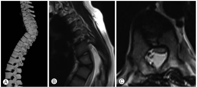
69.5°. The computerised tomography (CT) scan revealed a kyphoscoliotic spine with T7 hemivertebra (Fig. 1A). Her paraparesis had worsened and the neurologic examina-tion revealed some weakness in the ankle dorsilfexors of both the sides (left > right). Knee jerk was exaggerated on both the sides. Sensations were preserved in both the lower limbs. She had a left sided spastic gait and had difficulty in balancing while walking. The MRI revealed a flattened cord with signal changes over the apex of kyphotic defor-mity with T7 hemivertebra (Fig. 1B and 1C). In the wake of these developments, she was advised a deformity correction surgery along with anterior decompression to which the par-ents of the child agreed to.
A surgical protocol in the form of posterior correction of
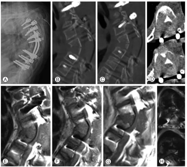
the kyphoscoliotic curve with instrumentation (monoaxial pedicle screw system), posterior decompression, posterior to anterior excision of T7 hemivertebra, spondylodesis and anterior interbody fusion was planned for her. After facetec-tomy from T3 to T10, posterior instrumentation was done from T3 to T10 sparing T7. The instrumentation was fol-lowed by posterior decompression by partial hemilaminec-tomy T6 and bilateral total laminechemilaminec-tomy T7. While carrying out the T7 hemivertebra excision, a trans cranial electrical motor evoke potential evaluation was carried out and it showed a near 100% drop in both the lower extremities (Fig. 2). Following the condition, the surgery was stopped, and a calculated dose of methyl prednisolone was administered intravenously and a wake up test was called for. Wake up test elicited weak (less than grade 5) movements of both the ankles and great toes. In the wake of a weakly positive wake up test, the surgery was not finished to its conclusion. The hemivertebra excision was discontinued and the deformity was fixed in situ with short rods (T5, T6, and T8) without further correction of the deformity or anterior interbody fu-sion (Fig. 3A).
Patient was shifted to intensive care unit, where the steroid therapy was continued besides routine supportive medication. On first postoperative day, CT scan revealed an incompletely decompressed T7 hemivertebra with its pos-terior cortex intact and kyphotic deformity persisting (Fig. 3B–3D). Thereafter, a gradual decline in the neurologic status was observed. On second postoperative day, power in all the muscle groups of right lower extremity was grade 4 B
A C
Fig. 1. Preoperaive computed tomography scan (A) showing 3D reconstrution of whole spine wherein kyphotic
deformity at T7 hemivertebra can be visualized. The T2 weighted magnetic resonance imaging (B, C) shows an overstretched flattened cord with signal changes over kyphotic deformity with T7 hemivertebra in sagittal and axial cut.
Fig. 2. The trans cranial electrical motor evoke potential
re-cordings while carrying out excision of hemivertebra reveal a significant drop to 6 µV on left side and to 7 µV on right side.
in the neurologic status. Three weeks later, the short rods were replaced with proper length rods (from T3-T10) and anterior column reconstruction and interbody fusion (T6-T8)
to this gradual neurologic deterioration. Direct mechanical trauma to the cord during the surgery could have triggered off the neuronal apoptotic changes, which we tried to
re-F A G B H C D E
Fig. 3. Immediate postoperative radiograph (A) shows a T3 to T10 instrumented spine with short rods with
in-complete excision of T7 hemivertebra. Postoperative computed tomography scan (B–D) on first postoperative day confirms a partially excised T7 hemivertebra with persisting kyphotic deformity. The magnetic resonance imaging (E–H) done on third postoperative day shows a tense flattened cord with signal changes over intact posterior cortex of T7 and a local hematoma posterior to the cord.
verse with a mega dose steroid therapy but the phenomenon of progressive damage to the neuronal tissue after the initial injury to the spinal cord could have resulted in the gradual deterioration on neurologic status and finally resulted in the paraplegia [3]. According to a study, it is suggested that severity of anterior angulation of spine is proportional to the pressure within the canal, which in turn may cause vascular insufficiency to the cord as it increases, thereby leading to an altered neurology [4]. Another experimental study sug-gests that kyphotic angle leads to flattening of the cord over the deformity thereby causing demyelination of anterior funiculus, neuronal loss and atrophy of anterior horn and decreased vascularity at the ventral side of compressed spi-nal cord [5]. Moreover, while attempting the deformity cor-rection and anterior decompression through single posterior approach, chances of injury to the neural structures increase due to over traction of the dura for better visualization of the structures [6]. In a review study, it was shown that ky-photic deformities compress the cord anteriorly and unless some confirmed posterior pathology exists; posterior canal exposure should be avoided as it renders the spine unstable and increases the kyphotic deformity [7]. A kyphotic spine should be decompressed anteriorly so that the tense flat-tened cord translates into the vertebral bodythereby achiev-ing decompression [8]. Compression to neurovascular struc-tures due to localized hematoma can be a possible reason for neurologic deterioration.
In this particular case, we believe that due to incomplete excision of the hemivertebra, the posterior cortex did not yield when the compression was applied to the screws
adjacent to the kyphotic apex. As a result, the correction hinge was shifted to the posterior cortex instead of it lying anteriorly. Moreover, the unyielding posterior cortex offered double hinges, as explained by our schematic representation (Fig. 5) and the compressive force caused the distraction at the adjacent vertebrae rather than collapsing the column. This distraction might have led to the stretching of the spinal cord thereby causing the neuronal damage and hence a dete-riorated neurology. The use of monoaxial screws aggravated the distraction, as the tulips were pulled towards the rod and thereby adding to the distraction. The postoperative restric-tion of patient being in supine posirestric-tion could have added on to the distraction as the fixation was over shorter segment and was not enough to hold the spine in a fixed position. Previously, it has been reported that mechanical stresses due to distraction, compression (more than acceptable limits) or translation etc may result in further neurologic damage [9]. Acute neurologic deterioration after kyphoscoliosis correc-tion surgery is a well-known phenomenon but gradual neu-rologic deterioration post kyphoscoliosis correction surgery though is a rare phenomenon. In a case report publication, Keyoung et al. [10] tried to evaluate the possible causes of delayed deterioration of neurology after thoracic kyphosis correction surgery and favoured the gradual vascular insult or a local hematoma as the possible causes of such outcome in their case. Our view about depressed neurology in this
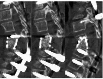
Fig. 4. The final surgery carried out three weeks later,
wherein short rods were removed and proper sized rods along with anterior interbody fusion T6-7-8 with titanium mesh cage and auto lamina bone graft were implanted.
Fig. 5. This drawing illustrates our postulate of cord
stretch-ing despite the cantilever beam fixation. In this case, the correction hinge, which ideally would have been at the anterior cortex of excised hemivertebra, was shifted to the posterior cortex. When compression force (C1, C2) was applied, the partially excised posterior cortex did not yield and offered two hinge points ‘a’ and ‘b’ and hence a distrac-tion (D1, D2) was observed at the adjacent vertebrae. This caused the stretching of the neural elements. Moreover, the use of monoaxial screws did not allow the translation of the screw tulips over the rod and hence the vertebral movement towards the rod added to the phenomenon of cord stretch-ing.
REFERENCES
1. Døssing KV, Christensen KS, Thomsen K, Bünger CE. Congenital kyphoscoliosis complicated by paraplegia. Ugeskr Laeger 1995;157:451-3.
2. McMaster MJ, Singh H. The surgical management of congenital kyphosis and kyphoscoliosis. Spine (Phila Pa 1976) 2001;26:2146-54.
3. Bramlett HM, Dietrich WD. Progressive damage after brain and spinal cord injury: pathomechanisms and treatment strategies. Prog Brain Res 2007;161:125-41. 4. Masini M, Maranhao V. Experimental determination of
the effect of progressive sharp-angle spinal deformity
cases. Spine (Phila Pa 1976) 1980;5:331-55.
8. Shenouda EF, Nelson IW, Nelson RJ. Anterior transver-tebral transposition of the spinal cord for the relief of paraplegia associated with congenital cervicothoracic kyphoscoliosis. Technical note. J Neurosurg Spine 2006;5:374-9.
9. Chang KW, Cheng CW, Chen HC, Chen TC. Correc-tion hinge in the compromised cord for severe and rigid angular kyphosis with neurologic deficits. Spine (Phila Pa 1976) 2009;34:1040-5.
10. Keyoung HM, Kanter AS, Mummaneni PV. Delayed-onset neurological deficit following correction of se-vere thoracic kyphotic deformity. J Neurosurg Spine 2008;8:74-9.