비소세포성 폐암에서 인슐린 양 성장
인자 결합 단백질-3 발현 조절 기전
연세대학교 대학원
의 과 학 과
비소세포성 폐암에서 인슐린 양 성장
인자 결합 단백질-3 발현 조절 기전
연세대학교 대학원
의 과 학 과
비소세포성 폐암에서 인슐린 양 성장
인자 결합 단백질-3 발현 조절 기전
지도 교수 김세규
이 논문을 박사 학위 논문으로 제출함
2004 년 6 월
일
연세대학교 대학원
의 과 학 과
장 윤 수
장윤수의 박사 학위논문을 인준함
심사위원
인
심사위원
인
심사위원
인
심사위원
인
심사위원
인
연세대학교 대학원
2004 년 6 월
일
감사의 글
논문이 완성되기까지 오랜 시간 동안 세심한 정성으로 이끌어 주시고 지도와 용기를 주신 김세규 교수님께 감사들 드립니다. 그리고 바쁘신 중에도 부족함을 일깨워 주시고, 학문적 충고와 자세한 자문을 해주신 여러 교수님께 깊은 감사를 드립니다. 장준 호흡기내과 과장님을 비롯하여 어려웠던 과정을 중도에 포기하지 않고 오늘날의 제가 있도록 이끌어 주신 호흡기 내과의 여러 교수님들께 감사 드립니다. 또한 실험 연구에 문외한이었던 저에게 실험의 논리적인 면과 세심한 과정을 일깨워 주시고 많이 위로해 주셨던 정현철 교수님과 다른 암전인 연구 센터 교수님께 깊이 감사드립니다. 그리고 이제는 연구실을 떠나고 없지만 실험적인 조언을 아끼지 않았던 임상 연구센터의 선생님들과 학생들에게도 고맙다는 말을 전합니다. 바쁘신 일과 중에서도 매달 시간을 내어주시어 지도해 주셨던 홍완기 교수님, 불안정한 상황에서도 항상 관심을 갖고서 조언을 아끼지 않았던 Dr. Mao, 그리고 세세한 부분까지 기술적 지도를 해주신 이호영 박사, 그리고 같은 실험실의 Luo Wang, Weiguo Wu, Shihua Sun 의 도움에 깊이 감사 드립니다. 또 타국에서 방황하던 저에게 정신적으로 많은 도움을 주신 문철소 교수님께도 감사 드립니다. 부족함이 많은 저이지만 항상 이해해 주시고 큰 사랑으로 보살펴 주신 어머님께 공경과 사랑을 드리며 이 논문을 드립니다. 어려웠던 과정을 곁에서 같이한 소중한 처와 딸의 사랑도 기억하겠습니다. 이 산물이 저의 무지함과 편견의 증거가 되지 않기를 바라며, 지식의 혼탁함만을 더했다는 평가를 받지 않았으면 좋겠습니다. 논문을 준비하는 과정에서 겪었던 수많은 시행착오, 수시로 엄습하였던 절망과 유혹 사이에서 갈등, 능력에 대한 회의감 등을 여러분의 격려와 함께 앞으로의 새로운 세상으로 나아가는 소중한 자산으로 생각하고 간직하며, 항상 겸손하고 성실한 마음 자세로 자신을 이끌어 나가겠습니다. 장윤수 드림차 례
국문요약․․․․․․․․․․․․․․․․․․․․․․․․․․․․․․․․․․․․․․․․․․․․․․․․․․․․․․․․․․․․․․․․․․․ 1
Ⅰ. 서론․․․․․․․․․․․․․․․․․․․․․․․․․․․․․․․․․․․․․․․․․․․․․․․․․․․․․․․․․․․․․․․․․․․ 3
II. 재료 및 방법 ․․․․․․․․․․․․․․․․․․․․․․․․․․․․․․․․․․․․․․․․․․․․․․․․․․․․․․․․․․ 6 1. NSCLC cell lines and culture .․․․․․․․․․․․․․․․․․․․․․․․․․․․․․․․․․․․․․․․․ 6 2. Western blot and northern blot analysis. ․․․․․․․․․․․․․․․․․․․․․․․․․․․․․ 6 3. Plasmid preparation and in vitro DNA methylation ․․․․․․․․․․․․․․․․․․․․․ 8 4. Luciferase assay and β-Gal assay․․․․․․․․․․․․․․․․․․․․․․․․․․․․․․..․․․․․․ 9 5. Bisulfite-PCR for restriction analysis and sequencing ․․․․․․․․․..․․․․․․ 9 6. Methylation-specific PCR ․․․․․․․․․․․․․․․․․․․․․․․․․․․․․․․․․․․․․․․․․․․․ 10 7. Electrophoretic mobility-shift assay ․․․․․․․․․․․․․․․․․․․․․․․․․․․․․․․․ 11 8. Chromatin immunoprecipitation analysis ․․․․․․․․․․․․․․․․․․․․․․․․․․․․․․ 12
III. 결과 ․․․․․․․․․․․․․․․․․․․․․․․․․․․․․․․․․․․․․․․․․․․․.․․․․․․․․․․․․․․․․․․․ 14 1. IGFBP-3 expression in NSCLC cell lines ․․․․․․․․․․․․․․․․․․․․․․․․․․․․․․ 14 2. Expression of IGFBP-3 reduced in NSCLC cell lines and restored by
5′-aza-dC ․․․․․․․․․․․․․․․․․․․․․․․․․․․․․․․․․․․․․․․․․․․․․․․․․․․․․․․․․․․․․․․․․․․․ 15 3. Sp-1/Sp-3 binding site as an important regulatory element of IGFBP-3 promoter ․․․․․․․․․․․․․․․․․․․․․․․․․․․․․․․․․․․․․․․․․․․․․․․․․․․․․․․․․․․․․․․․․․16 4. Detection of hypermethylation of the IGFBP-3 promoter in NSCLC cell lines ․․․․․․․․․․․․․․․․․․․․․․․․․․․․․․․․․․․․․․․․․․․․․․․․․․․․․․․․․․․․․․․․․․․․․18
5. Transcription factor Sp1-/Sp-3 binding altered according to methylation status .․․․․․․․․․․․․․․․․․․․․․․․․․․․․․․․․․․․․․․․․․․․․․․․․․․․․․․․․․․․․․․․․․․․20
6. Sp-1/Sp-3 consensus sequence binding factors in H1299 cells and effect of demethylation agent 5′-aza-dC ․․․․․․․․․․․․․․․․․․․․․․․․․․․․․․․․․․․․․․․․․․22
7. Luciferase activity decreased in SssI methylase catalyzed IGFBP-3 promoter, and coexpression of human MeCP2 cDNA provides additional repressesion ․․․․․․․․․․․․․․․․․․․․․․․․․․․․․․․․․․․․․․․․․․․․․․․․․․․․․․․․․․․․․․23
IV. 고찰 ․․․․․․․․․․․․․․․․․․․․․․․․․․․․․․․․․․․․․․․․․․․․․․․․․․․․․․․․․․․․․․․ 27
V. 결론 .․․․․․․․․․․․․․․․․․․․․․․․․․․․․․․․․․․․․․․․․․․․․․․․․․․․․․․․․․․․․․․․ 31
참고문헌 ․․․․․․․․․․․․․․․․․․․․․․․․․․․․․․․․․․․․․․․․․․․․․․․․․․․․․․․․․․․․․․․ 33
List of figures
Fig. 1. Analysis of the IGFBP-3 promoter. ․․․․․․․․․․․․․․․․․․․․․․․․․․․․․․․․․ 7
Fig. 2. IGFBP-3 expression in NSCLC cell lines ․․․․․․․․․․․․․․․․․․․․․․․․․․․ 14
Fig. 3. Effect of 5′-aza-dC on IGFBP-3 expression ․․․․․․․․․․․․․․․․․․․.․․․․ 15
Fig. 4. IGFBP-3 promoter activity in various NSCLC cell lines ․․․․․․․․․․․ 17
Fig. 5. Bisulfite-PCR/restriction, sequencing, and MSP in various NSCLC cell lines ․․․․․․․․․․․․․․․․․․․․․․․․․․․․․․․․․․․․․․․․․․․․․․․․․․․․․․․․․․․․․․․․․․․․ 19
Fig. 6. Binding of transcription factors Sp-1/Sp-3 alters according to methylation status․․․․․․․․․․․․․․․․․․․․․․․․․․․․․․․․․․․․․․․․․․․․․․․․․․․․․․․․ 21
Fig. 7. Sp-1/Sp-3 binding status of Sp-1, MeCP2, and HDAC and the effect of demethylating agent 5′-aza-dC .․․․․․․․․․․․․․․․․․․․․․․․․․․․․․․․․․․․․․․․․․․․ 23
Fig. 8. Expression level of MeCP2 in NSCLC cell lines and effect on in vitro methylated pGL2-∆1708 luciferase report plasmid ․․․․․․․․․․․․․․․․․․․․․․․․․․ 25
국문 요약
비소세포성 폐암에서 인슐린 양 성장 인자 결합 단백질-3 발현 조절 기전
인슐린 양 성장 인자 (IGF) 결합 단백질-3 (IGFBP-3)은 IGF와 결합하여 IGF의 세포분열촉진 및 항세포고사 기전을 억제할 뿐 아니라 IGF와는 독립적으로 세포고사를 유도함으로써 비소세포성 폐암 세포주의 성장을 억제한다. 이
연구에서는 비소세포성 폐암 세포주를 이용하여 IGFBP-3 promoter 내 CpG islands의 hypermethylation이 IGFBP-3 유전자의 발현에 어떠한 역할을 하는가와 유전자 발현을 억제하는 기전을 연구하였다. 15종의 비소세포성 폐암 세포주 중 7종 (46.7%)에서 IGFBP-3 promoter 내 CpG islands의 hypermethylation이 관찰되었다. 비소세포성 폐암 세포주에서 promoter 내 CpG islands의 methylation 상태는 IGFBP-3 유전자의 단백질 및 mRNA 발현 양상과 잘 일치하였으며, IGFBP-3의 발현이 억제되었던 비소세포성 폐암 세포주들 중 일부에서 demethylating 약제인 5′-aza-2-deoxycytidine (5′-aza-dC) 처리 후 그 발현이 회복되었다. IGFBP-3의 promoter 활성도에 중요한 역할을 하는 Sp-1/Sp-3 결합 요소 부위의 CpG islands는 IGFBP-3 단백질 발현이 억제된 비소세포성 폐암 세포주에서 hypermethylation 되어 있었으며, 이는 Sp-1 전사 인자의 결합을 억제하였다. ChIP assay에서 IGFBP-3
promoter내 CpG islands의 hypermethylation은 Sp-1/Sp-3 결합 요소에 Sp-1, methyl-CpG binding protein-2 (MeCP2), 그리고 histone deacetylase (HDAC)의 결합에 영향을 주었으며, 이는 5′-aza-dC 처리에 의하여 역전되었다. Sp-1/Sp-3 결합 요소를 포함하고 있는 IGFBP-3 promoter의 in vitro methylation은 promoter 활성도를 현저히 감소시켰으며 이는 MeCP2를 동시에 발현시켰을 때 더욱 억제되며 5′-aza-dC 처리 시 회복되었다. 이러한 결과들은 비소세포성 폐암에서 IGFBP-3 promoter의 hypermethylation이 IGFBP-3 발현을 억제하는 하나의 기전이며, HDAC과 MeCP2의 모집이 유도됨으로서 Sp-I의 결합이 억제되고 나아가 IGFBP-3 유전자의 발현이 억제됨을 보이는 것이다. 마지막으로 임상 시험중인 demethylation 약제로서 IGFBP-3의 발현을 유도할 수 있다는 것은 IGFBP-3의 세포의 성장과 고사에 미치는 중요한 역할을 고려해 볼 때 이들이 비소세포성 폐암의 치료에 사용될 수 있음을 시사하는 것으로 생각된다. ---핵심되는 말: 인슐린양 성장 인자, 인슐린 양 성장 인자 결합 단백질-3, CpG
비소세포성 폐암에서 인슐린 양 성장 인자 결합 단백질-3 발현 조절 기전
지도 교수 김세규 연세대학교 대학원 의과학과 장 윤 수 Ⅰ. 서론 폐암은 수술적 절제, 방사선 치료 및 항암 화학약물요법을 포함하여 사망률을 줄이기 위한 다양한 노력에도 불구하고 국내 및 미국을 비롯한 서구 남녀의 암 사망률 1위를 차지하고 있는 질환이다1. 5년-생존율은 1970년대의 7%에서 2000년에 14%로 향상되었으나 만족할 만한 수준에 도달하지 못하고 있으며, 또한 매년 1,000,000 예의 새로운 폐암 환자가 전세계적으로 생겨나고 있는 실정이다2. 국내에서도 약 5년 전부터는 위암 다음의 2 위의 발생 빈도를 보이고 있으나 치료 성적은 크게 호전되지 않은 상태이다. 이러한 암울한 통계적 수치는 폐암 발생 기전의 규명, 암 예방 및 조기 진단 방법의 개발이 시급하게 필요함을 시사하는 것이다.인슐린 양 성장 인자들 (insulin like growth factors; IGFs)은 사람의
세포들의 성장에 중요한 세포분열촉진 인자로서 세포고사를 억제할 뿐 아니라
정상 세포의 신생물성 변화와 전이에도 관여하는 것으로 알려져 있다3-5. 또한
세포분열을 유도하는 다른 성장 인자들 또는 스테로이드계 약물과는 상보적으로
작용하며 암의 성장을 억제하는 물질들에 대해서는 길항적으로 작용한다.
인슐린 양 성장 인자 결합 단백질-3 (IGF binding protein-3; IGFBP-3)은
IGFs가 세포 외부의 환경에서 수용체와 결합하는 것을 방해함으로써 이들의
세포분열촉진 및 세포고사를 억제하는 생물학적 활성에 길항하는 IGF-I 의존적
기능뿐 아니라5,6, IGFBP-3의 세포 표면 수용체가 관여될 것으로 생각되는 IGF-I
비의존적인 항증식성/세포 고사 촉진 기능을 가지고 있다7-9. 또한 IGFBP-3는
transforming growth factor-β, retinoic acid, tumor necrosis factor-α,
p53, anti-estrogens, flavonoid antioxidant silibinin, paclitaxel, 그리고
histone deacetylase (HDAC) 억제물인 trichostatin A와 같은 주요 물질들의
성장 억제 신호를 매개하는 것으로 밝혀져 있다9-16. 체내에 순환하는 IGF-I과 IGFBP-3의 혈장 및 혈청 농도는 정상인 사이에도 많은 차이를 보이고 있으며 대규모의 전향적 역학 연구 결과 IGF-I과 IGFBP-3은 개체간의 차이가 있어서 높은 IGF-I과 낮은 IGFBP-3의 농도가 폐암, 유방암, 대장 직장암, 전립선암 등을 비롯한 몇몇 주요 암 발생 위험과도 관련이 있는 것으로 알려지면서17-20 정상 범주의 개체간 농도 차이를 일으키는 기전에 대하여 관심이 증가하고
있다21. 따라서 IGF-I의 생물학적 기능을 조절하는 IGFBP-3의 발현 조절 기전을 밝히는 것이 중요하며, 비소세포성 폐암의 발생 및 성장 기전을 규명하고 나아가 폐암 치료에 응용될 수 있는 가를 확인할 수 있는 기초가 될 수 있다. 이 연구에서는 비소세포성 폐암에서 세포의 성장과 고사에 관여하고 IGF-I의 생물학적 활성을 조절하는 IGFBP-3의 발현 양상과 그 조절 기전 중의 하나인 상유전자적 변화를 비소세포성 폐암 세포주를 이용하여 관찰하였으며 나아가 상유전자적 변화를 통한 유전자 발현 억제 기전을 연구하였다.
Ⅱ. 재료 및 방법
1. NSCLC cell lines and culture. NCI-H1299, H661, H596, A549, H460, H441,
H358, H322, H1944, H292, H226B, SK-MES-1, Calu-6, Calu-1, H157 등 사람의 비소
세포성 폐암 세포주는 American Type Tissue Collection (Manassas, VA, USA)으로
부터 구입하였다. 이들은 10% fetal calf serum (FCS) (Life Technologies, Inc.,
Gaithersburg, MD, USA)이 첨가된 RPMI-1640 medium을 사용하여 배양하였다.
2. Western blot and northern blot analysis. IGFBP-3의 감지를 위해 30 µg의 단
백질을 sodium dodecyl sulfate (SDS)-polyacrylamide gel 상에서 전기 영동 분획
하였다. Polyvinylidene difluoride membrane (Bio-Rad Laboratories, Inc.,
Hercules, CA, USA) 으로 분획된 단백질을 옮긴 뒤 5% 탈지유를 포함하고 있는
TBS-T (Tris-buffered saline-Tween (20 mM Tris-HCl, pH 7.6; 150 mM NaCl; 0.1%
Tween-20))로 비특이 반응을 차단하였다. Membrane들은 goat polyclonal
anti-IGFBP-3 (DSL, Webster, TX, USA) 로 4°C 에서 16시간 동안 incubation한 다음
horseradish peroxidase-conjugation된 2차 항체 (Santa Cruz Biotechnology,
Santacruz, CA, USA)로 상온에서 1 시간 동안 반응시켰다. 면역 반응 단백질은
enhanced chemiluminescence (ECL) kit와 Hyperfilm ECL (Amersham Pharmacia
Biotech Inc., Piscataway, NJ, USA)을 이용하여 감지하였다. RNA의 분리는 약제
첨가하여 용해시킨 뒤 타 문헌에 기술되어 있는 대로 RNA를 분리하였다22. 20 ㎍의
RNA를 전기영동 한 후 Zeta-Probe membrane (Bio-Rad Laboratories) 옮긴 다음,
[α-32P] dCTP로 표지된 IGFBP-3 또는 glyceraldehyde-3-phosphate dehydrogenase
(GAPDH)의 cDNA 를 함유하고 있는 교잡 용액과 함께 처리하였다. 탐침은
Prime-ItR II Random Primer Kit (Stratagene, La Jolla, CA, USA)을 이용하여 표지한 뒤
MicroSpinTM S-300 HR Columns (Amersham Pharmacia Biotech Inc.)을 이용하여 정제
하였다. Membrane은 2회 세척 후 Hyperfilm ECL을 이용하여 감지하였다.
A
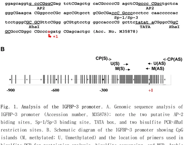
ggagcaggtg ccCGggCGag tctCGagctg caCGccccCG agctCGgccc CGgctgctca AP2 AP2
gggCGaagca CGggcccCGc agcCGtgcct gCGcCGaccC Gcccccctcc caacccccac Sp-1/Sp-3
tcctgggCGC GCGttcCGgg gCGtgtcctg ggccacccCG gcttctatat aCGggcCGgC
HhaI TATA HhaI
GCGccCGggc CGcccagatg CGagcactgc (Acc. No. M35878) +1
B
U(AS) M(AS) CP(S) U(S) M(S) +1 CP(AS) -900 -600 -300Fig. 1. Analysis of the IGFBP-3 promoter. A. Genomic sequence analysis of IGFBP-3 promoter (Accession number, M35878); note the two putative AP-2 biding sites, Sp-1/Sp-3 binding site, TATA box, and two bisulfite PCR-HhaI restriction sites. B. Schematic diagram of the IGFBP-3 promoter showing CpG islands (M, methylated; U, Unmethylated) and the location of primers used in bisulfite-PCR for restriction analysis, bisulfite sequencing, and MSP. Arabic
numbers indicate locations of nucleotides relative to the mRNA cap site. Vertical lines, position of CpG dinucleotides.
3. Plasmid preparation and in vitro DNA methylation. 1.9-kb IGFBP-3
promoter 가 luciferase reporter vector 의 일종인 pGL2-basic (Promega Corp.,
Madison, WI, USA)에 삽입된 pGL2-IGFBP-3 및 이의 삭제 변종인 pGL2-∆1600, ∆1675,
-∆1708, -∆1755, 및 -∆1795 는 Oregon Health Sciences University (Portland, OR,
USA) 의 오영만 교수로부터 제공 받았다 23. IGFBP-3 promoter 의 염기 서열은 Fig.
1A 와 같으며 이들의 성격을 간략하면 pGL2-∆1600 는 10 개의 p53 consensus 결합
부위와 2 개의 consensus AP-2 결합 부위를 포함하고 있으며, pGL2-∆1675 는 5 개의
p53 결합 부위와 하나의 consensus AP-2 결합 부위를 가지고 있다. 이 두 AP-2
결합 부위는 pGL2-∆1708 에서는 모두 삭제되어 있으며, pGL2-∆1755 에서는
Sp-1/Sp-3 결합 요소가 삭제되어 있다. MeCP2 (Accession No. AF158180)의 전체를 포함하는
cDNA 는 사람의 정상 폐 조직에서 얻은 cDNA library (Clontech, Palo Alto, CA,
USA)로부터 다음의 시발체를 이용 PCR 로 증폭하였다; sense primer
5′-GGGATCCATGGTAGCTGGGATG-3′, antisense primer 5′-GGAATTCAGCTAACTCTCTC-3′.
증폭된 PCR 산물은 TA cloning kit (Invitrogen, Carlsbad, CA, USA)를 이용하여
pCR-IIR plasmid 에 삽입, T7 및 M13R 시발체로 염기서열을 확인한 뒤, EcoRI
(Roche Molecular Biochemicals, Basel, Switzerland)로 절단하고 DNA polymerase
pCMX-mammalian expression vector 에 삽입하였다. In vitro DNA methylation 은
pGL2-∆1708 plasmid 를 5 mM S-adenosylmethionine (New England Biolabs, Beverly, MA,
USA)을 포함한 반응액 하에서 (methylation) 또는 포함하지 않은 반응액에서
(unmethylation) SssI methylase (New England Biolab)로 처리한 뒤 정제하여
얻었다. 이러한 plasmid 의 methylation 여부는 HhaI 제한 효소 (New England
Biolabs)로 처리한 뒤 전기영동 하여 확인하였다.
4. Luciferase assay and β-Gal assay. NCI-H1299 세포들을 포함한 비소세포성
폐암 세포주를 24 well plate 에 5X10⁴ cells/well 로 분주한 뒤 70% confluency 에
도달하였을 때 200 ng 의 reporter plasmid DNA 와 30 ng 의 CMV-β gal control
vector (GibcoBRL, Gaithersburg, MD, USA)를 serum-free 상태의 조건에서
lipofectamine (GibcoBRL)을 이용하여 transfection 을 시행하였다. 6 시간 뒤 10%
FCS 의 RPMI-1640 배지로 교환한 뒤 및 48 시간 더 배양 후 luciferase assay kit
(Promega)를 이용하여 luciferase 활성도를 측정하였다. 측정값은 동일 회사의
β-gal assay system 을 이용하여 측정된 CMV-β β-gal control vector 의 활성도로서
보정하였다.
5. Bisulfite-PCR for restriction analysis and sequencing. Herman 등에 의해
5′-methylation된 cytosine을 확인할 수 있다24. PCR에 사용된 시발체의 선택은
luciferase assay에서 확인된 promoter 활성도에 중요한 역할을 하는 부분인
Sp-1/Sp-3 결합 요소 부위로 IGFBP-3 promoter상의 위치는 Fig. 1B와 같다. Sense
primer 5′-TCGGGTATATTTTGGTTTTTGTAG-3′, antisense primer
5′-AAACATATAAAATCCAAACAAAAA-3′. 매 반응 시 SssI methylase로서 완전히
methylation 된 DNA를 positive control로 사용하였으며 annealing 온도는 60℃로
하였다. 25 ㎕의 PCR 산물 중 절반은 HhaI 제한 효소를 이용하여 절단한 뒤 2.5%
acrylamide gel에서 전기 영동 후 UV 하에서 확인하였으며 나머지 12.5 ㎕의 PCR
산물은 효소 반응 없이 2.5% acrylamide gel에서 전기 영동 하여 확인 뒤 QIAquick
Gel Extraction Kit (Qiagen, Valencia, CA, USA) 를 이용 분리, 정제하여 염기 서
열 확인에 사용하였다.
6. Methylation-specific PCR. 1 ㎍의 DNA를 동량의 salmon sperm DNA (Life
Technologies, Inc.)와 혼합한 뒤 Herman 등이 기술한 대로 DNA 화학 변성을 시행
하였다24. 간략하면 DNA를 2 M NaOH로 denature 한 뒤 10 mM hydroquinone과 3 M
sodium bisulfite (Sigma, St. Louis, MO, USA)로 처리하였다. 다음날 Wizard SV
Plus kit column (Promega)으로 분리 정제한 뒤 3 M NaOH로 처리하고 3배 부피의
100% ethanol과 1배 부피의 10 M NH4OAc로 상온에서 침전시켰다. 침전된 DNA는 70%
하여 증폭하였다. Methylation된 DNA의 증폭을 위해서는 Sense
5′-CGAAGTACGGGTTTCGTAGTCG-3′, 및 antisense 5′-CGACCCGAACGCGCCGACC-3′를 사용하였고
unmethylation된 DNA의 증폭을 위해서는 Sense 5′-TTGGTTGTTTAGGGTGAAGTATGGGT-3′
및 antisense 5′-CACCCAACCACAATACTCACATC-3′를 사용하였다. Bisulfite-PCR/제한 효
소 분석 [CP(S) 그리고 CP(AS)] 및 MSP [M(S), M(AS), U(S), 그리고 U(AS)]에 사용
된 시발체 조합은 Fig. 1B에 나와 있다. 매 반응시에는 DNA를 첨가하지 않은
negative control과 정상인의 림프구에서 얻은 DNA를 SssI methylase 처리 후
bisulfite 화학 변성을 시행한 positive control을 포함시켰으며 반응물은
ethidium bromide로 염색된 2.5% agarose gel에서 전기 영동 후 감지하였다.
7. Electrophoretic mobility-shift assay. 아래와 같이 methylation 되어 있지
않거나 (W.T.), oligomer 내 1개의 CpG island가 methylation (P.M.), 또는 3개의
CpG island가 모두 methylation 되어 있는 (C.M.) 3 쌍의 double-stranded 25-mer
oligonucleotide (Operon Technology Inc. Alameda, CA, USA)를 이용하였으며 그
염기서열은 다음과 같다. W.T.는 5′- TGCGCCGACCCGCCCCCCTCCCAAC-3′및 그 상보적
순서를 가진 oligomer이며, P.M. 및 C.M.은 각각 5′- TGCGCCGACCCM
GCCCCCCTCCCAAC-3′ 와 5′- TGCMGCCMGACCCMGCCCCCCTCCCAAC-3′ 및 그 상보적 순서를 가진 oligomer를
사용하였다. 이러한 oligomer는 T4 polynucleotide kinase (Promega)를 이용하여
[d(A-T)] (Roche Molecular Biochemicals); 20 mM HEPES (pH 7.6); 1 mM EDTA; 1
mM dithiothreitol; 0.2% Tween-20R; 30 mM KCl 을 포함한 20 ㎕의 반응액 하에서
표지된 oligomer와 함께 상온에서 15 분간 반응시켰다. 경쟁 반응에는 wild type
또는 mutant type Sp-1 consensus oligomer (Promega)를 추가하였다. 각 반응물들
은 0.5 X TBE buffer system을 이용하여 전기영동 한 뒤 gels을 건조하여
Hyperfilm ECL에 감광시켜 감지하였다.
8. Chromatin immunoprecipitation analysis. ChIP 분석은 Chen 등이 기술한 방
법을 이용하여 시행하였다25. 간략하면 비소세포성 폐암 세포는 2% FCS을 포함하는
RPMI-1640 배지에서 5′-aza-dC로 5일간 처리하였다. 이후 세포들은 37 ℃에서 10
분간 formaldehyde (1% final concentration)로 처리한 뒤 단백질 분해 효소 억제
제를 포함하고 있는 완충액 (25 mM Tris, pH 8.1; 10 mM EDTA; 1% SDS)으로 용해하
였다. 동일한 수의 세포들을 anti-Sp-1 (Santa Cruz Biotechnology, Santa Cruz,
CA, USA), anti-MeCP2, 또는 anti-HDAC (Upstate Biotechnology Inc.
Charlottesville, VA, USA) 항체로 처리하고 salmon sperm DNA/Protein A agarose
slurry (Upstate Biotechnology Inc.)로 면역 침전을 시행하였다. 단백질과 결합
하여 침전된 DNA는 LiCl wash buffer 와 Tris-EDTA로 세척한 뒤 elution buffer
(1% SDS, 0.1 N NaHCO3)를 추가하여 elution을 시행하였다. Cross−links는 20 mM
ethanol 침전법으로 DNA를 추출하고서 다음과 같은 시발체를 사용하여 증폭 후
[α-32P] dCTP로 표지된 IGFBP-3 promoter 탐침으로 southern blotting을 시행하여
분석하였다. (Sense: 5′–CGTGAGCACGAGGAGCAGGTG-3′: antisense:
5′-CAGGAGTGGGGGTTGGGAG-3′). 전체 세포 단백질−DNA 복합체 중 면역 침강된 양의 2%는
Ⅲ. 결과
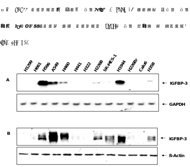
1. IGFBP-3 expression in NSCLC cell lines. 비소세포성 폐암세포주에서 IGFBP-3
발현 정도를 관찰하기 위해 IGFBP-3 항체와 α-32P로 표지된 IGFBP-3 탐침을 이용하여 Western 및 Northern blot 을 시행하였다. IGFBP-3의 mRNA는 NCI-H1299, H661, H441, H322, H226Br, 그리고 Calu-6 세포주에서는 관찰되지 않았으며 H226B, SK-MES-1 및 H358 세포주에서는 약하게 관찰되었다. 반면 H596, A549, H460, 그리고 H1944 세포주에서는 강하게 발현되었다. IGFBP-3 유전자의 단백질 발현 정도는 사용된 비소세포성 폐암 세포주에서 mRNA의 발현 정도와 잘 일치하였다 (Fig. 2A, B). IGFBP-3 ß-Actin B H1 299 H6 61 H59 6 A5 49 H3 22 H4 60 H4 41 H2 26 B H2 26 Br Ca lu6 H3 58 SK -M ES -1 H1 944 IGFBP-3 GAPDH A
Fig. 2. IGFBP-3 expression in NSCLC cell lines. The expression of IGFBP-3 in a panel of NSCLC cell lines was examined by A, Northern blot analysis
using full-length IGFBP-3, or GAPDH cDNA probe as a control; and by B, Western analysis using anti-human IGFBP-3 antibody, with expression of β-actin examined as a loading control.
2. Expression of IGFBP-3 Reduced in NSCLC cell lines and restored by 5′-aza-dC.
IGFBP-3의 발현이 관찰되지 않는 비소세포성 폐암 세포주들을 demethylating 약제인 5′
-aza-dC 처리 후 Northern blot을 시행하여 IGFBP-3의 발현 양상의 변화를 관찰하였다.
5′-aza-dC를 1 µM 또는 그 이상을 처리한 경우 대부분 IGFBP-3의 발현이 복원되었다 (Fig. 3A, B).
-
0.1 1 5 (µM 5’-aza-dc) IGFBP-3 GAPDH IGFBP-3 GAPDH A BFig. 3. Effect of 5′-aza-dC on IGFBP-3 expression. Effect of 5′-aza-dC on IGFBP-3 expression was studied by Northern blot analysis in H1299 NSCLC cells and H226B treated with 5′-aza-dc at concentrations of 0, 0.1, 1, and 5 µM for
5 days in RPMI 1640 medium supplemented with 2% FCS. The expression of GAPDH was analyzed as a control.
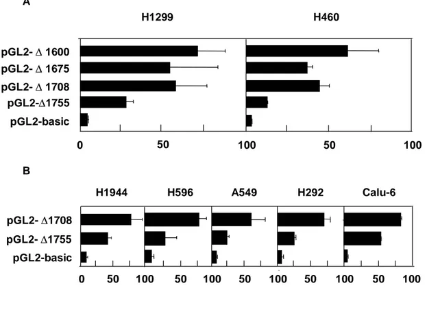
3. Sp-1/Sp-3 binding site as an important regulatory element of IGFBP-3 promoter. IGFBP-3 promoter의 활성도를 조절하는 주요 전사 인자 결합 부위를 규명하
기 위하여 pGL2-∆1600, -∆1675 -∆1708, 및 -∆1755 등의 IGFBP-3 promoter의 절단 변
이를 H1299, H460, H1944, H596, A549, H292, 및 Calu-6 세포주에 임시 transfection 시
킨 후 48 시간 뒤 luciferase 활성도를 측정하였다. H1299 및 H460 세포주에서
pGL2-∆1675 또는 -∆1708을 transfection 시킨 후 측정한 luciferase 활성도는 pGL2-∆1600을
이용하여 얻은 값과 비교 시 같거나 약간 낮았다. 반면 pGL2-∆1755를 transfection 시킨
후 얻은 luciferase 활성도는 pGL2-∆1600를 transfection 시켜 얻은 값보다 67~75% 정
pGL2-basic pGL2-∆ 1600 pGL2-∆ 1675 pGL2-∆ 1708 pGL2-∆1755 H1299 H460 A H1944 H596 A549 H292 Calu-6 pGL2-basic pGL2-∆1708 pGL2-∆1755 50 0 50 100 50 50 100 B 0 100 50 100 50 100 100 100 50
Fig. 4. IGFBP-3 promoter activity in various NSCLC cell lines. A. H1299 cells, which have methylated IGFBP-3 promoter, and H460 cells, which have unmethylated IGFBP-3 promoter, were transiently transfected with pGL2-basic, ∆1600, -∆1675 -∆1708, or -∆1755 luciferase reporter plasmids. B. H1944, H596, and A549 cells, which have unmethylated IGFBP-3 promoter, and H292 and Calu-6 cells, which have methylated IGFBP-3 promoter, were transiently transfected with pGL2-basic, -∆1708, and -∆1755 luciferase reporter plasmids. Luciferase activity was measured following incubation for 48 h. There was no difference in luciferase activity according to endogenous IGFBP-3 promoter methylation status. Data are shown as means (±S.D.) values of six different wells relative to cells transfected with pGL2-basic vector. The luciferase values were measured for at least three separate experiments.
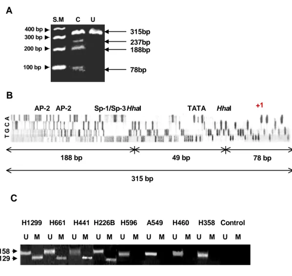
4. Detection of Hypermethylation of the IGFBP-3 Promoter in NSCLC Cell Lines. H1299, H661, H441, H322, H226Br, H226B, 및 Calu-6 세포주와 같이 낮은 IGFBP-3 발
현을 보이는 비소세포성 폐암 세포에서 bisulfite-PCR 및 HhaI을 제한 효소 절단법은
mRNA cap site로부터 –66 및 –16 번째의 cytosine이 methylation 되어 있을 때 관찰되는 237 bp, 188 bp 또는 78 bp 크기의 2개 이상의 절단 bands를 보였다. 188 bp 및 78 bp
크기의 bands의 경우 mRNA cap site로부터 –66 및 –16 번째 cytosine이 methylation 된 경우 관찰될 수 있으며 188 bp 및 315 bp 크기의 bands 는 각각 부분적으로 절단되었거나
절단되지 않은 경우 관찰될 수 있었다. 49 bp의 bands는 절단된 조각이 작아 시발체 및
시발체 이분자체와 구분되지 않아 확인이 어려웠다 (Fig. 5A). 이러한 부위의 methylation
상태를 확인하기 위하여 염기서열 분석을 H1299, Calu-6, 그리고 H661 세포주에서 얻어진
bisulfite-PCR 산물을 이용하여 시행하였다 (Fig. 5B). 2개의 putative AP2 결합 위치 (– 183 및 –145 번째 cytosine), Sp-1/Sp-3 결합 위치 (–96 번째 cytosine), 그리고 2개의 HhaI 절단 위치를 포함하여 promoter 활성도에 주요 역할을 하는 부위 내 대부분의 CpG
islands는 methylation되어 있었다. IGFBP-3 promoter의 methylation 상태를 다량의 검체
에서 확인할 수 있도록 MSP를 시행하였다. 129 bp 및 158 bp 크기의 PCR 산물은
methylation 및 unmethylation된 DNA가 증폭되었을 때 각각 관찰되었다. IGFBP-3 mRNA
transcripts가 관찰되지 않거나 매우 약하게 관찰되었던 H1299, H661, H441, H322, 및
B
T G C A +1 49 bp 188 bp 78 bp 315 bpAP-2 AP-2 Sp-1/Sp-3HhaI TATA HhaI
A
315bp 237bp 188bp 78bp 400 bp 300 bp 200 bp 100 bp S.M C UC
U M U M U M U M U M U M U M U M U M H1299 H661 H441 H226B H596 A549 H460 H358 Control 129 158Fig. 5. Bisulfite-PCR/restriction, sequencing, and MSP in various NSCLC cell lines. A. Bisulfite-PCR for HhaI restriction analysis of H1299 cells. When the methylated DNA was modified by bisulfite, two HhaI restriction sites were generated in the middle of the 315 bp PCR product. SM = DNA size marker; C = DNA digested with HhaI; U = undigested DNA. B. Sequence analysis of bisulfite-treated genomic DNA from H1299 cells. The PCR products were generated with outer primers CP (S) and CP (AS) and then sequenced with inner primer M (AS) that is specific for hypermethylated sequence. All the cytosines in the CpG islands remained as cytosine. C. MSP analysis of IGFBP-3
promoter methylation status in NSCLC cell lines. U = PCR products using primer set specific for unmethylated promoter; M = PCR products using primer set specific for hypermethylated promoter. SM = DNA size marker.
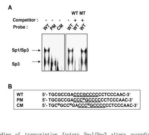
5. Transcription factor Sp1-/Sp-3 binding altered according to methylation status.
IGFBP-3 promoter 내 Sp-1/Sp-3 결합 요소의 methylation이 전사 인자의 결합에 미치는
영향을 관찰하기 위해 IGFBP-3 promoter의 Sp-1/Sp-3 결합 부위를 포함하는 mRNA cap
site로 부터 –108에서 –83까지를 CpG islands의 cytosine의 methylation 상태를 달리하여 3쌍의 25 bp oligomer를 제작하였다. W.T.는 5′-methylated cytosine이 CpG islands에 포
함되어 있지 않으며, P.M은 Sp-1/Sp-3 결합 요소 내 하나의 CpG island가 5′-methylated
cytosine을 포함하고 있으며, C.M.의 경우 내부의 모든 CpG islands 내 cytosine이
methylation 되게 제작하였다 (Fig. 6B). H1299 세포주의 핵 추출물과 W.T.를 사용한 경우
2개의 DNA-단백질 복합체가 관찰되었다. 그러나 이런 결합 양상은 CpG islands를
methylation 시킨 oligomer를 사용한 경우에는 변화되어 Sp-1/Sp-3 결합 요소 내부의 모
든 cytosine을 methylation 시킨 C.M.의 경우 두 DNA-단백질 복합체가 모두 형성되지 않
았으며, 중심부의 하나의 CpG island 내 cytosine을 methylation 시킨 P.M.의 경우 Sp-3
DNA-단백질 복합체의 형성에는 영향을 미치지 않는 반면 Sp-1/Sp-3 DNA-단백질 복합체
의 형성이 억제 되었다. DNA-단백질 결합의 특이도를 확인하기 위해 표지 되지 않은
결합 복합체는 완전히 사라졌으나, Sp-1 결합 부위를 돌연 변이시킨 oligonucleotide를 사 용한 경우에는 이러한 현상이 관찰되지 않았다 (Fig. 6A). WT 5’- TGCGCCGACCCGCCCCCCTCCCAAC-3’ PM 5’- TGCGCCGACCCMGCCCCCCTCCCAAC-3’ CM 5’- TGCMGCCMGACCCMGCCCCCCTCCCAAC-3’ WT WT WT WT MT WT PM CM Sp1/Sp3 Sp3
A
B
Probe : Competitor : - - - - + +Fig. 6. Binding of transcription factors Sp-1/Sp-3 alters according to methylation status. A. Gel−shift assays were performed using nuclear extracts isolated from H1299 cells by using wild−type (WT), partially methylated (PM), and completed methylated (CM) probes. Two major DNA-protein complexes were observed in the lane where WT oligomer was used; however, binding patterns of these two complexes were altered when methylated oligomers were used. Specificity of binding was verified as each complex was successfully competed away with increasing concentrations of unlabelled wild− type Sp-1 consensus oligonucletides, whereas they were not competed away with unlabelled mutant (Mut) Sp-1 consensus oligonucleotide. B. Schematic showing
wild−type 25-mer oligonucleotide from -105 to -82 of the IGFBP-3 promoter containing the consensus binding sequences for Sp-1/Sp-3. Wild−type and 5′-methylated deoxy-cytosine modified probes were used in gel−shift assays.
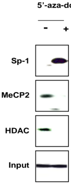
6. Sp-1/Sp-3 consensus sequence binding factors in H1299 cells and effect of demethylation agent 5′-aza-dC. IGFBP-3 promoter의 methylation이 Sp-1, MeCP2,
그리고 HDAC의 결합에 미치는 영향과 demethylation 약제인 5′-aza-dC의 역할을 알
아보기 위해 methylation된 IGFBP-3 promoter를 가지고 있는 H1299 세포주를 이용
하여 chromatin immunoprecipitation (ChiP) assay를 시행하였다. methyl-CpG 결합
단백질의 하나인 MeCP2는 Sp-1에 의해 활성화되는 leukosialin 유전자의 전사를
promoter가 methylation16 되면 HDAC와 같은 다른 억제인자들을 모집합으로서 전사
억제를 유도하는 것으로 알려져 있다26. MeCP2 와 HDAC은 동시에 Methylation된
IGFBP-3 promoter에 결합하며 gel-shift assay에서의 결과처럼 Sp-1의 결합은 억제
되었다. 1 µM 의 5′-aza-dC 로 5 일간 처리한 경우 IGFBP-3 promoter에 Sp-1의 결
Sp-1 MeCP2 HDAC Input
-
+ 5’-aza-dcFig. 7. Sp-1/Sp-3 binding status of Sp-1, MeCP2, and HDAC and the effect of demethylating agent 5′-aza-dC. ChIP analysis was performed to examine whether methylation status and treatment with demethylating agent 5′-aza-dC affected Sp-1, MeCP2, and HDAC binding on the IGFBP-3 promoter in H1299 NSCLC cells, which have methylated IGFBP-3 promoter. Methylated promoter containing the Sp-1/Sp-3 binding element did not recruit Sp-1, while MeCP2 and HDAC were recruited concurrently. After 5 days of treatment with 1 µM of 5′-aza-dC in 2% of fetal bovine serum, the binding affinity of Sp-1 on the IGFBP-3 promoter was increased, whereas the binding of MeCP2 and HDAC was significantly decreased.
7. Luciferase activity decreased in SssI methylase catalyzed IGFBP-3 promoter and coexpression of human MeCP2 cDNA provides additional repressesion.
IGFBP-3 promoter의 methylation이 promoter의 활성도에 미치는 영향을 확인하기 위하여
Sp-1/Sp-3 결합 요소를 포함하고 있는 pGL2-∆1708를 in vitro methylation 시킨 후
luciferase 활성도를 H1299 세포주에서 측정하였다. pGL2-∆1708 plasmid 내 CpG islands
의 methylation은 전사 활성도를 유의하게 감소시켰다 (data not shown). Methylation 되
어 있는 plasmid로 transfection된 세포주에 5′-aza-dC를 처리하였을 때 약물 처리 농도에
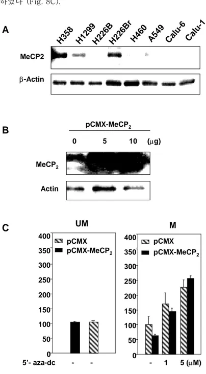
비례하여 전사 활성도가 회복되는 것을 확인할 수 있었다. 1 µM 및 5 µM 의 5′-aza-dC를 처리하였을 때, CpG methylated promoter의 전사 활성도는 약물 처리 전에 비하여 약 1.7 및 2.3 배 증가함을 확인할 수 있었다. MeCP2와 같이 내인적 methyl-결합 단백질들은 methylation된 주형에서 구조적인 전사의 억제를 하는 것으로 알려져 있다16. Methylation 된 promoter에서 전사 활성도의 조절에 대한 methyl-CpG 결합 단백질의 역할을 확인하기 위하여 비소세포성 폐암에서 내인성 methyl-CpG 결합 단백질 중의 하나인 MeCP2 의 발 현 양상을 확인하였다. 세포주 중 강한 IGFBP-3의 발현을 보이는 세포주는 IGFBP-3
promoter가 methylation된 세포주에 비하여 상대적으로 낮은 MeCP2 발현 양상을 보였다
(Fig. 8A). SssI 처리된 pGL2-∆1708과 pCMX mammalian expression vector에 삽입된 MeCP2 cDNA로 cotransfection을 시행하였다. MeCP2 발현의 유도는 pCMX-MeCP2
vector를 COS 세포주에 transiently transfection을 시행하여 확인하였다 (Fig. 8B).
Luciferase 분석에 따르면 methylation되지 않은 pGL2-∆1708를 pCMX-MeCP2과 cotransfection 시킨 경우에는 전사 활성도에 영향을 미치지 못하는 반면 SssI 처리된
pGL2-∆1708 에서는 pCMX-MeCP2의 cotransfection이 전사 활성도를 약 37.4% 정도 감 소하였다 (Fig. 8C). H35 8 H12 99 H22 6B r H22 6B Cal u-6 Cal u-1 H46 0 A54 9 MeCP2 β-Actin
A
0 5 10 (µg) MeCP2 ActinB
pCMX-MeCP2 5’- aza-dc - - - 1 5 (µM) 0 50 100 150 200 250 300 pCMX pCMX-MeCP2 350 400 pCMX-MeCP2 pCMX UM M 0 50 100 150 200 250 300 350 400C
Fig. 8. Expression level of MeCP2 in NSCLC cell lines and effect on in vitro methylated pGL2-∆1708 luciferase report plasmid. A. Expression levels of MeCP2 vary in human NSCLC cell lines. Cell lines that have high IGFBP-3 expression level with unmethylated IGFBP-3 promoter, expressed lower level of
MeCP2, compared to NSCLC cell lines that have hypermethylated IGFBP-3 promoter. B. 1.4−kb full−length MeCP2 cDNA was cloned in mammalian expression vector pCMX and transfected into the COS cell line. Note the strong expression of a 75−kD band in pCMX-MeCP2−transfected cells. C. H1299 NSCLC cells, which have methylated IGFBP-3 promoter, were transiently transfected with SssI methylase−catalyzed pGL2-∆1708 luciferase reporter plasmid. pCMX-MeCP2 (100 ㎍) was co-transfected with pCMX empty vector as a control. The cells were treated twice with 5′-aza-dC on the day of transfection, and 24 h later at the indicated doses. Luciferase activity was measured following incubation for 48 h. In unmethylated pGL2-∆1708 there was no difference with co-transfection of pCMX-MeCP2 (left). Methylation of all CpG islands of the IGFBP-3 promoter with SssI methylase had shut down reporter vector activity, with greater decreases in those cells co-transfected with pCMX-MeCP2. This suppression of luciferase activity was partially restored by demethylating agent 5′-aza-dC in the methylated reporter gene plasmid. This restoration partially interfered, however, with co-transfection with the MeCP2 expression vector.
Ⅳ. 고찰
이번 연구의 몇몇 결과들은 IGFBP-3 promoter내 CpG islands의 methylation이 비소
세포성 폐암 세포주에서 IGFBP-3 발현을 억제하는 한 기전임을 보이고 있다. 실험 에서 사용된 비소세포성 폐암 세포주의 절반 가까이에서 IGFBP-3 발현이 소실되어 있으며, IGFBP-3의 발현이 소실된 비소세포성 폐암 세포주에서 얻어진 DNA의 bisulfite 화학 변성 후 염기서열 분석 및 제한 효소 절단 분석, MSP의 결과는 promoter 활성도에 중요한 역할을 하는 Sp-1/Sp-3 결합 요소 및 주변의 CpG islands가 methylation 되어 있음을 보이고 있다. H226B, H1299, H661, H441, H322, H226Br, 및 Calu-6 세포주 등 IGFBP-3의 발현이 억제되어 있는 세포에서 단
백질 발현 양상이 promoter 내 CpG islands의 methylation 상태와 잘 일치하였다.
이전의 보고에서처럼 DNA의 methylation이 인식 염기서열에 전사 인자들의 결합을
억제한다는 보고와 같이 Sp-1/Sp-3 결합 요소와 주변 부위 CpG islands의
hypermethylation은 Sp-1 전사 인자의 결합을 억제하였다27-30. 과거 보고 중 Sp-1
은 인식 염기 서열의 methylation 상태와는 상관없이 결합한다는 보고가 있으나31,
32, anti-sense strand 내 두 개의 cytosine이 methylation되면 Sp-1의 결합을 억제
한다는 보고와 같이 Sp-1/Sp-3 결합 요소 내부 및 주변 부위의 CpG islands의
methylation에 의해 Sp-1의 IGFBP-3 promoter 에 대한 결합이 억제되는 것을 관찰
vitro methylation은 promoter 활성도를 감소시켰다. 더욱이 IGFBP-3 promoter의
활성도는 methylation 된 IGFBP-3 promoter를 가진 비소세포성 폐암 세포주에서,
demethylation 약제인 aza-dC를 처리함으로서 복원되었다. 그러나 이러한
5′-aza-dC 처리는 실험에 사용된 promoter의 methylation 이 확인된 7 종의 비소세포
성 폐암 세포주 중 4 가지 세포주에서만 IGFBP-3의 발현이 회복되어, IGFBP-3 단백
질 발현의 소실에 promoter hypermethylation 이외의 다른 기전이 작용할 것으로
생각되고 있다. 예로서 혈청 IGFBP-3 농도와 유전학적 다형성과의 연관성을 조사
한 임상 연구에서처럼, mRNA cap site로부터 –202 site의 다형성이 IGFBP-3
promoter 활성도를 조절하는 또 다른 요소로 생각되고 있다34. 이러한 소견들은
IGFBP-3의 발현을 조절하는 기전들이 다양하고 복잡함을 의미하는 것이다. IGFBP-3
promoter에는 최소한 4개의 대칭적으로 배열된 methylation된 CpG dinucleotide로
이루어진 MeCP2 결합 염기 서열로 여겨지는 요소가 Sp-1/Sp-3 결합 요소 인접한 곳
에 위치하고 있으며, MeCP2는 promoter 활성을 갖는 부위에서 멀리 떨어진 곳에서
도 유전자 발현을 억제할 수 있는 것으로 알려져 있다35. MeCP2와 같은 methyl-CpG
결합 단백질들이 methylation된 유전자의 발현 억제와 chromatin의 구조에 영향을
준다는 근거가 증가하고 있어36-38 MeCP2가 methylation된 IGFBP-3 promoter의 활성
도를 억제하는 데 주요한 역할을 할 것으로 가정하고서, methylation된 promoter를
MBD 단백질들 중 처음으로 발견된 것으로, methyl-CpG-binding domain (MBD)을 가
지고 있는 single polypeptide와 transcription repression domain (TRD)으로 구성
되어 있다39. MeCP2의 TRD는 전사 억제인자인 Sin-3와 HDAC co-repressor 복합체와
결합하는 것으로 알려져 있다40-41. 이는 MeCP2 와 HDAC이 methylation된 IGFBP-3
promoter에 모집된다는 결과와 잘 일치하며 나아가 MeCP2와 methylation된 DNA에
의한 유전자 발현의 소실은 5′-aza-dC 와 HDAC 억제제인 sodium butyrate에 의해
회복되었다. HDAC 억제제인 trichostatin A 또는 sodium butyrate에 의한 IGFBP-3
promoter의 활성도 증가는 Sp-1 결합의 증가에 의한다는 것이 사람 유방암 세포주
와 간암 세포주에서 증명되었다23,42. 이는 MeCP2가 methylation된 promoter가 전사
억제 기구를 모집하는데 주요한 역할을 하며 나아가 IGFBP-3의 전사 활성도를 억제
하는 것으로 시사되는 소견이다.
결론적으로, 상당수의 비소세포성 폐암 세포주에서 IGFBP-3 promoter의 주요 활성
을 갖는 부위의 CpG islands가 methylation 되어 있음을 확인하였다. 이들 비소세
포성 세포주에서 IGFBP-3 promoter의 methylation이 IGFBP-3의 전사를 억제하는 중
요한 기전임을 관찰할 수 있었다. 또한 IGFBP-3 promoter 활성도에 주요한 역할을
하는 Sp-1/Sp-3 결합 요소가 methylation 되어 있으며 이 부위의 methylation이
MeCP2와 HDAC의 결합을 유도하고 Sp-1 전사 인자의 결합을 억제함으로 methylation
양상과는 잘 일치하지 않으며 이는 다른 MBD를 가지고 있는 단백질이 methylation 된 promoter를 가지고 있는 비소세포성 폐암 세포주에서 IGFBP-3 발현 억제에 영향 을 줄 수 있기 때문이라 생각된다. 마지막으로 임상 시험중인 demethylation 약제 로서 IGFBP-3의 발현을 유도할 수 있다는 것은 IGFBP-3의 세포의 성장과 고사에 미 치는 중요한 역할을 고려해 볼 때 이들이 비소세포성 폐암의 치료에 중요한 약제로 사용될 수 있음을 시사하는 소견으로 생각된다.
Ⅴ. 결론
1. 인슐린 양 성장 인자 결합 단백질-3 (IGFBP-3)의 promoter는 상당수의 비소세포
성 폐암 세포주에서 주요 조절 인자 결합 부위 중 하나인 Sp-1/Sp-3 consensus
sequence 및 주변부의 CpG islands가 hypermethylation 되어 있다.
2. IGFBP-3의 promoter 내 Sp-1/Sp-3 결합 요소 및 주변부의 CpG islands가
hypermethylation되어 있으면, 이 부위에 methyl CpG binding protein-2 (MeCP2)와
histone deacetylalse (HDAC)가 모집되어 결합되는 반면, Sp-1의 결합은 억제되었
으며, demethylating 약제인 5′-aza-2-deoxycytidine (5′-aza-dC) 처리시 이러한 현
상은 역전되어 promoter 내 CpG islands의 hypermethylation이 IGFBP-3 발현을 억
제하는 기전 중의 하나로 추정된다.
3. SssI CpG methylase를 이용하여 in vitro methylation을 유도하였을 때 IGFBP-3
promoter의 CpG islands의 hypermethylation은 유의하게 promoter activity를 감소
시켰으며, 이는 동일 조건에서 MeCP2를 coexpression 시켰을 때 더욱 감소하여,
promoter 내 CpG islands가 hypermethylation되면 methyl-CpG 결합 단백의 결합이 유
4. 이상의 결론을 종합하면 5′-aza-dC와 같은 demethylation 약제가 IGFBP-3의 발
현을 유도할 수 있다는 것은 IGFBP-3의 세포의 성장과 고사에 미치는 중요한 역할
을 고려해 볼 때 이들이 비소세포성 폐암의 치료 약제로 사용될 수도 있음을 시사
Ⅵ. 참고문헌
1. Greenlee RT, Hill-Harmon MB, Murray T, Thun M. Cancer statistics, 2001. CA
Cancer J Clin 2001;51:15-36.
2. Parkin DM, Pisani P, Ferlay J. Estimates of the worldwide incidence of 25
major cancers in 1990. Int J Cancer 1999;80:827-41.
3. Jones JI, Clemmons DR. Insulin-like growth factors and their binding
proteins: biological actions. Endocr Rev 1995;16:3-34.
4. Dunn SE, Hardman RA, Kari FW, Barrett JC. Insulin-like growth factor-1
(IGF-1) alters drug sensitivity of HBL 100 human breast cancer cells by
inhibition of apoptosis induced by diverse anticancer drugs. Cancer Res
1997;57:2687–93.
5. Stewart CE, Rotwein P. Growth, differentiation and survival: multiple
physiological functions for insulin-like growth factors. Physiol Rev
1996;76:1005-26.
6. Baxter RC. Signaling pathways involved in antiproliferative effects of
2001;54:145-8.
7. Rajah R, Valentinis B, Cohen P. Insulin-like growth factor (IGF)-binding
protein-3 induces apoptosis and mediates the effects of transforming growth
factor-ß1 on programmed cell death through a p53- and IGF-independent
mechanism. J Biol Chem 1997;272:12181-8.
8. Hwa V, Oh Y, Rosenfeld RG. The insulin-like growth factor-binding protein
(IGFBP) superfamily. Endocr Rev 1999;20:761–87.
9. Oh Y, Muller HL, Lamson G, Rosenfeld RG. Insulin-like growth
factor-independent action of IGF-binding protein-3 in Hs578T human breast cancer
cells. Cell surface binding and growth inhibition. J Biol Chem
1993;268;14964-71.
10. Valentinis B, Bhala A, DeAngelis T, Baserga R, Cohen P. The human
insulin-like growth factor (IGF) binding protein-3 inhibits the growth of
fibroblasts with a targeted disruption of the IGF-I receptor gene. Mol
Endocrinol 1995;9:361–7.
11. Gucev ZS, Oh Y, Kelley KM, Rosenfeld RG. Insulin-like growth factor
β2-induced growth inhibition in human breast cancer cells. Cancer Res
1996;56:1545–50.
12. Martin JL, Coverley JA, Pattison ST, Baxter RC. Insulin-like growth
factor-binding protein-3 production by MCF-7 breast cancer cells: stimulation
by retinoic acid and cyclic adenosine monophosphate and differential effects
of estradiol. Endocrinology 1995;136:1219–26.
13. Buckbinder L, Talbott R, Velasco-Miguel S, Takenaka I, Faha B, Seizinger
BR, et al. Induction of the growth inhibitor IGF-binding protein-3 by p53.
Nature 1995;377:646-8.
14. Zi X, Zhang J, Agarwal R, Pollak M. Silibinin up-regulates insulin-like
growth factor-binding protein 3 expression and inhibits proliferation of
androgen-independent prostate cancer cells. Cancer Res 2000;60:5617–20.
15. Fowler CA, Perks CM, Newcomb PV, Savage PB, Farndon JR, Holly JM.
Insulin-like growth factor binding protein-3 (IGFBP-3) potentiates
paclitaxel-induced apoptosis in human breast cancer cells. Int J Cancer
2000;88:448–53.
expression by trichostatin A: potential therapeutic role in the treatment of
hepatocellular carcinoma. Int J Mol Med 2000;5:33–41.
17. Yu H, Spitz MR, Mistry J, Gu J, Hong WK, Wu X. Plasma levels of
insulin-like growth factor-I and lung cancer risk: a case–control analysis. J Natl
Cancer Inst 1999;91:151-6.
18. Hankinson SE, Willett WC, Colditz GA, Hunter DJ, Michaud DS, Deroo B, et
al. Circulating concentrations of insulin-like growth factor-I and risk of
breast cancer. Lancet 1998;351:1393–6.
19. Chan JM, Stampfer MJ, Giovannucci E, Gann PH, Ma J, Wilkinson P, et al.
Plasma insulin-like growth factor-I and prostate cancer risk: a prospective
study. Science 1998;279:563–6.
20. Ma J, Pollak MN, Giovannucci E, Chan JM, Tao Y, Hennekens CH, et al.
Prospective study of colorectal cancer risk in men and plasma levels of
insulin-like growth factor (IGF)-I and IGF-binding protein-3. J Natl Cancer
Inst 1999;91:620–5.
21. Burroughs KD, Dunn SE, Barrett JC, Taylor JE. Insulin-like growth
22. Kim YH, Dohi DF, Han GR, Zou CP, Oridate N, Walsh GL, et al. Retinoid
refractoriness occurs during lung carcinogenesis despite functional retinoid
receptors. Cancer Res 1995;55:5603–10.
23. Walker GE, Wilson EM, Powell D, Oh Y. Butyrate, a histone deacetylase
inhibitor, activates the human IGF binding protein-3 promoter in breast
cancer cells: molecular mechanism involves a Sp1/Sp3 multiprotein complex.
Endocrinology 2001;142:3817–27.
24. Herman JG, Graff JR, Myohanen S, Nelkin BD, Baylin SB.
Methylation-specific PCR. A novel PCR assay for methylation status of CpG islands. Proc
Natl Acad Sci U S A 1996;93:9821-6.
25. Chen H, Lin RJ, Xie W, Wilpitz D, Evans RM. Regulation of hormone-induced
histone hyperacetylation and gene activation via acetylation of an acetylase.
Cell 1999;98:675-86.
26. Kudo S. Methyl-CpG-binding protein MeCP2 represses Sp1-activated
transcription of the human leukosialin gene when the promoter is methylated.
Mol Cell Biol 1998;18:5492-9.
enhancer/promoter sequence TGACGTCA abolishes specific factor binding as well
as transcriptional activation. Genes Dev 1989;3:612-9.
28. Comb M, Goodman HM. CpG methylation inhibits proenkephalin gene
expression and binding of the transcription factor AP-2. Nucleic Acids Res
1990;18:3975-82.
29. Tate PH, Bird AP. Effects of DNA methylation on DNA-binding proteins and
gene expression. Curr Opin Genet Dev 1993;3:226-31.
30. Kovesdi I, Reichel R, Nevins JR. Role of an adenovirus E2 promoter
binding factor in E1A-mediated coordinate gene control. Proc Natl Acad Sci U
S A 1987;84:2180-4.
31. Samson SL, Wong NC. Role of Sp1 in insulin regulation of gene
expression. J Mol Endocrinol 2002;29:265-79.
32. Macleod D, Charlton J, Mullins J, Bird AP. Sp1 sites in the mouse aprt
gene promoter are required to prevent methylation of the CpG island. Genes
Dev 1994;8:2282-92.
33. Clark SJ, Harrison J, Molloy PL. Sp1 binding is inhibited by (m)Cp(m)CpG
34. Deal C, Ma J, Wilkin F, Paquette J, Rozen F, Ge B, Hudson T, et al. Novel
promoter polymorphism in insulin-like growth factor-binding protein-3:
correlation with serum levels and interaction with known regulators. J Clin
Endocrinol Metab 2001;86:1274-80.
35. Nan X, Meehan RR, Bird A. Dissection of the methyl-CpG binding domain
from the chromosomal protein MeCP2. Nucleic Acids Res 1993;21:4886-92.
36. Kass SU, Pruss D, Wolffe AP. How does DNA methylation repress
transcription? Trends Genet 1997;13:444-9.
37. Ng HH, Bird A. DNA methylation and chromatin modification. Curr Opin
Genet Dev 1999;9:158-63.
38. Razin A. CpG methylation, chromatin structure and gene silencing-a
three-way connection. EMBO J 1998;17:4905-8.
39. Lewis JD, Meehan RR, Henzel WJ, Maurer-Fogy I, Jeppesen P, Klein F, et al.
Purification, sequence, and cellular localization of a novel chromosomal
protein that binds to methylated DNA. Cell 1992;69:905-14.
40. Nan X, Ng HH, Johnson CA, Laherty CD, Turner BM, Eisenman RN, et al.
histone deacetylase complex. Nature 1998;393:386-9.
41. Jones PL, Veenstra GJ, Wade PA, Vermaak D, Kass SU, Landsberger N, et
al. Methylated DNA and MeCP2 recruit histone deacetylase to repress
transcription. Nat Genet 1998;19:187-91.
42. Choi HS, Lee JH, Park JG, Lee YI. Trichostatin A, a histone
deacetylase inhibitor, activates the IGFBP-3 promoter by upregulating Sp1
activity in hepatoma cells: alteration of the Sp1/Sp3/HDAC1 multiprotein
Abstract
Regulatory mechanism of insulin-like growth factor binding
protein-3 in non-small cell lung cancer
Chang, Yoon Soo
Department of Medical Science The Graduate School, Yonsei University
(Directed by Professor Kim, Se Kyu)
Insulin-like growth factor (IGF)—binding protein-3 (IGFBP-3) inhibits the proliferation of non-small cell lung cancer (NSCLC) cells by inducing apoptosis. We investigated whether hypermethylation of CpG islands in IGFBP-3 promoter play an important role in the loss of IGFBP-3 expression and regulatory mechanism that mediate the silencing of IGFBP-3 expression in the NSCLC cell lines. The IGFBP-3 promoter has CpG islands hypermethylation in 7 of 15 (46.7%) NSCLC cell lines. The methylation status correlated with the level of expression of protein and mRNA in NSCLC cell lines and reduced expression of IGFBP-3 was restored by the demethylating agent 5 ′-aza-2-deoxycytidine (5′-aza-dC) in a subset of NSCLC cell lines. The Sp-1/Sp-3
binding element in the IGFBP-3 promoter, important for promoter activity, was methylated in a subset of NSCLC cell lines and the methylation of this element suppressed the binding of the Sp-1 transcription factor. A ChIP assay showed that the methylation status of the IGFBP-3 promoter influenced the binding of Sp-1, methyl-CpG binding protein-2 (MeCP2), and histone deacetylase (HDAC) to Sp-1/Sp-3 binding element, which were reversed by 5′-aza-dC. In vitro methylation of the IGFBP-3 promoter containing the Sp-1/Sp-3 binding element significantly reduced promoter activity, which was further suppressed by the overexpression of MeCP2 and, however, this reduction in activity was rescued by 5′-aza-dC. These findings indicate that hypermethylation of the IGFBP-3 promoter is one of the possible mechanisms by which IGFBP-3 expression is silenced and MeCP2, with recruitment of HDAC, may play a role in silencing of IGFBP-3 expression. Considering critical biologic activities of IGFBP-3 on cell growth and apoptosis, demethylation agents, which recovered repressed IGFBP-3 expression, could be used as a new therapeutic modality in NSCLC.
Key Words: Insulin-like growth factor (IGF), insulin−like growth factor binding protein-3 (IGFBP-3), methylation of CpG islands, methyl CpG binding protein-2 (MeCP2), non-small cell lung cancer (NSCLC).