Mesenchymal Stem Cells Improve Wound Healing In Vivo via
Early Activation of Matrix Metalloproteinase-9 and Vascular
Endothelial Growth Factor
We investigated the effects of mesenchymal stem cells (MSCs) on wound healing using a three-dimensional (3D) collagen gel scaffold. Three circular full-thickness skin defects were created on the back of Sprague-Dawley rats. One site was covered with a 3D collagen gel containing 2 × 10⁶ MSCs (MSCs+/3D collagen+). Another site was replaced with a 3D collagen gel without MSCs and the third site was left empty. The wound size was
significantly reduced in the MSCs+/3D collagen+ sites. MSCs+/3D collagen+ sites exhibited the most neovascularization. FISH showed that Y-chromosome possessing cells were found within the dermis of MSCs+/3D collagen+ sites. Gelatin zymography revealed that the most intense expression of MMP-9 was detected early in the MSCs+/3D collagen+ sites. Our results indicate that MSCs upregulate the early expression of MMP-9 which induces the early mobilization of VEGF. Thus, MSCs appear to accelerate significantly wound healing via early activation of MMP-9 and VEGF.
Key Words: Mesenchymal Stem Cells; Wound Healing
Chul Han Kim1, Jang Hyun Lee2,
Jong Ho Won3 and Moon Kyun Cho4
Departments of 1Plastic and Reconstructive Surgery, 3Stem Cell Therapy Center and Institute for Clinical
Molecular Biology Research, and 4Dermatology,
Soonchunhyang University College of Medicine, Seoul; 2Department of Plastic and Reconstructive
Surgery, Hanyang University Guri Hospital, Guri, Korea
Received: 28 October 2010 Accepted: 2 May 2011 Address for Correspondence: Jong Ho Won, MD
Stem Cell Therapy Center and Institute for Clinical Molecular Biology Research, Soonchunhyang University College of Medicine, 657 Hannam-dong, Yongsan-gu, Seoul 140-743, Korea
Tel: +82.2-709-9283, Fax: +82.2-796-3543 E-mail: [email protected]
DOI: 10.3346/jkms.2011.26.6.726 • J Korean Med Sci 2011; 26: 726-733
INTRODUCTION
Wound healing requires the complex biomolecular processes of cell migration, proliferation, angiogenesis, and extracellular ma-trix remodeling (1, 2). This process involves various cells, growth factors, and the extracellular matrix (ECM). During wound heal-ing, cell migration and tissue remodeling necessitate degrada-tion of ECM and release of growth factors (3). The proteolytic degradation of ECM is achieved by extracellular proteases in-volving the matrix metalloproteinases (MMPs). These MMPs, particularly MMP-9, are thought to play a critical role in degrad-ing ECM to facilitate the necessary initial steps for angiogenesis. The high expression of MMPs stimulates the angiogenic response, subsequently promoting the process of wound healing. Angiogenesis is subjected to a complex control system with pro-angiogenic and anti-angiogenic factors. One of the most important pro-angiogenic factors is vascular endothelial growth factor (VEGF). During the early period of wound healing, angio-genic capillary sprouts penetrate into the provisional matrix and create microvascular networks (2). Neovascularizaton involves the dynamic interactions of cells, growth factors, and the matrix. VEGF stimulates and increases neovascularization.
Bone marrow-derived mesenchymal stem cells (BM-MSCs)
have the capacity to differentiate along three essential mesen-chymal lineages: osteoblasts, adipocytes, chondroblasts (4, 5). Also, BM-MSCs can express the phenotypic genes of hepato-cytes, cardiomyohepato-cytes, astrohepato-cytes, neurons, and skin-related tis-sue (6-10). There have been reports showing wound healing ef-fectively achieved with the application of BM-MSCs (11, 12). However, despite increasing knowledge of MSCs, their contribu-tion to wound healing is not understood, and many quescontribu-tions remain.
We selected the 3D collagen allograft model, which is most similar to the skin dermis structure, to apply rat MSCs in the col-lagen gel scaffold composed of rat tail type I colcol-lagen in an at-tempt to maintain the functions of stem cells for a prolonged period. The objective of our study was to investigate the effects of MSCs on wound healing using 3D collagen gel scaffold in vivo.
MATERIALS AND METHODS
Experimental animal model
Eighteen adult female Sprague-Dawley rats, weighing 200 g, were used at 6 weeks of age. All animal procedures were approved under the guidelines approved by the institutional animal care and use committee from Soonchunhyang University. This study
was carried out from March 2006 to May 2007 at the Soonchun-hyang University. Each rat had free access to both sterile water and standard rodent soft chow ad libitum.
Harvest and culture of MSCs
The bone marrow was harvested from specific pathogen-free, 6-week-old male Sprague-Dawley rats (Charles River Technol-ogy). In brief, the rats were killed, and femurs and tibias were removed aseptically. A hole was then created in the knee joint end of each bone with a 26-gauge needle, and marrow was flush ed with phosphate buffered saline (PBS, Gibco BRL, Grand Island, NY, USA) containing 2% fetal bovine serum (FBS, Gibco BRL). The marrow solution was washed by centrifugation at 400 × g for 5 min. The supernatant was aspirated, and the pellet was resuspended in Dulbecco’s modified Eagle’s medium-low glu-cose (DMEM-LG, Gibco BRL) containing 10% FBS and 1% pen-icillin-streptomycin (Gibco BRL). Cells were seeded into 75 cm2 tissue culture flask and cultured in humidified 5% CO2 incuba-tor at 37°C for 72 hr. Non-adherent cells were removed. When adherent cells reached subconfluence, they were detached with 0.25% trypsin-EDTA solution, and re-seeded at 2 × 10³ cells/cm2. The rat MSCs were used for experiments after the passage 5. Differentiation of MSCs
For osteogenic differentiation of MSCs, MSCs were maintained with DMEM medium containing 0.1 μM dexamethasone (Sig-ma, St. Louis, MO, USA), 50 μM ascorbate (Sigma), 10 mM β- glycerol phosphate (Sigma), and 10% FBS for 3 weeks, with the increase of calcium examined using Von Kossa staining. For adipogenic differentiation of MSCs, MSCs were cultured in DMEM medium (Adipogenic induction, MDI + I) containing 0.1 μM dexamethasone (Sigma), 10 μg/mL insulin (Sigma), 100 mM indomethacin (Sigma), 0.5 mM methyl-isobuthylxanthine (Sigma), and 10% FBS for 72 hr. The medium was changed to DMEM medium (Adipogenic maintenance, AM), containing insulin (10 μg/mL) and 10% FBS for 24 hr. The cells were then re-treated with MDI + I for a second treatment. The cultures were then maintained in AM for 1 week before fixation. Adipogenic differentiation was demonstrated by the accumulation of lipid vesicles with Oil-Red O staining.
For chondrogenic differentiation of MSCs, MSCs were treat-ed with trypsin-EDTA. Then, 2 × 10⁵ cells were added to 500 μL of DMEM medium containing 10-7 M dexamethasone (Sigma), 1 μg/mL TGF-β (R&D Systems, Minneapolis, MN, USA), and 50 μM ascorbic acid 2-phosphate (Sigma), and centrifuged at 1,000 rpm for 5 min. The cells were grown as a pelleted micromass for 3 weeks. The cell pellet was stained with toluidine blue.
Flow cytometric analysis of MSCs
MSCs were trypsinized, washed with PBS, and incubated with antibodies against fluorescein isothiocyanate
(FITC)-conjugat-ed CD44H (Pgp-1, BD Biosciences Pharmingen), CD90 (Thy-1, BD), CD11b (Integrin, BD), phycoerythrin (PE)-conjugated CD106 (VCAM-1, BD), and CD45 (BD). Analysis was performed with a flow cytometer (Beckman Coulter, Inc., CA, USA). Preparation of the three-dimensional collagen gel scaffold Collagen gel scaffolds were prepared in a sterile tube, by mixing an appropriate volume of rat tail type I collagen (BD Biosciences) to yield a final concentration of 1 mg/mL with medium cocktail (DMEM, NaHCO3 [44 mM/L], L-glutamine [4 mM/L], and folic acid [9 mM/L], neutralized with 1N NaOH to pH 7.2). Collagen solution (1 mL per well of a 6-well plate) was placed in an incu-bator at 37°C for 30 min to allow polymerization of the collagen. Assembly of stem cell/collagen tissue construct implants Control scaffolds were prepared as above. Early passage prima-ry rat MSCs (2 × 10⁶/well) were placed on top of each collagen scaffold and allowed to attach. After attachment, collagen scaf-folds were covered with 1 mL of media (DMEM, 10% FBS) and incubated for 72 hr at 37°C, and 5% CO2. The circular MSCs/3D collagen gel scaffolds, at a density of 2 × 10⁶ cells for implanta-tion, were 20-mm diameter.
Surgical procedures
In an aseptic surgical room, the rats were anesthetized with in-traperitoneal ketamine (60 mg/kg) and xylazine (8 mg/kg). After induction of general anesthesia, the dorsal regions of the rats were shaved. The dorsal skin was disinfected with 10% povidone-iodine and 70% alcohol. Three circular full-thickness defects 20-mm in diameter, including the panniculus carnosus muscle, were created on the back of each rat. One site was covered with 3D collagen gel containing 2 × 10⁶ male rat MSCs (MSCs+/3D collagen+ site). Another site replaced with 3D collagen gel with-out MSCs (MSCs-/3D collagen+) and the third site was left empty (MSCs-/3D collagen-). All wounds were covered with a sterile transparent silicone membrane (Tegaderm, 3M Health Care, St. Paul, MN, USA) to prevent desiccation. Each animal was then housed in its own cage to avoid damage of the wound. Digital pictures were taken to visualize the wound. Wound healing was calculated based on the remaining wound area.
The rats were sacrificed with an overdose of sodium pento-barbital at either 3, 7, 14, or 21 days after wounding. Full-thick-ness wound tissues, including the adjacent skin, were then har-vested, fixed immediately in 10% formalin for 12 hr, embedded in paraffin and sliced into 6-micrometer-thick sections for sub-sequent immunohistochemistry. For positive controls, normal healthy rat tissues were harvested.
Measurement of wound size
The wound sizes in a standard prone position were photographed using a 334 million d.p.i. digital camera (Canon) and an image
analyzer (Image Pro Plus, ver. 5.1; Media Cybernetics) on a per-sonal Microsoft computer. The macroscopic sizes of the remain-ing wounds were calculated three times for each animal at 0, 3, 7, 14, and 21 days after initial wounding.
Immunohistochemical staining
Paraffin sections were deparaffinized with xylene, rehydrated with 10 mM citrate buffer (pH 6.0) solution, and then treated in a microwave oven for 15 min to restore antigens. To suppress the reaction of endogenous peroxidase within the tissues, the sam-ples were treated with 3% peroxide for 5 min and then with a blocking solution for 30 min. Slides were then placed in a hu-mid chamber and incubated for 60 min with the anti-VEGF an-tibody (sc-152; Santa Cruz Biotec., Santa Cruz, CA, USA). Tissue staining was visualized with a 3,3´-diaminobenzidine (ACK500; ScyTek, Logan, UT, USA) substrate chromogen solution. Gelatin zymography
Protein samples (20 μg of protein) were mixed with 2× sample buffer (62.5 mM Tris-HCl pH 6.8, 10% glycerol, 2% SDS, 0.0025 bromphenol blue) and added to the gel without boiling. MMP-2 and MMP-9 activities were analyzed using 10% zymogram gel-atin gel (EC6175Box, Invitrogen). Following electrophoresis, gels were washed twice, first with a 1 × renaturing buffer (1 M Tris-HCl [pH 7.4], Triton X-100) for 30 min, after the gel was washed with distilled water for 30 min. Next, gels were reacted with a 1× developing buffer (50 mM Tris base, 40 mM HCl, 200 mM NaCl, 5 mM CaCl2, 0.02% [w/v] Brij35) for 30 min, by incubating over-night at 37°C. Gels were treated with 0.5% Coomassie blue R-250 in destaining solution (40% ethanol, 8% acetic acid) for 3 hr and destained in the same solution without Coomassie blue. X and Y chromosome fluorescence in situ hybridization X and Y chromosome fluorescence in situ hybridization (FISH) was performed in frozen tissues in order to confirm the existence of stem cells. Probes for rat DNA (ID Labs Inc.) were applied. Briefly, six-micrometer thick frozen tissue sections were rehy-drated for 5 min each in 100% EtOH, 90% EtOH, and 70% EtOH, and washed in 4 × standard saline citrate (SSC)/0.5% Tween 20
for 30 min in rotating shaker. Pre-fixated slides were incubated in a solution of protease (32-801260, VYSISTM) for 20 min at 37°C, and washed in PBS, PBS/MgCl2, and formaldehyde/PBS/MgCl2. Slides were dehydrated in 70%, 90%, and 100% EtOH and dried in air. Ten μL probe was dropped onto the slide, and sealed with a cover slip and rubber cement. Pre-treated sections and probes were denatured at 80°C for 5 min and incubated in an incubator for 4 hr at 37°C. Post-hybridization washes were performed for 2 min in a 0.4 × SSC solution and 30 sec in 2 × SSC/0.05% Tween 20. Tissue sections were counter-stained with 7 μL of 4´,6-diamid-ino-2-phenylindole (DAPI). Results were analyzed under a flu-orescent microscope and VYSISTM direct-labeled FISH probe (VYSISTM, USA).
Statistical analyses
Data were evaluated by analysis of variance (Stacel, add-in soft-ware to Microsoft® Excel 2000). Statistical analyses were per-formed using the one-way ANOVA followed by Scheffé’s multi-ple-comparison test, and P value of less than 0.05 was consid-ered statistically significant.
RESULTS
Characterization of cultured rat MSCs
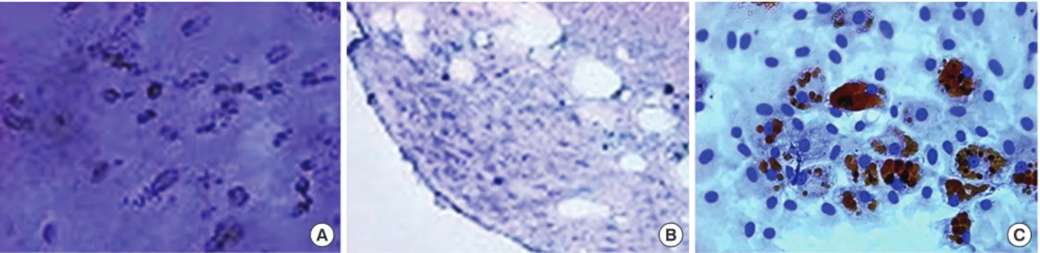
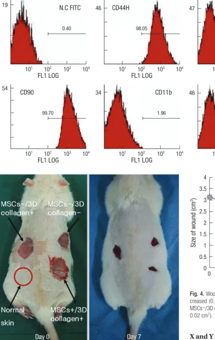
When cultured in osteogenic, chondrogenic, and adipogenic medium, they differentiated into osteocytes, chondrocytes, and adipocytes (Fig. 1). Flow cytometer analysis confirmed expres-sion of stem cell-related surface markers. Cultured adherent cells expressed CD44H and CD90, but not hematopoietic markers such as CD11b, CD45, and CD106 (Fig. 2). Thus, we confirmed that the adherent cells were mesenchymal stem cells.
Wound size measurements
Each experimental site was relatively of the same size at day 3 due to excision of the panniculus carnosus muscle. The most effective wound healing was observed at day 7 (Fig. 3). At day 7, the wound size of MSCs+/3D collagen+ sites was significantly decreased (0.48 ± 0.01 cm2: P < 0.05), which were significantly smaller than the MSCs-/3D collagen- sites (0.92 ± 0.01 cm2) or
A B C
Fig. 1. Characterization of MSCs in vitro. Differentiation of MSCs. (A) Cultured in appropriate differentiate media, MSCs differentiated osteocytes. (B) Chondrocytes in pellet cul-ture which were positive for toluidine blue. (C) Adipocytes were demonstrated by the accumulation of lipid vesicles with oil red O staining (original magnification, × 40).
the MSCs-/3D collagen- sites (0.66 ± 0.02 cm2). Also, there was no significant difference between MSCs-/3D collagen+ sites and MSCs-/3D collagen- sites (Fig. 4). MSCs+/3D collagen+ sites demonstrated accelerated wound healing. Since there was a pos-sibility that the rate of wound healing could vary with the defect position, we repeated the same procedure by changing the po-sitions of the defect sites, which yielded similar results.
Immunohistochemistry
The tissue samples from each experimental group at day 7, when stained with VEGF antibody, showed that MSCs+/3D collagen+ sites exhibited the most distinct neovascularization compared to other sites (Fig. 5).
X and Y chromosome fluorescence in situ hybridization Y-chromosomes were probed with spectrum red (Cy3), where-as X-chromosomes with spectrum green (FITC). FISH analysis showed that many Y-chromosome positive cells were identified within the dermis of MSCs+/3D collagen+ sites at day 7 (Fig. 6). Gelatin zymography
We used the gelatin zymography to assess the expression status of MMP-2 (72-kDa/gelatinase A) and MMP-9 (92-kDa/gelatin-ase B). MMP-2 was expressed in all experimental sites, includ-ing normal rat skin tissue, at day 3 and day 7. MMP-9 was not detected in normal tissue, but expressed in MSCs-/3D collagen+ sites, MSCs-/3D collagen- sites, and MSCs+/3D collagen+ sites. Interestingly, MMP-9 was expressed most intensively early in MSCs+/3D collagen+ sites at day 3. However, by day 7, MMP-9 was weakly expressed in MSCs+/3D collagen+ sites compared to MSCs-/3D collagen+ and MSCs-/3D collagen- sites (Fig. 7). FL1 LOG N.C FITC 0.40 101 102 103 104 19 FL1 LOG N.C PE 1.41 101 102 103 104 47 FL1 LOG CD44H 98.05 101 102 103 104 46 FL1 LOG CD90 99.70 101 102 103 104 54 FL1 LOG CD106 3.54 101 102 103 104 46 FL1 LOG CD11b 1.96 101 102 103 104 34 FL1 LOG CD45 2.00 101 102 103 104 46
Fig. 2. Flow cytometer analysis of rat mesenchymal stem cells. MSCs were analyzed by flow cytometer after stain-ing with FIFC- or PE-conjugated anti-bodies against indicated cell surface proteins. MSCs did not produce CD11b, CD45, CD106 antigens, but produced CD44H, CD90 antigens.
Fig. 3. Effects of MSCs. The wound size was significantly smaller in the MSCs+/3D collagen+ site at day 7.
Day 0 Day 7 Si ze o f w ou nd (c m 2)
Days after surgery
MSC-/3D collagen+ MSC-/3D collagen-MSC+/3D collagen+ * 0 3 7 14 21 4 3.5 3 2.5 2 1.5 1 0.5 0
Fig. 4. Wound size measurement. MSCs+/3D collagen+ sites were significantly de-creased (0.48 ± 0.01 cm2: *P < 0.05), which were significantly smaller than the
MSCs-/3D collagen+ sites (0.92 ± 0.01 cm2) or MSCs-/3D collagen- sites (0.66 ±
DISCUSSION
Wound healing is a complex process requiring the proper ex-pression of signaling molecules and receptors, cellular adhesion molecules, and ECM proteins. These molecules are processed by MMPs and regulate many processes of wound healing (13).
A B
C D
Fig. 5. Immunohistochemistry. VEGF was not detected in the normal rat skin tissue. The MSCs+/3D collagen+ site was showed the most expression of VEGF than MSCs-/3D collagen+ site and MSCs-/3D collagen- site (A, Normal rat skin; B, MSCs-/3D collagen+ site; C, MSCs-/3D collagen- site; D, MSCs+/3D collagen+ site; A to D, × 400 mag-nification).
A B
Fig. 6. Fluorescence in situ hybridization. (A) Positive control. Probe (Whole chromosome Painting Probe): Rat X/Y (FITC/Cy3) Probe. (B) Y-chromosome positive cells were iden-tified within the dermis of MSCs+/3D collagen+ site at day 7. Y-chromosomes were probed with spectrum red (Cy3), X-chromosomes with spectrum green (FITC).
MMPs contribute too many aspects of tissue regeneration and angiogenesis by promoting the release of ECM-bound cytokines or angiogenesis-modulating ECM fragments (14). Recent stud-ies have reported that implanted MSCs accelerate the restora-tion of damaged tissue (15-17). With regard to tissue regenera-tion, this potency of MSCs may play a more principal role than
trans-differentiation (18). In the present study, MSCs+/3D col-lagen+ sites significantly reduced the wound size and exhibited the most intense expression of VEGF. VEGF is a potent angio-genic factor that stimulates angiogenesis in vivo (19-21). Angio-genesis is a complex and dynamic process which is affected by many factors including growth factors, cytokines, and ECM (22). The expression of VEGF is thought to be potentiated in response to the activation of bioactive molecules, and VEGF is considered to mediate angiogenic activity during the early period of wound healing.
There are interesting differences between 2 and MMP-9 by gelatin zymography. MMP-2 was expressed in all experi-mental sites including normal rat skin tissue. This finding indi-cates that MMP-2 plays a less important role in early tissue me-tabolism. MMP-9 was expressed in MSCs+/3D collagen+ sites, MSCs-/3D collagen+ sites, and MSCs-/3D collagen+ sites, but not in normal rat skin tissue. Interestingly, during the early heal-ing period (day 3), MMP-9 was most intensively expressed in MSCs+/3D collagen+ sites. In the later period (day 7), however, MMP-9 was more weakly expressed in MSCs+/3D collagen+ sites, compare to MSCs-/3D collagen+ sites and MSCs-/3D col-lagen- sites. This suggests that MMP-9 is involved in the early period of wound healing which coincides with enhanced wound healing process via angiogenesis. This is in good agreement with the findings that many processes are involved in the early peri-od of wound healing require the action of proteases to facilitate cell movement, tissue degradation, granulation tissue formation and angiogenesis (1, 2). Hence, MMP-9, which is expressed ear-ly in MSCs+/3D collagen+ sites, appears to play critical roles in initiating angiogenesis.
There are reports that MMP-9, but not MMP-2, stimulates the production and secretion of VEGF in retinal pigment epithelial cells (23), and that inhibition of MMP-9 attenuates VEGF-in-duced intracerebral hemorrhage (24). Upon secretion, VEGF becomes bound to the ECM (25), and the interaction of VEGF with ECM proteins is mediated via ECM-binding domain. VEGF is encoded by a single gene and the coding portion contains
eight exons (26). Exon 6 and 7 have been demonstrated to en-code the ECM-binding domain of the protein. This domain can bind matrix proteins and is considered to account for the seques-tration of VEGF within the ECM (27, 28). Thus, VEGF becomes liberated by ECM degradation through MMPs. Their effects are due to the mobilization of VEGF release through the degrada-tion of ECM proteins by MMPs. In this study, VEGF and MMP were expressed in MSCs+/3D collagen+ sites. These factors may have an effect in a single manner or rather interact with each other. Early expressed MMP-9 promotes the release of extracel-lular matrix-bound cytokines, VEGF, which regulates angiogen-esis. In the present study, MSCs upregulate the early expression of MMP-9, not MMP-2. This early upregulation of MMP-9 seems to be the crucial steps in angiogenesis.
In the present study, we used a three-dimensional collagen gel scaffold. In vivo, cells within the tissue are surrounded by complex three-dimensional microenvironments. Three-dimen-sional scaffolds possibly give an appropriate cell density and mimic ECM microenvironments in vivo. The main structural element of the ECM is collagen type-I which is why 3D collagen scaffolds have been frequently used. Up to our knowledge, there is no prior study dealing with 3D collagen and MSCs in wound healing. The 3D collagen gel scaffold is constitutionally suited, therefore, stably introduces MSCs to the experimental sites. There have been reports stating that adult stem cells demon-strate a low level of engraftment (29, 30). At day 7, within the dermis of MSCs+/3D collagen+ sites, we detected Y-chromo-some possessing cells in FISH and VEGFs were expressed in large amounts by immunohistochemistry. These Y-chromo-some possessing MSCs and increased VEGFs, are expressed early in MSCs+/3D collagen+ sites, are most likely to promote dermal remodeling, since they were observed in the dermis of MSCs+/3D collagen+ sites. MSCs+/3D collagen+ site enhances pro-angiogenic properties by early induction of MMP-9 and VEGF.
In conclusion, early expression of MMP-9 and VEGF, as part of an underlying mechanism of accelerated wound healing, play
150 100 75 50 Day 3 HT1080 Norma l rat sk in MSCs+ /3D col lagen+ Norma l rat sk in MS Cs-/3D col lagen+ MS Cs-/3D col lagen+ MS Cs-/3D col lagen+ MS Cs-/3D col lage n-MSCs+ /3D col lagen+ Day 7
Fig. 7. Gelatin zymography. The level of MMP-9 (92-kDa/gelatinase B) was de-tect early in MSCs+/3D collagen+ site at day 3. MMP-2 (72-kDa/gelatinase A) was expressed in all experimental sites (HT-1080, positive control: Human fibrosar-coma cell line).
crucial roles in increasing angiogenesis and dermal remodeling.
REFERENCES
1. Martin P. Wound healing-aiming for perfect skin regeneration. Science 1997; 276: 75-81.
2. Singer AJ, Clark RA. Cutaneous wound healing. N Engl J Med 1999; 341: 738-46.
3. Vassalli JD, Saurat JH. Cuts and scrapes? Plasmin heals! Nat Med 1996; 2: 284-5.
4. Rubio D, Garcia-Castro J, Martín MC, de la Fuente R, Cigudosa JC, Lloyd AC, Bernad A. Spontaneous human adult stem cell transformation. Can-cer Res 2005; 65: 3035-9.
5. Pittenger MF, Mackay AM, Beck SC, Jaiswal RK, Douglas R, Mosca JD, Moorman MA, Simonetti DW, Craig S, Marshak DR. Multilineage poten-tial of adult human mesenchymal stem cells. Science 1999; 284: 143-7. 6. Sato Y, Araki H, Kato J, Nakamura K, Kawano Y, Kobune M, Sato T,
Mi-yanishi K, Takayama T, Takahashi M, Takimoto R, Iyama S, Matsunaga T, Ohtani S, Matsuura A, Hamada H, Niitsu Y. Human mesenchymal stem cells xenografted directly to rat liver are differentiated into human hepa-tocytes without fusion. Blood 2005; 106: 756-63.
7. Toma C, Pittenger MF, Cahill KS, Byrne BJ, Kessler PD. Human mesen-chymal stem cells differentiate to a cardiomyocyte phenotype in the adult murine heart. Circulation 2002; 105: 93-8.
8. Kopen GC, Prockop DJ, Phinney DG. Marrow stromal cells migrate throughout forebrain and cerebellum, and they differentiate into astro-cytes after injection into neonatal mouse brains. Proc Natl Acad Sci USA 1999; 96: 10711-6.
9. Kondo T, Johnson SA, Yoder MC, Romand R, Hashino E. Sonic hedge-hog and retinoic acid synergistically promote sensory fate specification from bone marrow-derived pluripotent stem cells. Proc Natl Acad Sci USA 2005; 102: 4789-94.
10. Wu M, Yang L, Liu S, Li H, Hui N, Wang F, Liu H. Differentiation poten-tial of human embryonic mesenchymal stem cells for skin-related tissue. Br J Dermatol 2006; 155: 282-91.
11. Han SK, Yoon TH, Lee DG, Lee MA, Kim WK. Potential of human bone marrow stromal cells to accelerate wound healing in vitro. Ann Plast Surg 2005; 55: 414-9.
12. Falanga V, Iwamoto S, Chartier M, Yufit T, Butmarc J, Kouttab N, Shray-er D, Carson P. Autologous bone marrow-dShray-erived cultured mesenchymal stem cells delivered in a fibrin spray accelerate healing in murine and human cutaneous wounds. Tissue Eng 2007; 13: 1299-312.
13. Gill SE, Parks WC. Metalloproteinases and their inhibitors: regulators of wound healing. Int J Biochem Cell Biol 2008; 40: 1334-47.
14. Heissig B, Hattori K, Friedrich M, Rafii S, Werb Z. Angiogenesis: vascu-lar remodeling of the extracelluvascu-lar matrix involves metalloproteinases. Curr Opin Hematol 2003; 10: 136-41.
15. Kinnaird T, Stabile E, Burnett MS, Shou M, Lee CW, Barr S, Fuchs S, Ep-stein SE. Local delivery of marrow-derived stromal cells augments col-lateral perfusion through paracrine mechanisms. Circulation 2004; 109: 1543-9.
16. Gnecchi M, He H, Liang OD, Melo LG, Morello F, Mu H, Noiseux N,
Zhang L, Pratt RE, Ingwall JS, Dzau VJ. Paracrine action accounts for marked protection of ischemic heart by Akt-modified mesenchymal stem cells. Nat Med 2005; 11: 367-8.
17. Redmond DE Jr, Bjugstad KB, Teng YD, Ourednik V, Ourednik J, Wake-man DR, Parsons XH, Gonzalez R, Blanchard BC, Kim SU, Gu Z, Lipton SA, Markakis EA, Roth RH, Elsworth JD, Sladek JR Jr, Sidman RL, Snyder EY. Behavioral improvement in a primate Parkinson’s model is associated with multiple homeostatic effects of human neural stem cells. Proc Natl Acad Sci USA 2007; 104: 12175-80.
18. Prockop DJ. “Stemness” does not explain the repair of many tissues by mesenchymal stem/multipotent stromal cells (MSCs). Clin Pharmacol Ther 2007; 82: 241-3.
19. Flamme I, von Reutern M, Drexler HC, Syed-Ali S, Risau W. Overexpres-sion of vascular endothelial growth factor in the avian embryo induces hypervascularization and increased vascular permeability without alter-ations of embryonic pattern formation. Dev Biol 1995; 171: 399-414. 20. Hippenstiel S, Krüll M, Ikemann A, Risau W, Clauss M, Suttorp N. VEGF
induces hyperpermeability by a direct action on endothelial cells. Am J Physiol 1998; 274: L678-84.
21. Bernatchez PN, Soker S, Sirois MG. Vascular endothelial growth factor effect on endothelial cell proliferation, migration, and platelet-activating factor synthesis is Flk-1-dependent. J Biol Chem 1999; 274: 31047-54. 22. Arnold F, West DC. Angiogenesis in wound healing. Pharmacol Ther 1991;
52: 407-22.
23. Hollborn M, Stathopoulos C, Steffen A, Wiedemann P, Kohen L, Bring-mann A. Positive feedback regulation between MMP-9 and VEGF in hu-man RPE cells. Invest Ophthalmol Vis Sci 2007; 48: 4360-7.
24. Lee CZ, Xue Z, Zhu Y, Yang GY, Young WL. Matrix metalloproteinase-9 inhibition attenuates vascular endothelial growth factor-induced intra-cerebral hemorrhage. Stroke 2007; 38: 2563-8.
25. Park JE, Keller GA, Ferrara N. The vascular endothelial growth factor (VEGF) isoforms: differential deposition into the subepithelial extracel-lular matrix and bioactivity of extracelextracel-lular matrix-bound VEGF. Mol Biol Cell 1993; 4: 1317-26.
26. Vincenti V, Cassano C, Rocchi M, Persico G. Assignment of the vascular endothelial growth factor gene to human chromosome 6p21.3. Circula-tion 1996; 93: 1493-5.
27. Houck KA, Ferrara N, Winer J, Cachianes G, Li B, Leung DW. The vascu-lar endothelial growth factor family: identification of a fourth molecuvascu-lar species and characterization of alternative splicing of RNA. Mol Endocri-nol 1991; 5: 1806-14.
28. Houck KA, Leung DW, Rowland AM, Winer J, Ferrara N. Dual regula-tion of vascular endothelial growth factor bioavailability by genetic and proteolytic mechanisms. J Biol Chem 1992; 267: 26031-7.
29. Dai W, Hale SL, Martin BJ, Kuang JQ, Dow JS, Wold LE, Kloner RA. Al-logeneic mesenchymal stem cell transplantation in postinfarcted rat myo-cardium: short- and long-term effects. Circulation 2005; 112: 214-23. 30. Noiseux N, Gnecchi M, Lopez-Ilasaca M, Zhang L, Solomon SD, Deb A,
Dzau VJ, Pratt RE. Mesenchymal stem cells overexpressing Akt dramati-cally repair infarcted myocardium and improve cardiac function despite infrequent cellular fusion or differentiation. Mol Ther 2006; 14: 840-50.
AUTHOR SUMMARY
Mesenchymal Stem Cells Improve Wound Healing In Vivo via Early Activation of
Matrix Metalloproteinase-9 and Vascular Endothelial Growth Factor
Chul Han Kim, Jang Hyun Lee, Jong Ho Won and Moon Kyun Cho
We investigated the effects of mesenchymal stem cells (MSCs) on wound healing using a three- dimensional (3D) collagen gel scaffold. Full-thickness skin defects in Sprague-Dawley rats were effectively recovered with neovascularization by MSCs+3D collagen. The transplanted MSCs were found within the dermis of wound sites. Gelatin zymography revealed intense expression of MMP-9 in the MSCs+3D collagen-treated sites. We suggest that the early expression of MMP-9 and VEGF as part of an underlying mechanism of accelerated wound healing by MSCs+3D collagen.