저작자표시 2.0 대한민국 이용자는 아래의 조건을 따르는 경우에 한하여 자유롭게 l 이 저작물을 복제, 배포, 전송, 전시, 공연 및 방송할 수 있습니다. l 이차적 저작물을 작성할 수 있습니다. l 이 저작물을 영리 목적으로 이용할 수 있습니다. 다음과 같은 조건을 따라야 합니다: l 귀하는, 이 저작물의 재이용이나 배포의 경우, 이 저작물에 적용된 이용허락조건 을 명확하게 나타내어야 합니다. l 저작권자로부터 별도의 허가를 받으면 이러한 조건들은 적용되지 않습니다. 저작권법에 따른 이용자의 권리는 위의 내용에 의하여 영향을 받지 않습니다. 이것은 이용허락규약(Legal Code)을 이해하기 쉽게 요약한 것입니다. Disclaimer 저작자표시. 귀하는 원저작자를 표시하여야 합니다.
A THESIS
FOR THE DEGREE OF MASTER OF SCIENCE
COMPARATIVE ANALYSIS OF A CYSTEINE PROTEASE
INHIBITOR, CYSTATIN B FROM A TELEOST
Oplegnathus fasciatus AND TWO MOLLUSK SPECIES
Haliotis discus discus AND Ruditapes philippinarum
H.K. Ajith Premachandra
Department of Marine Life Sciences
GRADUATE SCHOOL
JEJU NATIONAL UNIVERSITY
COMPARATIVE ANALYSIS OF A CYSTEINE PROTEASE
INHIBITOR, CYSTATIN B FROM A TELEOST Oplegnathus
fasciatus AND TWO MOLLUSK SPECIES Haliotis discus
discus AND Ruditapes philippinarum
H.K. Ajith Premachandra
(Supervised by Professor Jehee Lee)
A thesis submitted in partial fulfillment of the requirement for the degree of MASTER OF SCIENCE
February 2013
This thesis has been examined and approved by
Thesis director, Choon Bok Song, Professor of Marine Life Sciences
Seungheon Lee, Professor of Marine Life Sciences
Jehee Lee, Professor of Marine Life Sciences
Date
Department of Marine Life Sciences
GRADUATE SCHOOL
JEJU NATIONAL UNIVERSITY
i CONTENTS 요약문 iii SUMMARY vi LIST OF FIGURES x LIST OF TABLES xi ABBRIVIATIONS xii 1. INTRODUCTION 1
2. MATERIALS AND METHODS 11
2.1. Experimental animals 11
2.1.1. Rock bream (Oplegnathus fasciatus) 11
2.1.2. Disk abalone (Haliotis discus discus) 11
2.1.3. Manila clam (Ruditapes philippinarum) 11
2.2. Identification of cystatin B sequences from three species 12 2.3. Bacterial artificial chromosome (BAC) library construction and screening 13
2.4. In silico analysis of DNA and Protein sequences 14
2.5. Cloning and over-expression of recombinant cystatin Bs 15
2.6. Cysteine protease inhibition assay 16
2.7. Isolation of tissues from experimental animals 17
2.8. Immune challenge experiment for temporal mRNA expression analysis 18
2.9. Total RNA extraction and cDNA synthesis 19
2.10. Quantitative real-time PCR (qPCR) analysis 19
2.11. Statistical analysis 20
3. RESULTS 21
3.1. Identification and characterization of cystatin B sequences 21 3.2. In silico analysis and comparison of cystatin B sequences 24
3.2.1. Promoter region analysis 24
3.2.2. Genomic structure comparison 26
3.2.3. Pairwise and multiple sequence alignment analysis 27
3.2.4. Computer based molecular modeling 29
3.3. Phylogenetic analysis of cystatin B 30
ii
3.5. Cysteine protease inhibition assay 32
3.6. Tissue specific transcriptional profile of cystatin B 33
3.7. Immune-regulated transcriptional profile of cystatin B 35
4. DISCUSSION 40
CONCLUSIONS 50
REFERENCES 51
iii 요약문
Cystatin 은 cysteine proteinase inhibitor 에 속하는 단백질로 항상성 유지, 염증 반응, 항체 생성, 암세포 전이, 면역반응, cathepsin-의존적 세포 사멸 등의 생체내의 생리학적 및 병원체에 관련된 다양한 생체공정에 관여하는 많은 단백질들이 이에 포함된다. 이러한 cystatin superfamily 는 살아있는 모든 생물에서 존재하는 것으로 알려져 있다. Cystatin 중에서 cystatin B 는 intracellular cysteine proteinases inhibitor 로 papain 과 같은 cysteine proteinase 에 결합하여 그 활성을 억제하는 역할을 하는 것으로 알려져 있으며, lysosome 으로부터 유출된 proteinases 로부터 보호 및 외부 침입으로부터 생물학적 방어에 관여하는 것으로 생각되고 있다. 상염색체 열성 질환인 신경성 발적의 원인인 cystatin B 의 결함은 인간에서 근간대성경련, 강직간대 발작등을 일으킨다고 알려져 있으며, 이러한 이유로 경제적으로 중요한 해양 생물에서 면역학적 관점에서의 기능적 특성 및 구조분석을 통한 Cystatin B 의 역할에 대한 이해는 매우 중요하다고 할 수 있다.
이 연구에서는 돌돔 (RbCyt B), 까막전복 (AbCyt B), 바지락 (McCyt B)으로부터 각각 cystatin B 유전자를 확인하였고, transcriptome 분석을 통하여 cDNA 서열을 확인 하였고, BAC library 를 제작하고 Cystatin 을 포함하는 BAC clone 의 Sequencing 을 통하여 genomic DNA 의 서열을 확인하였다. 돌돔과 까막전복의 genomic DNA 서열은 Spiedy 를 이용하여 exon 과 intron 및 구조를 확인하고 TF search online tool 을 이용하여 프로모터 부위의 예상되는 Transcription factor 결합부위를 분석하였다. Complete cDNA 확인 하고 단백질 암호화 부위를 확인하였다. 이를 이용하여 다른 종의 cystatin B 단백질들과 ClustalW pairwise alignment 와 sequence analysis 를 수행하였다. Cystatin B 의 orthologue 간의 관계를 확인 하기 위하여 보고된 cystatin family 의 아미노산 서열을 이용하여 계통분류학적 분석을 수행하였고, 구조 및 기능에 대한 이해를 위하여 I-TASSER sever for protein structure and function prediction 을 이영하여 3 차원 구조 modeling 을 수행하였다. 이 세 종으로부터 얻어진 cystatin 유전자의 단백질 암호화 부위를 pMAL-c2X expression vector 에 cloning 하고 재조합 단백질의 overexpression 및 정제 후
iv
cysteine proteinase inhibitory assay 를 수행하였다. 또한 quantitative real-rime RT-PCR 을 통하여 조직특이적 발현과 면역 자극후의 발현변화를 관찰 하였다.
RbCyt B 와 AbCyt B 의 genomic sequence 는 프로모터 부위를 포함하여 약 3.85 Kb 와 8.4 Kb 를 나타내었다. RbCyt B 와 AbCyt B 의 Genomic 구조 분석은 다른 종의 cystatin 에서 나타나는 특징처럼 3 개의 exon 과 2 개의 intron 으로 구성되어 있는 것을 확인하였다. RbCyt B 와 AbCyt B 모두의 프로모터 부위에서 외부스트레스나 병원체 등에 의하여 활성화 되는 여러 전사인자 결합 부위를 찾을 수 있었다. RbCyt B 의 cDNA 전체 서열 길이는 614 bp, AbCyt B 는 1967 bp 그리고 McCyt B 는 509 bp 로 각각 300 bp, 303 bp, 297 bp 의 open reading frame (ORF)를 가지고 있어 각각 100, 101, 그리고 99 개의 아미노산 서열을 암호화 하고 있다. 세 개의 모든 Cyst B 분자들은 type I cystatin 그룹에 속하는 시스테인 분해효소 억제자의 특징인 Glycine 잔기, QxVxG motif 와 PW motif 가 잘 보존된 conserved cystatin-like 도메인을 포함하고 있으며, 약 11 kDa 정도의 분자량을 가질 것으로 예측되었다. 또한 이 세 종으로부터 확인된 3 개의 단백질 모두에서 type I cystatin 에서 보여지는 전형적인 특징이 모두 관찰되었다. RbCyt B 는 European sea bass 와 가장 높은 identity 와 similarity 를 나타내었으며, AbCyt B 와 McCyt B 는 각각 human 과 pacific oyster 와 가장 높은 identity 를 나타내었다. 계통분류학적 분석결과 이 연구에서 확인하고자 하는 RbCyt B, AbCyt B, McCyt B 들은 다른 cystatin B 단백질들과 함께 그룹을 형성하는 결과를 나타내었으며, 돌돔의 cystatin B 는 경골어류의 cystatin B 와 그룹을 형성하였고, 까막전복과 바지락의 Cystatin B 는 연체동물 군과 함께 그룹을 형성하였다. 3 차원 구조 모형화를 통하여 Cystatin B 의 stefin family proteinases 의 전형적인 특징을 확인하였다. 특히 첫 번째 hairpin loop 의 penta peptide conserved motif (QxVxG)와 PW motif, N-terminal 부위의 glycine 잔기는 papain-like cysteine protease 의 active site 에 highly compatible 한 wedged-shaped edge 형태를 나타내었고, 이는 구조적, 기능적으로 cystatin B 에 속함을 입증하고 있다. Cystatin B 의 단백질 암호화 부위 서열을 pMAL-c2X expression vector 에 클로닝하여 MBP 와 fusion 된 단백질을 발현시키고 생화확적 기능분석을 하였다. 돌돔,
v
까막전복 바지락의 세가지 재조합 단백질들은 azo-casein 의 가수분해에 대한 papain 의 proteolytic 활성에 대하여 눈에 띄는 protease inhibitory activity 를 보였다. Papain 과 cystatin B 가 1:1 로 존재하는 상태에서 papain activity 는 재조합 cystatin B 에 의하여 80% 이상의 저해 활성을 나타내었다. 돌돔, 까막전복, 바지락으로부터 확인한 Cystatin B 는 모든 조직에서 조직 특이적 발현이 관찰되었다. 돌돔에서는 다른 조직에 비하여 간, 비장, 아가미에서 유의적으로 많은 발현을 나타내었으며, 전복과 바지락에서는 hemocyte, 아가미, mantle 그리고 소화관에서 높은 발현을 나타내었다. 이는 외부와 자주 접촉하는 기관으로 미생물과의 잦은 접촉으로 인한 면역반응 조절에 따른 것으로 생각되었다.
하지만 박테리아와 pathogen associated molecules (PAMs)를 통한 면역 자극 후에는 면역조직에서 유의적인 발현 증가를 나타내었다. 돌돔의 신장과 비장에서는 Edwardsiella tarda 의 감염에 따라 유의적인 발현 증가를 나타내었다. Vibrio parahaemolyticus 를 처리한 전복과 Vibrio tapetis 를 처리한 바지락에서 모두 아가미와 hemocyte 에서 mRNA 의 발현이 증가하는 것을 나타내었으나, 바지락에서는 LPS 를 처리하였을 때 gill 과 hemocyte 모두에서 발현량이 상대적으로 감소하는 결과를 나타내었다. Poly I:C 를 처리한 바지락에서는 Cystatin B 의 mRNA 가 up-regulation 과 down-regulation 이 반복적으로 일어나는 복합적인 발현양상을 나타내었다. 결론적으로, 이 연구에서는 돌돔, 까막전복, 바지락의 세가지 종으로부터 획득한 cystatin B 분자의 비교분석에 의한 접근을 통하여 일반적인 구조와 기능적 특성이 진화적으로 잘 보존되어 있음을 확인 하였다. 이런 이유로, 이 연구는 면역학적 관점에서 경골어류와 해양 무척추동물의 세포생물학적인 cystatin B 의 특성에 대한 이해를 하는데 매우 가치 있는 자료로 사용될 수 있을 것이다.
vi
SUMMARY
Cystatins are a large family of cysteine proteinase inhibitors which are involved in vast array of physiological and pathological processes where the cysteine proteinases participate in, including protein homeostasis, inflammatory responses, antigen processing, metastasis, immune responses, and cathepsin dependent apoptosis. The cystatin superfamily is consisted of the members representing all living organisms studied to date. Among cystatins, cystatin B is an intracellular cysteine proteinase inhibitor which is a tightly-binding reversible inhibitor of papain-like cysteine proteases. Cystatin B is also thought to play a role in protection against the proteinases leaking from lysosomes, and in biological defense system against invaders. Defects in cystatin B cause progressive myoclonic epilepsy type 1 (EPM1), which is an autosomal recessive disorder characterized by severe, stimulus-sensitive myoclonus, and tonic–clonic seizures in human. Hence, it is important to understand the role of cystatin B in economically important marine species also with respect to its structural and functional properties in immunological perspectives.
In the present study, three cystatin B genes were isolated from three different marine species, rock bream (Oplegnathus fasciatus), disk abalone (Haliotis discus discus) and Manila clam (Ruditapes philippinarum) either by transcriptome, cDNA and/or BAC library screening and analysis. The full-length cDNA sequences from all three species were identified from respective transcriptome or cDNA libraries and additionally, genomic sequences from rock bream cystatin B (RbCyt B) and disk abalone cystatin B (AbCyt B) were identified in respective BAC libraries. Genomic DNA sequences from both rock bream and abalone were analyzed using Spiedy and TFSEARCH online tools in order to identify the exon-intron architecture and transcription factor (TF) binding sites in the promoter regions, respectively. Complete cDNA sequences were used to identify the coding regions and both amino acid and nucleotide sequences were compared with other known cystatin B sequences
vii
from different taxonomic classes using ClustalW pairwise and multiple sequence analysis. Phylogenetic analysis was performed considering all reported cystatin families in order to establish the relationship between orthologous. In order to understand and compare the structural and functional relationship, 3D structural modeling was performed using I-TASSER server for protein structure and function prediction. The coding regions of three genes cloned individually into pMAL-c2X expression vector and the purified recombinant proteins were subjected to cysteine proteinase inhibitory assay under in vitro conditions. Finally, quantitative real-time PCR was performed in order to determine the tissue specific mRNA expressions in healthy animals and regulated transcriptional behavior after induction of animals with immune stimulants and live pathogens.
Genomic sequences of RbCyt B and AbCyt B were approximately 3.85Kb and 8.4 Kb in length including promoter regions, respectively. Genomic structure analysis indicated common structural architecture characterized by three exons and two introns in all compared cystatin B sequences from different taxonomic classes including RbCyt B and AbCyt B. In addition, some exons exhibited identical length in all species. Several transcriptional factor binding sites, which are activated by physiological and pathological stimuli, were found in both RbCyt B and AbCyt B promoter regions. The full-length cDNA sequences of RbCyt B (614 bp), AbCyt B (1967 bp) and Manila clam cystatin B (McCyt B; 509 bp) contain open reading frame (ORF) of 300 bp, 303 bp and 297 bp which encodes for a 100, 101 and 99 amino acid polypeptides, respectively. All three cystatin B molecules indicated the predicted molecular mass around 11 kDa and three polypeptides contained conserved cystatin-like (CY) domain characterized by cysteine protease inhibitor signature, conserved Gly, QxVxG motif and PW motif, confirming the resemblance of type 1 cystatins. In addition, characteristic features of the type 1 cystatins were found in all three polypeptides. The RbCyt B exhibited highest identity and similarity to that of European seabass (Dicentrarchus labrax) while
viii
AbCyt B and McCyt B exhibited highest identity with the corresponding orthologous from human (Homo sapiens) and Pacific oyster (Crassostrea gigas), respectively. Phylogenetic tree analysis indicated clustering of three novel cystatin B members in the sub cluster of cystatin B, where RbCyt B positioned in teleost clade while AbCyt B and McCyt B positioned in mollusk clade. The 3D structural models of cystatin Bs, demonstrated the typical features of the stefin family proteinases. Importantly, the penta peptide conserved motif (QxVxG) in the first hairpin loop, PW motif, and the glycine residue located close to the N-terminal of the molecule manifest that the potential of forming a wedged-shaped edge which is highly compatible to the active site of the papain-like cysteine proteases, substantiating its structural and functional relationship with known cystatin B members.
In order to characterize the biochemical properties of cystatin B proteins, the coding sequences were cloned into the pMAL-c2X expression vector and expressed as a fusion protein with MBP. The recombinant proteins showed remarkable protease inhibitory activity against the proteolytic activity of papain on azo-casein hydrolysis. Over 80 % of the papain activity was inhibited by recombinant cystatin Bs at the concentration ratio of 1:1 with papain.
In the present study, we observed that all three cystatin Bs were universally expressed in different tissues examined where, significantly higher expressions were detected in liver, spleen and gills from rock bream as well as in hemocytes, gills, mantle and digestive tract from abalone and Manila clam, which are believed to be either function in immune regulation or frequently contact with microbes. However, after the immune stimulations with either live bacteria or pathogen associated molecules (PAMs) indicated significant induction of relative mRNA expression in immune tissues examined. Both head kidney and spleen from rock bream indicated significant up-regulation of relative mRNA expression after infection with live bacterial (Edwardsiella tarda) pathogen. Although, bacterial infections (Vibrio
ix
indicated up-regulation of relative mRNA expression in gill and hemocytes, induction with LPS indicated down-regulated transcriptional profile in both gill and hemocytes from Manila clam. In contrast, poly I:C-stimulated clams showed a complex behavior of its McCyt B transcriptional profile along with two early and late phase positively regulated responses in both gill and hemocytes.
In conclusion, the present approach in comparative analysis of cystatin B molecules from three species demonstrated common structural and functional features conserved in the evolutionary process regardless of species difference. Hence, this study will be valuable and make significant contribution to understand the properties of cystatin B gene in cellular biology of marine teleost and mollusk species in immunological perspectives.
x
LIST OF FIGURES
Figure 1: Three marine species used in the research Figure 2: Oryzacystatin three-dimensional structure Figure 3: Diverse biological functions of the cystatins
Figure 4A: Nucleotide and deduced amino acid sequences of RbCyt B Figure 4B: Nucleotide and deduced amino acid sequences of AbCyt B Figure 4C: Nucleotide and deduced amino acid sequences of McCyt B Figure 5A: Putative promoter region and 5'-UTR region of the RbCyt B gene Figure 5B: Putative promoter region and 5'-UTR region of the AbCyt B gene
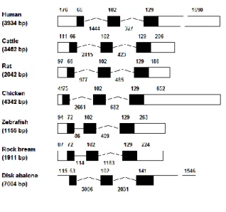
Figure 6: Schematic representation and comparison of rock bream and disk abalone cystatin B genomic structures with that of other species
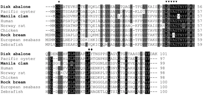
Figure 7: Alignment of the RbCyt B, AbCyt B and McCyt B amino acid sequences with selected known cystatin B sequences
Figure 8: Predicted three-dimensional structure of RbCyt B, AbCyt B and McCyt B Figure 9: Phylogenetic tree of cystatin superfamily members from different species Figure 10: SDS-PAGE analysis of over-expressed and purified recombinant fusion
proteins
Figure 11: Papain inhibitory activity profile of recombinant fusion proteins Figure 12A: Tissue-specific mRNA expression of RbCyt B in healthy rock breams Figure 12B: Tissue specific mRNA expression of AbCyt B in healthy disk abalones Figure 12C: Tissue specific mRNA expression of McCyt B in healthy Manila clams Figure 13: RbCyt B mRNA expression in E. tarda-infected head kidney and spleen Figure 14: AbCyt B mRNA expression after bacterial challenge in gill and hemocytes Figure 15: Expression profile of McCyt B in gill and hemocytes upon V. tapetis, LPS and
xi
LIST OF TABLES
Table 1: Description of the primers used in the study
Table 2A: Amino acid identity and similarity of RbCyt B to other known cystatin B proteins
Table 2B: Amino acid identity and similarity of AbCyt B to other known cystatin B proteins
Table 2C: Amino acid identity and similarity of McCyt B to other known cystatin B proteins
xii
ABBREVIATIONS
‰ Parts per thousand
3D Three dimension
AbCyt B Disk abalone cystatin B
AIDS Acquired immunodeficiency syndrome
Am Adductor muscle
ANOVA Analysis of variance
BAC Bacterial artificial chromosome BLAST Basic local alignment search tool
bp Base pair(s)
cDNA Complementary deoxyribonucleic acid CFU Colony forming unit(s)
C-terminal Carboxyl terminal, COOH-terminus
CY Cystatin-like domain
DNA Deoxyribonucleic acid
dNTP Deoxynucleotide-triphosphate
Dt Digestive tract
ECM Extra cellular matrix
EPMI Progressive myoclonic epilepsy type 1 EST Expressed sequence tag(s)
FAO Food and Agriculture Organization of the United Nations
Ft Foot
Gl Gill
GS-FLX Genome sequencer FLX
Hc Hemocytes
HMWK High molecular weight kininogens
Hp Hepatopancreas
IFN Interferon
IL Interleukin
IPTG Isopropyl β-D-1-thiogalactopyranoside
Kb Kilobase(s)
kDa Kilo Dalton
KGP Lys-gingipain
LB Luria-Bertani
LMWK Low molecular weight kininogens
LPS Lipopolysaccharides
MBP Maltose binding protein McCyt B Manila clam cystatin B
xiii MHC Major Histocompatibility Complex MMP Matrix metalloproteases
mRNA Messenger RNA
Ms Muscle
Mt Mantle
NCBI National Center for Biotechnology Information
NC-IUBMB Nomenclature Committee of the International Union of Biochemistry and Molecular Biology
nm Nanometer(s)
NO Nitric oxide
N-terminal Amino terminal, NH2-terminus
OD Optical density
ORF Open reading frame
p.i. Post infection/ post induction PAGE Polyacrylamide gel electrophoresis PBS Phosphate buffered saline
Poly I:C Polyinosinic:polycytidylic acid PPB Potassium phosphate buffer
qPCR Quantitative real-time polymerase chain reaction rAbCyt B Recombinant disk abalone cystatin B fused with MBP RbCyt B Rock bream cystatin B
RCSB Research Collaboratory for Structural Bioinformatics
RGP Arg-gingipain
rMcCyt B Recombinant Manila clam cystatin B fused with MBP rProtein Recombinant protein(s) fused with MBP
rRbCyt B Recombinant rock bream cystatin B fused with MBP
SDS Sodium dodecyl sulfate
Sp Siphon
STAT Signal transducer and activator of transcription
Taq Thermus aquaticus
TF Transcription factor
TNF Tumor necrosis factor
TSS Transcription start site
Tt Testis
UTR Untranslated region
α Alpha
β Βeta
γ Gamma
- 1 -
1. INTRODUCTION
Importance of the Three Experimental Marine Organisms
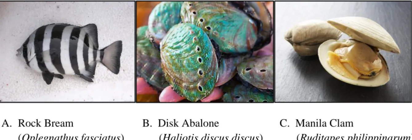
Over the past decade, marine food sources have overtaken a large portion of the world food market, representing a promising supply of nutritional resources to compensate for the decrease in terrestrial sources that has resulted from overconsumption. This trend has promoted mariculture to the status of an economically important component of the global economy. Aquaculture industry has been developed with the advancing scientific technology and mariculture is one of the rapid developing industries in worldwide which provide essential fatty acids and essential amino acids along with enriched source of other nutrients. Many fish and shellfish species are used either as cultured or capture fisheries, where rock bream (Oplegnathus fasciatus) (Fig. 1A) is one of the most economically important and widely consuming fish species, mostly in eastern Asia. However, recently the decrease production was associated with disease outbreaks, especially the viral diseases (irido virus), bacterial diseases (Edwardsiella tarda) and unfavorable environmental conditions with the growing intensified industry. In contrast, shellfish culture also makes huge contribution to the industry and to the national economy especially in East Asian countries including Korea, Japan and China. Disk abalone (Haliotis discus discus) (Fig. 1B) is a valuable marine gastropod species (Phylum: mollusks), widely distributed and cultured in Asia (McBride, 1998). The flesh of abalones is considered to be a desirable and highly nutritious food, and is consumed either in raw or cooked form in a variety of different dishes. Manila clam (Ruditapes philippinarum) (Fig. 1C) also is an edible marine bivalve mollusk species, which is harvested in large scale by the mariculture industry, and naturally distributed in intertidal zones of Yellow sea bordering countries such as Korea, Japan, China and Philippines. Later, it was introduced as commercially valuable species to many parts of the world (Goulletquer, 1997) and according to the FAO fishery statistics current global aquaculture production of
- 2 -
Manila clam exceeds 3.5 million tons per year (FAO Fishery Statistics, http://www.fao.org/fishery/culturedspecies/Ruditapes_philippinarum/; accessed on 27/10/2012). However, significant mortality is present in marine mollusks including disk abalone and Manila clam aquaculture due to many reasons, including pathogens, stressful environments, pollutants, and disease outbreaks (Villena, 2003; Hooper et al., 2007). Recently, disease management is taken the most important and critical factor to minimize the production losses which target basically either by pathogen control with chemotherapeutics or by host protection with vaccines and immune-stimulants (Park, 2009). As such, there is significant interest in gaining a detailed understanding of the host-pathogen interactions and immune modulations in these mariculture creatures to prevent drastic losses of their harvest due to different pathogenic infections and the studies on immunogenetics will provide more precise approaches to develop new strategies of efficient disease control.
A. Rock Bream B. Disk Abalone C. Manila Clam
(Oplegnathus fasciatus) (Haliotis discus discus) (Ruditapes philippinarum)
Fig. 1. Three marine species used in the research.
Cysteine Proteases and Their Multifunctional Role in Cellular Biology
Proteolysis is a universal mechanism mediated by proteases (also termed as peptidase and proteinase) where amide bonds (peptide bonds) hydrolyzed in protein catabolism. Proteases occur naturally in all organisms and are involved in multifaceted roles of physiological reactions from simple food protein digestion to complicated immune signaling
- 3 -
pathways (e.g., the blood-clotting cascade, the complement system, apoptosis pathways, and the invertebrate prophenoloxidase-activating cascade) (Vasiljeva et al., 2007; Brix et al., 2008). With the completion of human genome study, recently it was reported that about 2% of the all gene products are proteases in human genome (Turk and Turk, 2008).These proteases are currently categorized into six broad groups depending on the reactive groups at the active site involved in the catalysis by the International Union of Biochemistry and Molecular Biology, into serine proteases (EC 3.4.21), cysteine proteases (EC 3.4.22), aspartate (EC 3.4.23), metalloproteases (EC 3.4.24), threonine proteases (EC 3.4.25) and another unknown type of proteases (Nomenclature Committee of the International Union of Biochemistry and Molecular Biology (NC-IUBMB)). Cysteine proteases, the most well studied protease is the thiol (-SH)-dependent group, which utilizes cysteine residues as nucleophiles to attack the amide bonds of a target protein (Chapman et al., 1997; Potempa et al., 2005), have been found in viruses, fungi, bacteria, protozoa, plants and mammals (Barrett, 1986; Mizuno et al., 1987; Sharma et al., 1989). The cysteine proteases classified into 21 families (C1–C21) according to distinctive sequences and tertiary structures; however, the majority of cysteine proteases belong to the C1 family which are largely known as papain-like proteases including the lysosomal cysteine proteases, cathepsins (Dubey et al., 2007). Traditionally, cysteine proteases had been known as the lysosomal mediators of terminal protein degradation; however recent findings in human biology have been illustrated more expanded role of cysteine proteinase including apoptosis, MHC class II immune responses, prohormone processing, and extracellular matrix remodeling which is important to the bone development (Chapman et al., 1997). Cysteine cathepsins are lysosomal proteases with housekeeping as well as highly specialized functions, for an example, matrix metalloproteases (MMP) are primarily responsible for the homeostasis of the extracellular matrix, whereas under disease conditions, cysteine proteases contribute to destruction of
- 4 -
extracellular matrix (Cudic and Fields, 2009; Bromme and Wilson, 2011). In the pathogenic micro-organisms including bacteria, fungi, viruses and parasites, cysteine proteases can act as virulence factors, which promote diseases in the host organisms (Takahashi et al., 1994; Mottram et al., 2004; Rudenskaya and Pupov, 2008). For an example, proteolytic enzymes produced by Porphyromonas gingivalis are important virulence factors, produced in large quantities to cause periodontal disease. It has been shown that these proteases can directly or indirectly degrade constituents of the periodontal tissues, destroy host defense elements, dysregulate coagulation and complement kallikerin-kinin cascades (Dubey et al., 2007). Furthermore, parasite derived cysteine proteases were reported to play key roles in immunoevasion, enzyme activation, virulence, tissue and cellular invasion as well as excystment, hatching and moulting (Sajid and McKerrow, 2002). Thus, many of imbalanced activity of cysteine proteinases may lead to wide range of human diseases including cancer (Joyce et al., 2004; Turk et al., 2004), rheumatoid arthritis, osteoarthritis, osteoporosis (Yasuda et al., 2005; Vasiljeva et al., 2007), tumorigenesis and multiple sclerosis (Berdowska and Siewinski, 2000) along with neurological disorders. Therefore, certainly the eukaryotic systems have evolved precise regulatory mechanisms for cysteine proteinases, modulating their expression, secretion, and maturation through specific degradation of mature enzymes and targeted inhibition largely through interaction with their endogenous inhibitors called cystatins (Rzychon et al., 2004).
Cysteine Protease Inhibitors
The activity of the cysteine proteinases are regulated by naturally occurring inhibitory proteins such as α2-macroglobulin and cystatins (Bobek and Levine, 1992). These inhibitors protect the cells from destructive endogenous or external proteolysis and/or could be involved in the control mechanism responsible for intracellular or extracellular protein
- 5 -
degradation by cysteine proteinases of host, bacterial, fungi, parasites and viral origin. The first cysteine proteinase inhibitor to be isolated and characterized was obtained from chicken egg white in 1963 and was shown to inhibit ficin, papain (Fossum and Whitaker, 1968; Sen and Whitaker, 1973) and cathepsin B and C (Keilova and Tomasek, 1974). Later, the name "cystatin" was applied to this protein (Barrett, 1981), and it was shown to be a very potent inhibitor of other cysteine proteinases of the papain superfamily, such as cathepsin H and L (Anastasi et al., 1983). Subsequently, cystatins have been isolated from many tissues and body fluids of humans and different animal species (Turk et al., 1985; Barrett et al., 1986) as well as from plants such as rice (Abe et al., 1988) and soybean (Brzin et al, 1990).
Cystatin Superfamily
The cystatins are competitive, reversible, tight binding cysteine proteinase inhibitors which display structural and functional similarities. Cystatins are considered as the largest and the best described group of natural exosite binding cysteine proteinase inhibitors, which obstruct the access of substrate without interacting with the enzyme catalytic center (Bode and Huber, 2000). Cystatins predominantly interact with cysteine proteases, such as the plant-derived papain and human lysosomal cathepsins B, H, and L (Barrett, 1987; Turk and Bode, 1991). The cystatins are now considered as cystatin superfamily, which is mainly divided into three families on the basis of their location, size and complexity of polypeptide chains as type 1 cystatins (Stefins) (Stefins A and B, also known as cystatins A and B), type 2 cystatins (Cystatins), and type 3 cystatins (Kininogens). Family 1 cystatins are comprised of cystatin A and B, which are unglycosylated inhibitors and characterized by low molecular weight (∼11 KDa) and a single cystatin-like (CY) domain structure. This group of cytoplasmic proteins is composed of ∼100 amino acid residues that lack of disulfide bond, signal peptide, and carbohydrate side chain (Barrett et al., 1986; Turk and Bode, 1991).
- 6 -
Family 2 cystatins characterized with molecular masses in the range of 13–14 kDa, and contain signal peptide and disulfide bonds at the carboxy terminus. However, some members may also be glycosylated or phosphorylated. Examples of these are cystatins E/M (glycosylated on N108) and cystatins S and SN (consensus phosphorylation sites at S2 and S98, respectively). The family 2 cystatins include cystatins C, D, E/M, F, G, S, SA, and SN (Cornwall and Hsia, 2003). In contrast to stefins, cystatins contain a signal sequence for secretion through the cell membrane to the extracellular space. Family 3 cystatins (kininogens) exhibit high structural complexity with more than one CY domain, which are large multifunctional glycoproteins with inhibitory activities (Kellermann et al., 1987). Kininogens are found in the plasma and secretions of mammalian species. Three types of kininogens are identified to date as the low-molecular-weight (LMWK), high-molecular-weight (HMWK), and T-kininogens which are single-chain glycoproteins with the molecular weight of ~50 to 120 kDa. They are comprised of three CY domains that resulted from gene duplication. They contain additional disulfide bonds and are also glycosylated. Kininogens have an additional polypeptide at the C terminus containing the bradykinin sequence, which can be cleaved by kallikrein (Bobek and Levine, 1992). Recently, another category of the cystatin superfamily, family 4, was reported. This new family includes cystatins with invertebrate origin and its members are mostly from parasitic nematodes (Khaznadji et al., 2005; Li et al., 2010).
Structure and Function of Cystatins
All cystatins contain a binding site that binds to the catalytic site of papain-like proteinases, and inhibits them reversibly. This site is created by several conserved regions of the protein, including a glycine in the N-terminal region, a central glutamine-valine-glycine (QxVxG) motif in first hairpin loop of the protein, and a PW motif in the
- 7 -
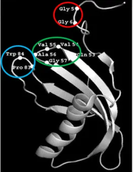
second hairpin loop in the C terminal region (Björk and Ylinenjärvi, 1989; Margis et al., 1998; Rzychon et al., 2004). Each cystatin structure has a core of five-stranded anti-parallel β-sheets wrapped around a central five turn α-helix as shown by the ribbon model of the structure (Fig. 2).
Fig. 2. Oryzacystatin three-dimensional structure. Ribbon
representation of Oryzacystatin shows a five turn α-helix and five stranded antiparallel β-pleated sheet. The amino acids of the three functional domains of cystatins are encircled. (Source: Rodriguez-Mahillo et al., 2007).
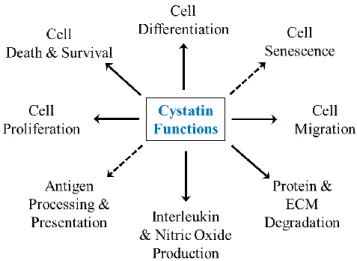
Like cysteine proteinases, cystatins have been found in diverse organisms. Cystatins are involved in both biological and pathological processes which cysteine proteinases participate in, including protein homeostasis, inflammatory responses, antigen processing, metastasis, immune responses, and cathepsin dependent apoptosis (Synnes, 1998; Abrahamson et al., 2003; Kopitar-Jerala, 2006; Lefebvre et al., 2008; Shah and Bano, 2009). Additionally, there are many diseases observed with decreased cystatin levels, such as cancer, inflammatory diseases, osteoporosis, diabetes, neurodegenerative diseases and renal failure (Fi. 3) (Turk and Turk, 2008).
- 8 -
Fig. 3. Diverse biological functions of the cystatins. The cytoplasmic stefins and cell-secreted cystatins have been implicated in a multitude of biological processes, including cell differentiation, proliferation, survival, migration and interleukin production, leading under some instances to the increased production of NO. (Source: Keppler, 2006).
Cystatin in health and diseases
A large number of normal and pathological processes in the cellular environment are controlled by the balance between proteases and their inhibitors. The general physiological role of cystatins is believed to be the protection of cells from inappropriate endogenous or exogenous proteolysis by regulating the activity of cysteine proteinases both of host and of microbial origin. An increase level in local proteinases and proteinase inhibitors has been reported in the presence of inflammation (Keilova and Tomasek, 1977; Jarvinen, 1978), acquired immunodeficiency syndrome (AIDS) (Alavaikko et al, 1985), and malignancy (Okumichi et al., 1984; Sloane and Honn, 1984). It was also suggested that the cysteine proteinase, cathepsin B play a role in the cancer cell migration from their site of origin to secondary or metastatic sites by where they dissolve the extracellular matrix proteins to pave the way; however cystatins play a significant role at here to inhibit the cathepsin activity (Sloane and Honn, 1984). Cystatins have been shown to possess a wide range of effects in immune cells. For an example, cystatins involve in nitric oxide production by induction of tumor necrosis factor alpha (TNFα) and interleukin (IL) 10 synthesis through the interferon
- 9 -
gamma (IFNγ) activated murine macrophages. In turn, nitric oxide has inhibitory activity on cysteine proteases, especially those from parasitic protozoa (Vray et al., 2002). Furthermore, in the neurodegenerative disorder known as progressive myoclonus epilepsy of Unverricht-Lundborg type 1 (EPMI), a significant reduction in cystatin B activity and increased levels of cathepsin B, L, and S were observed in lymphoblastoid cells of EPMI (Rinne at al., 2002).
Cystatin B
Among cystatins, cystatin B is an intracellular cysteine proteinase inhibitor which is a tightly-binding reversible inhibitor of cathepsins by forming a dimer stabilized by non-covalent forces to inhibit C1 family cysteine proteases; papain and cathepsin B, H, and L (Anastasi et al., 1983; Cimerman et al., 2001; Lefebvre et al., 2008). Cystatin B (also known as stefin-B), was originally discovered as an inhibitor of cathepsin B (Lenney et al., 1979) and showed wide distribution among different cell types and tissues (Turk and Bode, 1991). Apart from its anti-protease activity, cystatin B has been shown to be involved in immune responses to bacterial and fungal infections, and in anti-apoptotic processes in the cerebellum (Takahashi et al., 1994; Di Giaimo et al., 2002). Cystatin B was also thought to play a role in protection against the proteinases leaking from lysosomes, and in biological defense system against invaders (Lefebvre et al., 2004; Li et al., 2010; Xiao et al., 2010). In contrast, defects in cystatin B gene cause progressive myoclonic epilepsy type 1 (EPM1), which is an autosomal recessive disorder characterized by severe, stimulus-sensitive myoclonus, and tonic–clonic seizures in human (Pennacchio et al., 1996; Riccio et al., 2005). Furthermore, the genes involve in proteolysis, apoptosis and glial activation including, cathepsin S, C1q B-chain of complement, β2-microglobulin, glial fibrillary acidic protein, apolipoprotein D, fibronectin 1 and metallothionein II were found to elevate the transcript level in neurological tissues from cystatin B knockout mice (Lieuallen et al., 2001). Hence, cystatin B is unique
- 10 -
among the members in cystatin superfamily which is involved in variety of pathological processes and having a unique structure containing a free cysteine in the N-terminal segment of the proteinase-binding region that facilitates the tight binding of target proteinases. Cystatin B reversibly inhibits the papain-like cysteine proteinases, and was found to play a protective role in organisms due to its omnipresent distribution in cells and tissues (Rahman et al., 1983; Abrahamson, 1994). It most likely prevents inappropriate proteolysis caused by the action of lysosomal cysteine proteinases, primarily the cathepsins B, H, K, L, and S (Turk et al., 1997). In addition, cystatin B has been implicated in innate immune responses to bacterial infections in vertebrate and invertebrate lineages including human, turbot fish (Scophthalmus maximus) and leech (Theromyzon tessulatum) (Suzuki et al., 2000; Lefebvre et al., 2004; Xiao et al., 2010), as well as in responses against viral infections (Luciano-Montalvo and Melendez, 2009). These observations may reflect the potential inhibitory action of cystatin B toward pathogen-derived cysteine proteinases which are secreted as virulent factors or as suppressors of host cysteine proteinases to protect the cells from inappropriate proteolysis.
Hence, this study has been focused on the molecular characterization of the cystatin B gene from three different marine species, including one teleost and two mollusk species; to investigate the role of cystatin B-like proteins in marine organisms from genomic to protein level. Furthermore, to understand the role in innate immune response, the temporal mRNA expression analysis was employed after different pathological infections.
- 11 -
2. MATERIALS AND METHODS
2.1. Experimental animals
2.1.1. Rock bream (Oplegnathus fasciatus)
Healthy rock breams with an average body weight of 45-50 g were provided by the National Fisheries Research and Development Institute (Republic of Korea), and they were maintained in 40 L tanks at 24 ˚C with sand-filtered, aerated seawater (salinity of 34 ± 1 ‰) at the Marine Molecular Genetics Laboratory of Jeju National University. Two weeks of acclimatization was carried out before any experimentation conducted. The fish were fed daily with commercial feed.
2.1.2. Disk abalone (Haliotis discus discus)
Healthy disk abalones were purchased from ‘Youngsoo’ commercial abalone farm in Jeju Island, Republic of Korea. They were maintained as 40 animals per tank in flat bottomed tanks (250 L) with sand-filtered aerated seawater at a salinity of 34 ‰ at 18 ±1˚C during the experimental period at Marine and Environmental Research Institute of Jeju National University. Abalones were acclimatized for 7 days before the experiment, and they were fed with fresh sea weed, Undaria pinnatifida, during the acclimatization period.
2.1.3. Manila clam (Ruditapes philippinarum)
Manila clams with average shell length of 35±5 mm were collected from the Eastern coastal region of the Jeju Island (Republic of Korea) and thereafter maintained in 250 L fiberglass tanks with controlled conditions same as in abalones at Marine and Environmental Research Institute of Jeju National University.
- 12 -
2.2. Identification of cystatin B sequences from three species
Rock bream cystatin B (RbCyt B) was originally identified by pyrosequencing of normalized cDNA using Roche GS-FLX system (DNA Link, Republic of Korea) as described in a previous report (Whang et al., 2011a). The RbCyt B cDNA sequence was identified by applying the basic local alignment search tool (BLAST; National Center for Biotechnology Information (NCBI), http://www.ncbi.nlm.nih.gov/BLAST). One of the ESTs was found to be homologous to known cystatin B. This sequence was cloned by amplification with gene-specific primers designed to the oligonucleotide sequences (RbCytB F2 and RbCytB R2; Table 1). Similarly, the full length cDNA sequence of abalone cystatin B (AbCyt B) was identified by analyzing the previously constructed abalone expressed sequence tag (EST) database sequences (Munasinghe et al., 2006). BLAST analysis indicated that one of the ESTs was homologous to previously identified cystatin Bs. This sequence was selected to design the BAC library screening primers and quantitative real-time polymerase chain reaction (qPCR) primers used in the study (Table 1). Finally the complete cDNA sequence encoding for Manila clam cystatin B (McCyt B) was identified using the same alignment tool at NCBI from our previously established Manila clam cDNA sequence database (Lee et al., 2011).
- 13 -
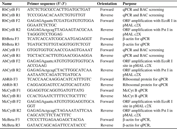
Table 1. Description of the primers used in the study
Name Primer sequence (5'-3') Orientation Purpose
RbCytB F1 ATCTCTGCGCCACTTGATGCTGAT Forward qPCR and BAC screening RbCytB R1 TCCCGGACACAATCTGTGTTGT Reverse qPCR and BAC screening RbCytB F2 GAGAGAgaattcTCGATGATGTGTGGA
GGAATCTCTGC
Forward ORF amplification with EcoR I in pMAL c2X
RbCytB R2 GAGAGActgcagTTAGAAGTACGCAA TAGGGTCCTGGAG
Reverse ORF amplification with Pst I in pMAL c2X
RbBeta F3 TCATCACCATCGGCAATGAGAGGT Forward β-actin for qPCR RbBeta R3 TGATGCTGTTGTAGGTGGTCTCGT Reverse β-actin for qPCR
AbCytB F1 GTGGTGGTGCAACCGAAGTGAAAT Forward qPCR and BAC screening AbCytB R1 TGCTACCACTTGTGAACGGAAGGA Reverse qPCR and BAC screening AbCytB F2 GAGAGAgaattcATGTGTGGTGGTGCA
ACCGAAG
Forward ORF amplification with EcoR I site in pMAL c2X
AbCytB R2 GAGAGActgcagCTACTTGGCATCAA AATAATCCAGATCTGATGCA
Reverse ORF amplification with Pst I in pMAL c2X
AbRib F3 TCACCAACAAGGACATCATTTGTC Forward Ribosomal protein for qPCR AbRib R3 CAGGAGGAGTCCAGTGCAGTATG Reverse Ribosomal protein for qPCR McCytB F1 GGAGGTGCAGGTGATGTTATG Forward McCyt B qPCR
McCytB R1 CCACTGAATCTTTTCCTGCTTTC Reverse McCyt B qPCR McCytB F2 GAGAGAgaattcATGTGTGGAGGTGCA
GGT
Forward ORF amplification with EcoR I site in pMAL c2X
McCytB R2 GAGAGActgcagCTAGAAATATTCAA CAGCATCTTCTACTTTC
Reverse ORF amplification with Pst I in pMAL c2X
McBeta F3 CTCCCTTGAGAAGAGCTACGA Forward β-actin for qPCR, McBeta R3 GATACCAGCAGATTCCATACCC Reverse β-actin for qPCR,
2.3. Bacterial artificial chromosome (BAC) library construction and screening
Two separate BAC libraries of O. fasciatus and H. discus discus were constructed by Lucigen® Co. (Middleton, Wisconsin) using randomly sheared genomic DNA from rock bream whole blood cells and disk abalone gill tissues. Briefly, genomic DNA was randomly sheared and blunt end of large inserts (>100 kb) was ligated into pSMART® BAC vector to obtain an unbiased, full coverage library. Around 92,160 clones for each, possessing an average insert size of 120 Kb were arrayed in 240 of 384-well microtiter plates separately in each library. BAC clones inoculated into 384-deep well plates were grown individually. Aliquots of the grown cultures were pooled with other clones from the same plate, row or column pools for DNA preparation. Screening of the BAC-library was carried out with a PCR-based method (TaKaRa Bio, USA) following the manufacturer’s instructions using gene-specific primers (Table 1). The identified clones were isolated from the corresponding
- 14 -
wells and confirmed by colony PCR with gene-specific primers. After confirmation, BAC DNA from positive clone was isolated and purified using QIAGEN Large-Construct Kit, following the manufacture’s protocol and was subjected to sequencing by Roche (454) Genome Sequencer FLX (GS-FLX™) system (DNA Link, Inc., Korea).
2.4. In silico analysis of DNA and protein sequences
The amino acid sequences corresponding to RbCyt B, AbCyt B and McCyt B coding sequences were derived using DNAssist 2.2 (version 3.0). The orthologous sequences of cystatin B were compared by the BLAST search program at NCBI. Pairwise sequence alignment and multiple sequence alignment were performed by the EMBOSS Needle and ClustalW2 programs, respectively (http://www.ebi.ac.uk/Tools) (Thompson et al., 1994). Prediction of characteristic protein domains and conserved regions was carried out by the ExPASy-prosite database (http://prosite.expasy.org), MotifScan scanning algorithm (http://myhits.isb-sib.ch/cgi-bin/motif_scan) and Simple Modular Architecture Research Tool (http://smart.embl-heidelberg.de/). Some of the physicochemical properties of amino acid
sequences were determined by the ExPASy ProtParam tool
(http://web.expasy.org/protparam). Phylogenetic analysis was carried out by the Neighbor-Joining method with bootstrapping values taken from 1000 replicates using Molecular Evolutionary Genetics Analysis (MEGA) software, version 5 (Tamura et al., 2011).
The identified BAC clone sequences from rock bream and abalone BAC libraries were used to analyze the genomic structures and promoter regions. The potential transcription factor (TF) binding sites in the RbCyt B and AbCyt B promoter regions were predicted using the TFSEARCH version 1.3 (http://www.cbrc.jp/research/db/TFSEARCH.html) online database with threshold score at 90.0. The exon-intron structure was predicted by aligning the cDNA sequence with genomic sequence using the Spidey program
- 15 -
(http://www.ncbi.nlm.nih.gov/IEB/Research/Ostell/Spidey). Genome structures of cystatin B from other species were obtained from the Ensembl Genome Browser (http://www.ensembl.org/index.html) and visualized using Gene Maper version 2.5 (http://genemaper.googlepages.com) in order to compare the structures.
In order to reveal the tertiary structure of the three cystatin B molecules, computer simulation model was generated using I-TASSER (Roy et al., 2010; Zhang, 2008) online server and visualized the three dimensional (3D) structure using PyMOL Molecular Graphic System 1.3 and POV-Ray for windows v3.62 softwares.
2.5. Cloning and over-expression of recombinant cystatin B
To generate the recombinant proteins (rproteins) fused with maltose binding protein (MBP), the complete open reading frames (ORFs) were amplified from respective cDNAs by PCR with respective cloning primers (Table 1) containing EcoR I and Pst I restriction sites. The PCR amplification was carried out in a TaKaRa thermal cycler (TaKaRa Korea Biomedical Inc., Korea) using a 50 µL reaction mixture composed of 5 U of ExTaq polymerase (TaKaRa), 5 µL of 10x ExTaq buffer, 8 µL of 2.5 mM dNTPs, 80 ng of template, and 20 pmol of each primer. The thermal cycling conditions were: initial incubation at 94 ˚C for 3 min, followed by 35 cycles of 94 ˚C for 30 s, 55 ˚C for 30 s and 72 ˚C for 30 s, and a final extension at 72 ˚C for 2 min. The amplified products and pMAL-c2X vector were digested with the appropriate restriction enzymes, and confirmed by agarose gel electrophoresis. Appropriate sized fragments were excised and purified using the Accuprep Gel Purification Kit (Bioneer Co., Daejeon, Korea) and ligated into the linearized pMAL-c2X MBP-fused expression vectors by incubation with Mighty Mix (TaKaRa) for overnight at 4 ˚C. Cloned vectors were transformed into Escherichia coli DH5α cells. The size of the construct and sequences of the ORFs were confirmed by electrophoresis and sequencing,
- 16 -
respectively (Macrogen Inc., Korea). The recombinant fusion constructs were transformed in to E. coli BL21 (DE3) cells and grown on agar plates. A single colony was selected, inoculated in 5 mL of Luria-Bertani (LB) broth supplemented with 100 μg/mL of ampicillin, and grown overnight at 37 ˚C. Then, 1 mL of the inoculum was transferred into 500 mL of LB broth containing ampicillin, and grown at 37 ˚C in a shaking incubator (200 rpm). When the optical density (OD) reached 0.6 (at 600 nm), cultures were induced with isopropyl-β-thiogalactopyranoside (IPTG) to a final concentration of 0.2 mM. After incubation at 20 ˚C for 8h, the cells were harvested by centrifugation and the pellet was re-suspended in column buffer (20 mM Tris-HCL, pH 7.4 and 200 mM NaCl). Recombinant cystatin Bs fused with MBP (rRbCyt B, rAbCyt B and rMcCyt B) were purified using the pMAL protein fusion and purification system (Maina et al., 1988). Samples collected at different steps in expression and purification process were analyzed on a 12% sodium dodecyl sulfate-polyacrylamide gel electrophoresis (SDS-PAGE) under reduced conditions, with low molecular weight protein marker (Enzynomics, Korea). The resulted gel was stained with 0.05% Coomassie blue R-250, followed by a standard destaining procedure. The purified rprotein concentration was measured according to the Bradford method (Bradford, 1976) using bovine serum albumin (BSA) as the standard.
2.6. Cysteine protease inhibition assay
To analyze the activity of purified recombinant proteins (rRbCyt B, rAbCyt B and rMcCyt B), in vitro papain inhibitory assay was performed as reported previously, with some modifications (Xiao et al., 2010). Briefly, papain (P3375; Sigma-Aldrich, USA) was dissolved in 0.1 M potassium phosphate buffer (PPB), pH 7.4, to obtain 1 mg/mL solution. Subsequently, different volumes of the rprotein and papain were mixed together to generate the following rProtein/papain concentration ratios in 125 µL of final volumes: 0/1 (control),
- 17 -
1/32, 1/16, 1/8, 1/4, 1/2, 1/1, 1.5/1. The mixtures were then incubated at 25 ˚C for 10 min, after which 125 µL of azo-casein (0.5% in PPB) was added to the mixture and was allowed to react by incubating at 37 ˚C for 30 min. The reaction was terminated by adding an equal volume of 10% trichloroacetic acid. The same procedure was followed simultaneously for the MBP as the control and both positive (without rprotein) and negative (without papain) controls were implemented. Finally, the mixture was centrifuged and the supernatant was subjected to measure the OD440. The relative activity was calculated as: 100 × [1-(OD440 of test sample /OD440 of control)] (Xiao et al., 2010). All the samples were tested in three replicates at each point.
2.7. Isolation of tissues from experimental animals
Healthy animals described in section 2.1 were selected to examine the tissue specific mRNA expression profile of cystatin B gene. Animals were carefully dissected on ice and tissues (blood, gills, liver, spleen, brain, intestine, kidney, head kidney, and muscle from rock bream; gills, mantle, muscle, hemolymph, hepatopancreas, digestive tract and testis from abalone and hemolymph, adductor muscle, mantel, siphon, gills and foot from clam) were isolated carefully from three replicate animals. The blood from rock breams was collected using a heparin contained syringe fitted with a 22-gauge needle from the caudal vein. Collected blood was immediately transformed into micro tubes and centrifuged at 3000× g for 10 min at 4 ˚C to collect the blood cells. The abalone hemolymph was withdrawn from the cephalic arterial sinus, accessed anteriorly at the angle between the foot and the head using a syringe fitted with a 22-gauge needle. Collected hemolymph was immediately transferred into micro tubes and was centrifuged as previous. The supernatant was removed and hemocytes were collected. Hemolymph (1-2 mL/clam) from clam was collected from the posterior adductor muscle using a 26 G syringe as same to the abalone to collect the
- 18 -
hemocytes. All the tissue samples were snap-frozen in liquid nitrogen, and stored at -80 ˚C until use for RNA extraction.
2.8. Immune challenge experiment for temporal mRNA expression analysis
Rock breams were immune-challenged with live Edwardsiella tarda bacterium. O.
fasciatus were intra-peritoneally (i.p) injected either with 100 μL live E. tarda (5 × 10³
CFU/μL) in phosphate buffered saline (PBS) or equal volume of PBS per fish as described previously (Whang et al., 2011b). Tissue samples from head kidney and spleen were collected at 0h (un-injected control), 3h, 6h, 12h, 24h and 48h post-infection (p.i.). Three replicates of fish were obtained from each group to isolate tissues at each time point and, pooled tissue samples were subjected to total RNA extraction. Tissue collection was conducted as described in section 2.7.
To determine the immune responses of the AbCyt B, immune stimulation/challenge experiment was devised and conducted using pathogenic Gram-negative bacterial species,
Vibrio parahaemolyticus. Animals were injected with 100 μL of 1×10⁴ CFU/mL of bacteria in saline (0.9% NaCl) and the same volume of saline injected group was used as a control. Gill and hemolymph tissue samples were collected at different time points (0, 3, 6, 12, 24 and 48 h) after the infection as described in section 2.7 and tissues were snap-frozen and stored at -80 ˚C. Four replicates were used to collect the tissues at each time point.
Clams were challenged with Vibrio tapetis (a Gram negative bacterial pathogen), LPS (an endotoxin of Gram negative bacterial cell walls), or poly I:C. As described previously, 100 µL of V. tapetis (1.9×10⁶ cells/mL in 0.9% saline), LPS (100 μg/clam in 0.9% saline), or poly I:C (100 μg/clam in 0.9% saline) was injected directly into adductor muscle (Umasuthan et al., 2012). A group of un-injected clams served as negative controls. Gills and hemocytes
- 19 -
tissues were collected at 0 (un-injected), 3, 6, 12, 24 and 48 h of p.i. from control and challenged groups, including five clams from each group. Total RNA was extracted from pooled tissue samples using at least five clams and used for cDNA synthesis.
2.9. Total RNA extraction and cDNA synthesis
Total RNA was isolated from pooled tissues at each time point from three different species separately, using the TRIzol reagent (Sigma-Aldrich) according to the manufacturer’s instructions. Concentration of purified RNA was determined by measuring at A260 on a spectrophotometer (BioRad, USA). Purified RNA samples were diluted to 1 μg/μL and cDNAs were synthesized from 2.5 µg of RNA by using the PrimeScriptTM First Strand cDNA Synthesis kit (TaKaRa Bio Inc., Japan), following the manufacturer’s protocol. The resultant cDNA was then diluted 40-fold (total 800 µL) before storage at -20 ˚C.
2.10. Quantitative real-time PCR (qPCR) analysis
Synthesized cDNA was used to quantify the mRNA expression level in different tissues at different time points in both the immune-challenged and control groups by the qPCR technique using gene-specific primers (Table 1). Each reaction was carried out in a 15 μL of reaction mixture containing 4 μL of 1:40 diluted template cDNA, 7.5 μL of 2× SYBR Green Mix, 0.6 μL of each primer (10 pmol/μL), and 2.3 μL of PCR-grade water, and by using the Thermal Cycler Dice Real Time System (Model TP800; TaKaRa Bio Inc.). The real time (RT)-PCR cycle program consisted of one cycle at 95 ˚C for 10 s, followed by 45 cycles of 95 ˚C for 5 s, 58 ˚C for 20 s, and 72 ˚C for 20 s, and finally, 1 cycle of 95 ˚C for 15 s, 60 ˚C for 30 s and 95 ˚C for 15 s. The baseline was automatically set to maintain consistency. The mRNA expression was determined by the 2−∆∆CT (Livak) method (Livak and Schmittgen,
- 20 -
2001) and was tested in triplicates. Beta-actin gene expression was detected as the internal control (Accession No. FJ975145) for the rock bream and all the RbCyt B expression values were normalized to the beta-actin values. The tissue specific expression values were compared with that of muscle and immune challenged values were normalized to the PBS control at each time point and compared with non-injected (0h) control. For abalones, the relative mRNA expression was calculated by first normalizing to expression values of the gene codes for abalone ribosomal protein L5 (Wan et al., 2011) as the internal control (Accession No. EF103443) and then was normalizing to the expression values of the saline injected control group at each time point and compared with un-injected (0h) control . Tissue specific values were compared with expression in muscle. In Manila clam, beta-actin gene expression was used as the internal control (Zhang et al., 2011). The expression level of adductor muscle was considered as the basal level, by which expression in all other tissues was compared. To determine the relative expressions, the observed expression after challenge was first normalized to the beta-actin and saline-injected controls and then compared with expression in the unchallenged (0h) group.
2.11. Statistical analysis
To determine the statistical significance between the experimental and control groups, all the mRNA expression analysis data were subjected to either Student's t-test or one-way analysis of variance (ANOVA) in SPSS 16.0 for Windows (SPSS, Chicago, IL, USA). Differences were considered statistically significant at P < 0.05 and all the data were represented as means ± standard deviation.
- 21 -
3. RESULTS
3.1. Identification and characterization of cystatin B sequences
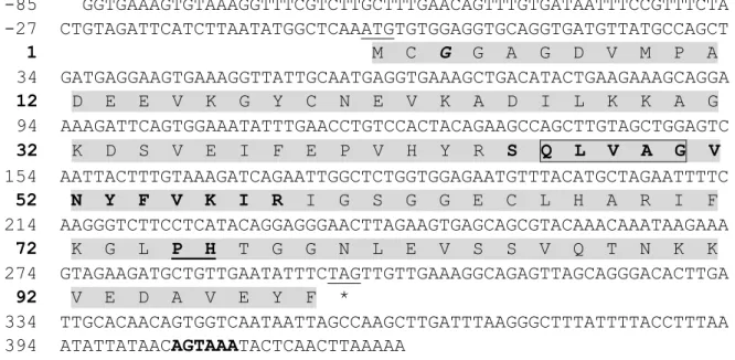
The cDNA sequence of RbCyt B was identified in rock bream multi-tissue normalized cDNA GS-FLX database by BLAST analysis, and this sequence was used to isolate the respective colony from a BAC library. The complete genomic sequence was determined and deposited in the GenBank database under accession number JQ287496. The full-length cDNA was found to contain a 5'-untranslated region (UTR) of 87 bp and a 3'-UTR of 224 bp with a polyadenylation signal sequence (AATAAA) that was found 11 bp upstream of the poly (A) tail (Fig. 4A). Its ORF was found to be 300 bp in length, encoding a polypeptide of 100 amino acids. Analysis of the amino acid sequence revealed the presence of a CY domain and a cysteine protease inhibitor signature, which suggests that the protein may belong to the cystatin superfamily. Analysis with the SignalP program indicated that there was no signal peptide, and the ExPASy Prosite database indicated that no carbohydrate side chains or disulfide bonds associated with the protein. The estimated molecular mass of RbCyt B is 11 kDa and its isoelectric point is 6.6. Collectively, these data suggest that the newly-identified RbCyt B is a member of the family 1 cystatin.
- 22 -
Fig. 4A. Nucleotide and deduced amino acid sequences of RbCyt B. The numbers of nucleotides and
deduced amino acid residues are shown in the left margin. The start codon (ATG) and the stop codon (TAA) are underlined. The cystatin-like domain is shaded, and the cysteine protease inhibitor signature is indicated in boldface letters. The 48QIVSG52 motif is boxed in the middle of domain and conserved N-terminal glycine residue (G6) is marked in bolted italics, and the P76C77 motif is indicated in underlined boldface. The poly (A) signal is boxed, and the poly (A) tail is underlined at the end of the nucleotide sequence.
The full length AbCyt B cDNA sequence of 1967 bp contains a 303 bp open reading frame (ORF) encodes for 101 amino acids, a 115 bp 5'-UTR and a 1546 bp 3'-UTR with a poly-adenylation signal (AATAAA). AbCyt B is comprised of a single CY domain with a conserved glycine residue near the N-terminal, a glutamine-valine-valine-alanine-glycine (QVVAG) motif at the middle of domain and a variant of proline-tryptophan (PW) motif with tyrosine (PY) substitution at C-terminal region. However, no signal peptide or disulfide bonds were identified. The predicted molecular mass of the derived protein was shown 11 kDa and the isoelectric point was predicted as 5.49. The complete cDNA and deduced amino acid sequences of AbCyt B are shown in Fig. 4B. The complete genomic sequence was deposited in GenBank under accession number JQ653304.
1 CCTGACTCAACCTGAACGCCTCACTGAAACAGACTCACTCGGTGTCTTTCGTCTTTTCGC 61 TCATTTTAAACCCTAAAAACTGTCAAAATGTCGATGATGTGTGGAGGAATCTCTGCGCCA 1 M S M M C G G I S A P 121 CTTGATGCTGATGAAGACATCCAGAAAATGTGTGATAACGTAAAACCTCATGCAGAGGAG 12 L D A D E D I Q K M C D N V K P H A E E 181 AAAGCAGGGAAGAAATATGATGTTTTCACAGCCAAGACATACACAACACAGATTGTGTCC 32 K A G K K Y D V F T A K T Y T T Q I V S 241 GGGACCAACTACTTCATAAAGATCCATGTGGGAGGAGACGATCATGTTCACCTTCGTGTT 52 G T N Y F I K I H V G G D D H V H L R V 301 TACAAAAAACTCCCCTGTCATGGAGGAGGCCTTGAGCTGAGTGGCATGCAGCACTCCAAG 72 Y K K L P C H G G G L E L S G M Q H S K 361 AGCCTCCAGGACCCTATTGCGTACTTCTAATGGGATCCCAAACCAAACAAAGCACCAGTG 92 S L Q D P I A Y F * 421 CCTACAGGCTGCCATCTCATTATAACTACTTATTATCAGATTATCCTGACTCATTACATA 481 AAACAATATTCTCATTCCTAACATGTCATAACATACTGTATACGTACTGTAGATCATGAT 541 GTTAGTATGTCAAAATGGAACTATTTTGCACAAGATGAATTAATAAAGATCAAAAAAAAA 601 AAAAAAAAAAAAAA
- 23 -
Fig. 4B. Nucleotide and deduced amino acid sequences of AbCyt B. The start (ATG) and stop (TAG)
codons are underlined. The cystatin-like domain is shaded and cysteine protease inhibitor signature sequence is in boldface. The conserved G3, 45QVVAG49 motif and P74Y75 motif are showed in bold italic, boxed and underlined boldface, respectively. The poly (A) signal (AATAAA) is in italic boldface at the end of nucleotide sequence.
The complete cDNA sequence of McCyt B was determined to consist of 509 nucleotides, comprising a 297 bp open reading frame (ORF) that encodes 99 amino acids, an 85 bp 5-UTR, and a 127 bp 3-UTR. The 3-UTR contains a polyadenylation signal
1 GAAGCAAACAAGTCA CATGAGGCGTTCCCC GCTGATTACGGAAAA CACACAGATATTTAG 61 GAGCTGCGGTTGTTG CTGTTGTTTGCTGTG AAAGTTTGATTAAAC GAAGATGTCG ATG
1 M
119 TGTGGTGGTGCAACC GAAGTGAAATCTGCA ACAGAAGAAGTGCAA AAACTCTGTAATGAG 2 C G G A T E V K S A T E E V Q K L C N E 179 GTTCGAGAGGCCTTG CAAACACAAGCAGGG AGAACGTTTGGAGCG TACAAAGCCATATCC
22 V R E A L Q T Q A G R T F G A Y K A I S
239 TTCCGTTCACAAGTG GTAGCAGGAACCAAC TACTTTGTTAAGGTC CAGGTGGATGAAAAC
42 F R S Q V V A G T N Y F V K V Q V D E N
299 GATGAACACTTTCAC CTGAGGATATTCGCC CCCCTCCCCTACACC AACTCCCCACCCTCC 62 D E H F H L R I F A P L P Y T N S P P S 359 CTTGCTGGCTATCAG ACTGGACACACCGCT GCATCAGATCTGGAT TATTTTGATGCCAAG 82 L A G Y Q T G H T A A S D L D Y F D A K 419 TAGTTGCCGTCCAAG TTTACACCATGTGCT GGACAAGCTCTTGAT AGCTTATGTTCATGG *
479 GATCTTTACATAATC TCTCAGTATAACATA TCAGTAGCTAAAGCT CACGCTTCATGTGTT 539 ATCTCCCTTGAATAT CTTCAGGAAATGATC TGTTCATAAATCATT TTGAAATAAAAAAAC 599 AAAACAGTTATCATG ATTAATCTGGTACAT AAAAACAACATCCAT TTAGTGTATTTTAAA 659 CAAATAAATGGATAT TTTTTACACTTTTTG TCTCGGTGGTGAGCG TCCCCTGCAGCCTGT 719 ATCAGTTGTATTGTG TACACTCAAGAAGAA CTGCCATTAACACCA TGGAACAACATCATG 779 GTGGTGAGATGCTTG TAAACACCAAAACAC AAGATTATGATGCAA GTATGCTCATTTCCT 839 TCCTCAGATCAGCAT CACGTCCGCCGCACA AGGATATGTCAAATT TGTGTTTGTATTGTT 899 TTGCTTCCCTTTGGG ACTACAGAATGTAAA AAAATGAATTTGGTT GACTATTGTCCCTAC 959 TACAACAGGTTGTTT TCAGAAATAGTATAT TTGCTGCTGTTTAGC GTTTCATAGTTCTCA 1019 TTTTGTAACCTACCA TTTTCTATTATACTT GTTTTGTCTATCAGT ATGTAGGCTGCTGCA 1079 GGAATGCCCATATCA TGTTATGTCACTCTG TTGTGACAATGCTTC CTAGAGAGCTGCCTG 1139 TATGATCTGCTCAGA GGGGTTACAGGGATA AATATATACAGCCAG TATCTTGCTTATGTG 1199 TATTCCCCGTGCTTG TGGGTGTCACCAGTG GTACCAGAAAACGTC CGAACCTGCAACACT 1259 TTGACTTTTCCGTCC ATATTAAGCTGACAC ACCATGATATAACTG GAATACTGTTCAAAG 1319 TGTTGTTAAACCCAA CTCGCTCACTCATAT CTATACAATATAAAA ATGGTTAAAGTTGCT 1379 GAAATTACAAATTGA TGTGTGCTGTTTGAA AAGCTGAAAACTTTC TTACATTTTCATGTG 1439 AAAATGCTATAAGGC TGCATTTGCAGATTC TGACATTCTTGATTG TCCATGAAATTCTTG 1499 AATGTTCATGTAAAA TGTCAGATTTCTTTG GAAACGAGGACCAAA AGTTACAGTGCAGGT 1559 GAATTTTGATAATTT ATTATTGTCCATGAT CTCCTGATCCTTTGC AAACGAACATTATAG 1619 CAGTTTCTGCACAGT AGATACATATTTAAT ATGAAAGTTACTGAT TCAGAGGAATCTGCT 1679 CTTGGGAGTGTTTGT AGTCGGTTCCACCTG ACAGACTTATCTTAG CTGCCGTCTCCGGTT 1739 GTTTCATCTCACTGC CACTGAACCCTTCAT TTCACGTGTTGGGAT CTGTGGACTTAATTA 1799 ATCAGCAGTTCTGCA TGAAAATGCAAGACA GGAATTGTGAAACGA CAAGACCTTTTATCA 1859 GTGTCTATTTGTTGA GCCATAGATGAAATT AAGGTAACAGTTGTG ATGTTGACTTTTTTA 1919 TCTGCTGTTGAAATT TTCACATAAACAACA ATAAACCATTCTTTT TACA