Anti-oxidant and Anti-inflammatory Effects of the
Fermented Rhododendron weyrichii Flower Extracts in
Shindari, a Traditional Jeju Fermented Drink
Nari Lee, Su Bin Hyun, Suk Hyun Yun, You Chul Chung, and Chang-Gu Hyun*
Department of Chemistry and Cosmetics, Jeju National University, Jeju 63243, Republic of Korea
Received: April 20, 2020 / Revised: July 7, 2020 / Accepted: July 28, 2020
Introduction
Nuruk is the Korean traditional fermentation starter. The shape, manufacturing process, and fermentation period of Nuruk varies depending on the unique climatic condition of each region [1−3]. In Jeju, red barley is used for the preparation of traditional Nuruk, because, Jeju province has very few rice paddies, Nuruk is prepared from barley rather than rice. Jeju barley Nuruk is also
used for brewing a traditional fermented drink called Shindari. It is made by fermenting barley Nuruk and steamed rice for a short period of time [4].
The process of fermentation has been reported to improve the biological properties of the raw material, including the antioxidant, anti-inflammatory, and skin-whitening properties by the production of new bioactive compounds [5−7]. Particularly, Nuruk contains various microorganisms that are involved in saccharification and alcohol fermentation. Therefore, these microorgan-isms have the potential to improve the biological proper-ties of raw materials.
Rhododendron weyrichii Maxim.(Ericaceae) is a decid-The aim of this study was to investigate the anti-oxidant and anti-inflammatory activities of the Rhododendron weyrichii flower extract fermented using Shindari, a traditional Jeju barley Nuruk-based fermentation. In this study, we examined the antioxidant potential of R. weyrichii flower extracts (RF) and R. weyrichii flower extracts fermented with Nuruk or Shindari (RFFN or RFFS, respectively) using various in vitro antioxidant assays including DPPH and ABTS radical scavenging assays, total phenol content and FRAP assays. We also evaluated the anti-inflammatory activity of the RF and RFFS on murine RAW 264.7 cells. The anti-inflammatory activity was evaluated by treating the RAW 264.7 cells with various concentrations (6.25, 12.5, 25, and 50 µg/ml) of RF or RFFS. As a result, we observed that the ABTS radical scavenging activ-ity and total phenol content of RFFS was higher than that of RF and RFFN. Additionally, lipopolysaccha-ride-induced nitric oxide (NO) production was significantly lower in RFFS-treated cells when compared to the LPS-treated control. In addition, RFFS-treated cells exhibited decreased expression of inducible NO synthase (iNOS) proteins and high-performance liquid chromatography (HPLC) fingerprinting showed that both the quercetin and quercetin glucoside (quercitrin and isoquercitrin) levels were affected by the fermentation process. In conclusion, our data suggests that traditional fermentation could be an important strategy in improving the biological properties of raw materials including their antioxidant and inflammatory activities. Finally, RFFS may be a candidate for developing topical antioxidant and anti-inflammatory agents.
Keywords: Fermentation, Rhododendron, inflammatory, Nuruk, Shindari
*Corresponding author
Tel: +82-64-754-3542 E-mail: [email protected]
uous shrub that is mainly found in the island of South Korea and the Japanese archipelago, which are tem-perate regions that experience high rainfall during summer. The shrub grows to a height of 3 to 6 m on the mountainside. The leaves present at the end of the branches are trilobed with dark green and brown hairs on their surface. The red-colored flowers have funnel-like shape (diameter 3.5−6.6 cm) and bloom in May [8]. Recently, our group reported that the leaf extract of R. weyrichii can be used as a skin-whitening agent as the extract suppresses melanogenesis [9]. Additionally, the flower extracts mitigated the UVB-induced inflamma-tion in human keratinocytes [10]. However, there are no studies on the fermented flower extract of R. weyrichii. The aim of this study was to investigate the antioxidant and anti-inflammatory potential of R. weyrichii flower fermented extracts. Additionally, the chemical constituent of the fermented extracts was evaluated fermentation characteristics of Shindari added with R. weyrichii flower by high-performance liquid chromatography (HPLC).
Material and Methods
Chemicals and reagents
The lipopolysaccharide (LPS) from Salmonella enter-ica serotype typhimurium and the Griess reagent (G4410) were purchased from Sigma-Aldrich Chemical Co. (USA). The anti-iNOS antibody was purchased from Millipore (USA), and anti-β-actin antibody was pur-chased from Santa Cruz Biotechnology (USA).
We procured 3-(4,5-dimethylthiazol-2-yl)-2,5-diphen-yltetrazolium bromide (MTT), ammonium pyrrolidin-edithiocarbamate (APDTC), protease inhibitor cocktail from Merck (Germany). Dulbecco’s modified Eagle medium (DMEM), fetal bovine serum (FBS), penicillin/ streptomycin, trypsin-ethylenediaminetetraacetic acid, BCA protein assay kit were purchased from Thermo Fisher Scientific (USA). Dimethyl sulfoxide (DMSO), radioimmunoprecipitation (RIPA) buffer, and enhanced chemiluminescence (ECL) kit were purchased from Biosesang (Korea) and Laemmli sample buffer (2X) was purchased from Bio-Rad (USA).
R. weyrichii flower extract
R. weyrichii flowers were supplied by Helios, Inc. (Jeju, Korea). The dried flowers (100 g) were incubated
with 600 ml of 70% ethanol solution on a shaker at room temperature for 24 h. This process was repeated three times. The extract solution was filtered through a filter paper and the solvent was evaporated. The extract was freeze-dried, and the powdered extract was used for experimental analysis. The yield of the R. weyrichii flower extract (RF) was 22%.
Subsequently, the fermented extract was prepared in two ways. First, the dried RF extract (1 g) and Nuruk (barley yeast, 1 g) was mixed with 1 g of sugar and added to 50 ml distilled water. Another one was mixed with 1 g of dried RF extract (1 g) and Nuruk (barley yeast, 1 g) with steamed rice and added to 50 ml distilled water. It was prepared by reference to Shindari methods. And then each mixture was fermented for 3 days in an incubator at 30℃.
After fermentation is over, 117 ml of 95% ethanol was added to each fermented mixture to prepare a 70% ethanol extract and stirred at room temperature for 24 h. The extracts were filtered and concentrated by a rotary evaporator. And then, freeze-dried for use in the experiment. The yield of the R. weyrichii flower extracts fermented with Nuruk (RFFN) was 34% while that of the R. weyrichii flower extracts fermented with Shindari (RFFS) was 10.95%.
DPPH radical scavenging activity
The antioxidant activity of the extracts was evaluated by 2,2-diphenyl-1-picrylhydrazyl (DPPH) free radical scavenging assay following the protocols of Blois et al. (1958) with minor modifications [11]. Briefly, 0.2 mM DPPH solution was prepared in ethanol and the absor-bance of the solution was adjusted to 0.94 ± 0.02 at 515 nm using ethanol. The extract (20 µl) was incubated with 180 µl of 0.2 mM DPPH solution at room tempera-ture for 10 min. The absorbance of the mixtempera-ture was mea-sured at 515 nm using a microplate reader (SUNRISE, TECAN Austria GmbH). The percentage of DPPH free radical scavenging activity was calculated using the fol-lowing formula:
Radical scavenging activity (%)
= [1 – ((Asample- Ablank) / Acontrol)] × 100 ABTS+ radical scavenging activity
following the methods of Re et al. (1999) with minor modifications [12]. Briefly, the ABTS+ radical was generated by incubating 7.4 mM ABTS (2,2′-Azino-bis (3-ethylbenzothiazoline-6-sulfonic acid) diammonium salt) and potassium persulfate at room temperature under dark condition for 16 h. The solution was diluted using ethanol to obtain an absorbance value of 0.78 ± 0.02 at 700 nm. The extract (20 µl) was incubated with 180 µl of the diluted ABTS+ solution at room tempera-ture for 15 min. The absorbance of the mixtempera-ture was mea-sured at 700 nm. The percentage of ABTS+ radical scavenging activity was calculated using the same for-mula used for calculating the DPPH radical scavenging activity.
Total phenolic content assay
The total polyphenol content was determined by colorimetric assay following the method described by Folin-Denis (1981) [13]. Briefly, the extract (100 µl) was diluted to a total volume of 1 ml using 900 µl distilled water. The extract was incubated with 100 µl Folin-Ciocalteu’s phenol reagent at room temperature for about 3 min. Further, 200 µl of Na2CO3 solution (7%, w/v) was added and mixed, followed by the addition of 700 µl distilled water. This mixture was incubated at room temperature for 1 h. The absorbance of the reac-tion mixture was measured at 700 nm using a micro-plate reader. The total polyphenol content was quantified using the gallic acid standard curve (R2 = 1). Ferric Reducing Ability of Plasma (FRAP) assay
The ferric reducing capacity of the extracts was evalu-ated following the method described by Benzie and Strain (1996) [14]. Briefly, the FRAP reagent was prepared using 300 mM sodium acetate buffer (pH 3.6), 10 mM 2,4,6-Tris (2-pyridyl)-s-triazine (TPTZ, dissolved in 40 mM HCl), and 20 mM ferric chloride solution in the ratio of 10:1:1. The extract (20 µl) was incubated with 180 µl of the FRAP reagent at 37℃ for 10 min. The absorbance of the reaction mixture was measured at 590 nm using a microplate reader. The ferrous (Fe2+) ion was quantified using the FeSO4· 7H2O standard calibra-tion curve (R2 = 0.9996).
Cell line and culture
The mouse monocyte cell line, RAW 264.7 was
obtained from the Korean Cell Line Bank (KCLB). The cells were sub-cultured in DMEM supplemented with 10% FBS and 1% penicillin/streptomycin every second day and maintained in a humid atmosphere of 5% CO2 at 37℃.
Cell viability assay
The cell viability was measured using 3-(4,5-dimeth-ylthiazol-2yl)-,5-diphenyltetra-zolium bromide (MTT) assay [15]. The RAW 264.7 cells (1 × 10⁵ cells/well) were seeded in the 24-well plates. Further, the cells were treated with various concentrations of the extract for 24 h. The MTT solution was added to each well. The for-mazan crystals were dissolved in dimethyl sulfoxide (DMSO) and the absorbance was measured at 570 nm using a microplate reader (SUNRISE, TECAN Austria GmbH).
Nitric oxide (NO) production assay
The RAW 264.7 cells (1 × 10⁵ cells/well) were seeded in the 24-well plates. The cells were then co-treated with RF or RFFS (6.25, 12.5, 25, and 50 µg/ml) and lipopoly-saccharide (LPS: 1 µg/ml) for 24 h. The culture superna-tant was incubated with an equal volume of griess reagent at room temperature (22−25℃) for 10 min. The NO production was measured at 540 nm using a micro-plate reader.
Western blot analysis
The RAW 264.7 cells were seeded in the 60-mm dish (2.5 × 10⁵ cells/dish) and cultured for 24 h. The cells were treated with various concentrations of RFFS (12.5, 25, and 50 µg /ml) or APDTC (15 µM) and LPS (1 µg/ml) for 24 h. The cells were subjected to trypsin treatment and lysed by vortexing every 10 min for 1 h in RIPA buf-fer containing 1% protease inhibitor cocktail. The cells were centrifuged at 21055 ×g for 20 min and the protein in the supernatant was quantified by bicinchoninic acid (BCA) protein assay. Equal amount of proteins (20 µg) was loaded into the polyacrylamide gel and the proteins were subjected to sodium dodecyl polyacrylamide gel electrophoresis (SDS-PAGE). The proteins were then transferred to a polyvinylidene difluoride (PVDF) mem-brane. The membrane was blocked using 5% non-fat skim milk (blocking buffer) prepared in tris-buffered saline containing 0.1% Tween 20 (TBS-T) for 3 h at room
temperature. The membrane was washed three times using TBS-T. The membrane was then probed with the primary antibody (1:1000) and incubated for 24 h. The membrane was 4 times washed at 10 min intervals with TBS-T. The membrane was then incubated with the sec-ondary antibody (1:3000) for 3 h. The membrane was washed again with TBS-T at 10 min intervals. The pro-teins were detected using the ECL kit and the images were captured and analyzed using Chemidoc (Fusion solo 6S.WL, France).
HPLC analysis
The quantitative analysis of the RF, RFFN, and RFFS extracts was performed on an HPLC instrument with a 2695 separation module (Waters, USA). The column used for the separation of RF, RFFN, and RFFS extracts was the YMC-Triart C18 analytical column (250 mm × 4.6 mm, 5 µm, 12 nm, Waters 2695), which was main-tained at 30℃. The mobile phase used for the efficient separation of the analytes was acetonitrile and 20 mM phosphoric acid. The flow rate was constantly main-tained at 1.0 ml/min and the injection volume was 10 µl. The analytes were detected using the PDA detector. The wavelength range used for the quantification of analytes was 280−370 nm. The data analysis was performed using the Waters Empower software. The RF, RFFN, and RFFS extracts were dissolved in 70% ethanol at a concentration of 10 mg/ml.
Statistical analysis
Cell viability and NO production assay were repeated
at least four times, and each experiment was performed in triplicate. All data are presented as mean ± standard deviation (SD). *p < 0.05, **p < 0.01 and ***p < 0.001 were considered statistically significant.
Results
Evaluation of antioxidant activity of RF, RFFN, and RFFS The antioxidant activity of the phenolic compounds is mainly due to their redox properties, which allow them to act as reducing agents, hydrogen donors, and singlet oxygen quenchers. The FRAP method is based on the reduction of ferric-TPTZ complex to ferrous-TPTZ in the presence of antioxidants [16−18].
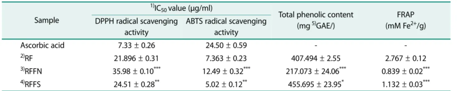
The total phenolic content, FRAP value, and radical scavenging activity of RF, RFFN and RFFS are pre-sented in Table 1. We have observed that RF, RFFN, and RFFS have high antioxidant efficacy. The FRAP value of RFFS was 1.4-fold higher than that of RFFN. Further, we observed that the phenolic content of RFFS was 1.1-fold, and 2.1-fold higher than that of RF, and RFFN. The IC50 value of RFFN and RFFS for ABTS scavenging activity was lower than that of the positive control, ascorbic acid. The IC50 value of RFFS was 4.88-fold lower than that of ascorbic acid and 1.4-4.88-fold lower than RF. Additionally, the radical scavenging activity of RFFN and RFFS was dose-dependent (Fig. 1). However, as can be seen from the experimental results, the fer-mentation process of R. weyrichii flowers does not always increase all antioxidant efficacy, but this indi-cated that Shindari fermentation is more efficient in
Table 1. Antioxidant effect of RF, RFFN, and RFFS.
Sample
1)IC
50 value (μg/ml)
Total phenolic content (mg 5)GAE/)
FRAP (mM Fe2+/g) DPPH radical scavenging
activity
ABTS radical scavenging activity Ascorbic acid 7.33 ± 0.26 24.50 ± 0.59 - -2)RF 21.896 ± 0.31 7.363 ± 0.23 407.494 ± 2.55 2.767 ± 0.12 3)RFFN 35.98 ± 0.10*** 12.49 ± 0.32*** 217.073 ± 24.06*** 0.839 ± 0.02*** 4)RFFS 24.51 ± 0.28** 5.02 ± 0.12** 455.695 ± 23.95* 1.132 ± 0.03*** 1)IC
50 : Concentration required to inhibit 50% of radicals 2)
RF : R. weyrichii flower extract
3)RFFN : R. weyrichii flower extracts fermented with Nuruk 4)
RFFS : R. weyrichii flower extracts fermented with Shindari 5)Gallic acid equivalent
extracting antioxidants from R. weyrichii flowers com-pared to Nuruk fermentation. Therefore, we comcom-pared the R. weyrichii flower extracts and R. weyrichii flower extracts fermented with Shindari in anti-inflammatory efficacy experiments.
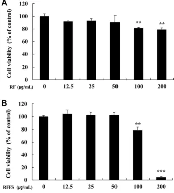
Effect of RF, and RFFS on the viability and NO production of RAW 264.7 cells
In this study, we investigated the effect of R. weyrichii flower extracts fermented with Shindari on the LPS-induced inflammation in RAW 264.7 cells. The cytotoxic-ity of various concentrations (6.25, 12.5, 25, and 50 µg/ ml) of RF, and RFFS extracts against RAW 264.7 cells was evaluated after 24 h treatment. As shown in Fig. 2, the viability of RAW 264.7 cells was more than 80% upon treatment with 50 µg/ml RF or RFFS. However, the viability of RAW 264.7 cells treated with 100 and 200 µg/ml of RF or RFFS was significantly lower than
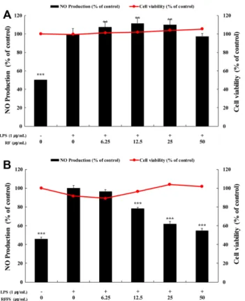
that of the untreated cells (control). Therefore, we used RF and RFFS at concentrations of 6.25, 12.5, 25, and 25µg/ml for further experiments. The LPS-induced monocytes are known to be activated through the pro-duction of inflammatory mediators, such as NO and cytokines. Thus, we evaluated the amount of NO released into the culture supernatant using the griess reagent. The NO production decreased by 2%, and 45% upon treatment (50 µg/ml) with RF and RFFS, respec-tively compared to the LPS-treated cells. Additionally, no cytotoxicity was observed at this concentration (Fig. 3). This indicated that the anti-inflammatory effect of RFFS was higher than that of RF at 50 µg/ml.
Effects of RFFS on iNOS and COX-2 expression of RAW 264.7 cells
Western blotting was performed to determine whether NO production was associated with the protein expres-sion of iNOS. The expresexpres-sion of iNOS in the
LPS-Fig. 1. Free radical scavenging activity of RF, RFFN, and RFFS. (A) DPPH, (B) ABTS radical scavenging activity. RF : R.
wey-richii flower extract, RFFN: R. weywey-richii flower extracts fermented
with Nuruk, RFFS: R. weyrichii flower extracts fermented with Shindari. The data are expressed as mean ± SD (n = 3).
Fig. 2. Cytotoxicity of various concentrations of RF and RFFS against RAW 264.7 cells. The cells were treated with
var-ious concentrations of (A) RF and (B) RFFS for 24 h, and the cell viability was measured by MTT assay. RF: R. weyrichii Flower extract, RFFS: R. weyrichii flower extracts fermented with Shindari. The data are presented as the mean ± standard devi-ation (n = 4). **p < 0.01, ***p < 0.001 vs control.
induced RAW 264.7 cells was significantly higher than that in the LPS-untreated cells. The co-treatment of RFFS (50 µg/ml) and LPS significantly reduced the expression of iNOS by 61% compared to the LPS-treated group. The negative control, APDTC inhibited the expression of iNOS by 80% at 15 µM compared to the LPS-treated group (Fig. 4). This indicated that RFFS suppresses the LPS-induced inflammation in the RAW 264.7 cells by inhibiting the NO production and iNOS expression.
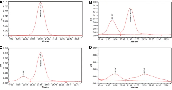
HPLC analysis of the RF, RFFN, and RFFS
HPLC fingerprinting was used to identify the chemi-cal components of R. weyrichii flower fermented extract that are involved in antioxidant and anti-inflammatory activities. The flavonoids, such as quercetin was
reported to exhibit cancer, inflammatory, anti-viral, and antioxidant properties [19−21]. The chemical structure of quercitrin and isoquercitrin contains quer-cetin, which is bound to a glycoside. Fermentation is a metabolic process in which the glycosyl group is con-verted into acids, gases, and alcohol (aglycone), which is
Fig. 3. Effect of RF and RFFS on NO production in LPS-stim-ulated RAW 264.7 cells. The cells were treated with various
concentrations (6.25, 12.5, 25, and 50 μg/ml) of (A) RF and (B) RFFS for 24 h. LPS-stimulated cells were used as a positive control. RF: R. weyrichii Flower extract, RFFS: R. weyrichii flower extracts fermented with Shindari, LPS: lipopolysaccharide, NO: Nitric oxide. The data are presented as the mean ± standard deviation (n = 4). **p < 0.01, ***p < 0.001 vs. positive control.
Fig. 4. Effect of RFFS on the protein expression of iNOS in RAW 264.7 cells. (A) The cells were treated with RFFS (12.5, 25,
and 50 μg/ml), LPS (1 μg/ml), and APDTC (15 μM) of iNOS syn-thetase inhibitor for 24 h. The cell lysates were subjected to western blotting. (B) Quantitation of the protein levels of iNOS. β-actin served as a loading control. RFFS: R. weyrichii flower extracts fermented with Shindari, LPS: lipopolysaccharide, iNOS: inducible NO synthase. The data are presented as the mean ± standard deviation (n = 3). ***p < 0.001 vs. positive control.
Table 2. Chemical constituents of RF, RFFN, and RFFS. The concentration of all standard materials was measured at 100 ppm.
Sample
(ppm) Quercitrin Isoquercitrin Quercetin
STD 100 100 100
1)RF 23.74 19.73 19.73
2)RFFN 17.07 8.54 51.16
3)RFFS - - 83.50
1)RF: R. weyrichii flower extract
2)RFFN: R. weyrichii flower extracts fermented with Nuruk 3)RFFS: R. weyrichii flower extracts fermented with Shindari
mediated by yeasts, bacteria, or fungi [22]. We observed a dramatic increase in the amount of aglycones in the R. weyrichii flower extracts fermented with Nuruk or Shindari. Further, RFFS were remarkably decreased
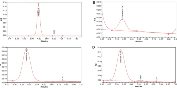
the quercitrin and isoquercitrin content. Additionally, RFFS enhanced the quercetin and content by 4-fold and 1.6-fold, respectively, compared to RF, and RFFN sam-ples (Table 2; Fig. 5−7).
Fig. 5. Quercitrin content analysis from various RF, RFFN, and RFFS by HPLC. (A) quercitrin, (B) RF, (C) RFFN, and (D) RFFS.
Discussion
Nuruk (yeast) is a traditional fermented starter that is manufactured in a wide variety of forms and recipes, depending on the geographic environment of the region. Jeju Island, a volcanic island, has been used for tradi-tional fermented foods by using barley to make Nuruk. In particular, Shindari is a traditional drink that is fer-mented for 3 to 4 days in a short time by adding barley Nuruk to the steamed rice [23]. Various beneficial micro-organisms are present in these Nuruk, and vitamins, essential amino acids, and organic acids produced through the fermentation process affect nutrition and flavor [24]. In addition, previous studies have been con-ducted on the effects of Nuruk in various aspects such as antioxidants and whitening [25, 26].
In this study, we demonstrated the antioxidant poten-tial of R. weyrichii flower fermentation extracts using various in vitro assays including DPPH and ABTS radi-cal scavenging, and FRAP assay, total phenol content. Further, the anti-inflammatory activity of RFFS decreased NO production by downregulating iNOS pro-tein expression in the LPS-stimulated RAW 264.7 cells.
HPLC fingerprinting suggest that deglycosylation by microorganisms during Shindari fermentation may degrade glycosides of quercitrin and isoquercitrin to
increase the content of quercetin, which is known to exhibit antioxidant and anti-inflammation activities.
Hence, Shindari fermentation has the potential to improve biological properties such as antioxidant and anti-inflammatory properties. Thus, we suggest that REFS is a potential antioxidant and anti-inflammatory candidate for topical application. In order to use these results, a complete understanding of the efficacy of the fermentation starter, barley Nuruk or Shindari, and the increased efficacy after fermentation of the extract is needed. Therefore, it is considered that it is necessary to further compare the efficacy of the fermentation starter and the fermentation extract under the same conditions.
Acknowledgments
This work was supported by the Academic and Research Institu-tions R&D Program (C0516709) funded by the Ministry of SMEs and Startups (MSS, Korea).
Conflict of Interest
The authors have no financial conflicts of interest to declare.
References
1. Ross RP, Morgan S, Hill C. 2002. Preservation and fermentation:
past, present and future. Int. J. Food Microbiol. 79: 3-16.
2. Lee JE, Kim JH. 2017. Concept of Nuruk on Brewing Technology. pp.123-134. Brewing Technology.
3. Ponnusamy K, Lee S, Lee CH. 2013. Time-dependent correlation of the microbial community and the metabolomics of traditional barley nuruk starter fermentation. Biosci. Biotechnol. Biochem. 77: 683-690.
4. Cha IT, Lee HW, Song HS, Yim KJ, Kim KN, Kim D, et al. 2014. Diver-sity of lactic acid bacteria in the Korean traditional fermented beverage shindari, determined using a culture-dependent method. Curr. Top. Lact. Acid Bact. Probiotics. 2: 34-37.
5. Chung YC, Ko JH, Kang HK, Kim SY, Kang CI, Lee JN, et al. 2018. Antimelanogenic effects of Polygonum tinctorium flower extract from traditional Jeju fermentation via upregulation of extracellu-lar signal-regulated kinase and protein kinase B activation. Int. J.
Mol. Sci. 19: 2895.
6. Hur SJ, Lee SY, Kim YC, Choi I, Kim GB. 2014. Effect of fermenta-tion on the antioxidant activity in plant-based foods. Food Chem.
160: 346-356.
7. Pérez-Gregorio MR, Regueiro J, Alonso-González E, Pastrana-Castro LM, Simal-Gándara J. 2011. Influence of alcoholic fermen-tation process on antioxidant activity and phenolic levels from mulberries (Morus nigra L.). LWT-Food Sci. Technol. 44: 1793-1801. 8. Yoichi W, Tamaki I, Sakaguchi S, Song JS, Yamamoto SI, Tomaru N. 2016. Population demographic history of a temperate shrub,
Rhododendron weyrichii (Ericaceae), on continental islands of
Japan and South Korea. Ecol. Evol. 6: 8800-8810.
9. Kim MJ, Kim SS, Yun SH, Kim SY, Hyun KH, Lee J, et al. 2016. Mela-nogenesis inhibitory activity of Rhododendron Weyrichii in mouse B16 melanoma cells. Orient. J. Chem. 32: 1899-1907. 10. Yang EJ, Yun SH, Ko JH, Kang HK, Lee JN, Park SM, et al. 2019.
Pro-tective effect of Rhododendron weyrichii flower extract against UVB-induced proinflammatory cytokine production in human keratinocytes. J. Appl. Pharm. Sci. 9: 15-19.
11. Blois MS. 1958. Antioxidant determinations by the use of a stable free radical. Nature 181: 1199.
12. Re R, Pellegrini N, Proteggente A, Pannala A, Yang M, Rice-Evans C. 1999. Antioxidant activity applying an improved ABTS radical cation decolorization assay. Free Radic. Biol. Med. 26: 1231-1237. 13. Srivastava M, Tiwari R, Sharma N. 2013. Assessment of phenol
and flavonoid content in the plant materials. J. New Biol. Rep. 2: 163-166.
14. Benzie IF, Strain JJ. 1996. The ferric reducing ability of plasma (FRAP) as a measure of “antioxidant power”: the FRAP assay. Anal.
Biochem. 239: 70-76.
15. Gerlier D, Thomasser N. 1986. Use of MTT colorimetricassay to measure cell activation. J. Immunol. Methods 94: 57-63. 16. Rice-Evans C, Miller N, Paganga G. 1997. Antioxidant properties
of phenolic compounds. Trends Plant Sci. 2: 152-159.
17. Ak T, Gülçin İ. 2008. Antioxidant and radical scavenging proper-ties of curcumin. Chem. Biol. Interact. 174: 27-37.
18. Pandey KB, Rizvi SI. 2012. Ferric reducing and radical scavenging activities of selected important polyphenols present in foods. Int.
J. Food Properties 15: 702-708.
19. Comalada M, Camuesco D, Sierra S, Ballester I, Xaus J, Gálvez J, et
al. 2005. In vivo quercitrin anti‐inflammatory effect involves
release of quercetin, which inhibits inflammation through down‐regulation of the NF‐κB pathway. Eur. J. Immunol. 35: 584-592.
20. ElAttar T, Virji AS. 1999. Modulating effect of resveratrol and quercetin on oral cancer cell growth and proliferation. Anticancer
Drugs 10: 187-193.
21. Mahat MYA, Kulkarni NM, Vishwakarma SL, Khan FR, Thip-peswamy B, Hebballi V, et al. 2010. Modulation of the cyclooxy-genase pathway via inhibition of nitric oxide production contributes to the anti-inflammatory activity of kaempferol. Eur.
J. Pharmacol. 642: 169-176.
22. Dajanta K. 2009. Enhanced aglycone production of fermented soybean products by Bacillus species. Acta Biologica Szegediensis
53: 93-98.
23. Oh YJ. 2009. Jeju Traditional Food Fermentation culture in envi-ronment of east asia. J. Cheju Studies 32: 157-203.
24. Lee JS, Lee TS, Park SO, Noh BS. 1996. Flavor components in mash of Takju prepared. Korean J. Food Sci. Technol. 28: 316-323. 25. Lee SJ, Cho SW, Kwon YY, Kwon HS, Shin WC. 2012. Inhibitory
effects of ethanol extracts from Nuruk on oxidative stress, mela-nogenesis, and photo-aging. Mycobiology 40: 117-123.
26. Jung DJ, Chang BS. 2019. Assessing the antioxidant activity and potential utility in cosmetics of nuruk, a traditional Korean Fer-mentation Starter. J. Invest. Cosmetol. 15: 17-23.