Vol. 46, No. 4, pp. 555 - 561, 2005
Tailgut cysts (TGCs) are rare congenital cysts that occur in the retrorectal or presacral spaces. Although most tailgut cysts have been reported as benign, there have been at least 9 cases associated with malignant change. We report herein on an unusual case of a 40-year-old woman with a carcinoembryonic antigen (CEA)-producing adenocarcinoma arising within a TGC who underwent surgical resection and local radiation therapy. Despite the complete resection, metastatic adenocarci-noma developed five months after surgery. CEA-producing adenocarcinoma from a TGC is extremely rare and only two cases, including this case, have been reported in the English medical literature. Besides CEA, the serum levels of CA 19-9 became markedly elevated in this patient. Given that the serum CEA level decreased to the normal range after complete resec-tion of tumor and that the tumor recurrence was associated with a rebound of the CEA serum level, our case shows that serial measurements of serum CEA can be used for treatment planning and for assessing the patient's treatment response for this rare disease.
Key Words: Tailgut cyst, developmental cyst, presacral space, adenocarcinoma, carcinoembryonic antigen
INTRODUCTION
The tailgut cyst (TGC) is a rare benign cystic lesion located in the retrorectal space, and it is believed that this cyst is derived from the per-sistent embryologic remnants of the postanal gut.1
These lesions typically present as a presacral mass, and they are frequently misdiagnosed, which may be due in part to the physician's un-familiarity with this entity.1,2 In a comprehensive
review of 53 cases by Hjermstad and Helwig,1 a
correct histologic diagnosis was made in only 2 cases. The incidence of malignancy in TGCs is extremely rare; besides our case, only 10 well-do-cumented cases of associated malignancy arising from within a tailgut cyst have been reported in the English literature.2-11 There have only been 6
cases of tailgut cysts, including one case of adeno-carcinoma, reported in Korea.11-16 We describe
here in this report the clinicopathologic and radio-logic features of adenocarcinoma arising within a TGC. Furthermore, we discuss and suggest the possible therapeutic implications of serum CEA and CA 19-9 for this rare disease.
CASE REPORT
A 40-year-old woman was admitted to our hos-pital because of a 1 month history of severe perianal pain. Her medical history included a wide excision of presacral cystic masses; this operation was performed in July, 2001, when she presented with abdominal pain. The operative specimen consisted of two well-circumscribed cystic masses that measured 3 cm and 5 cm in diameter, respectively. The cysts were unilocular and contained a yellowish, gelatinous material. Microscopically, the cyst lining consisted of vari-ous epithelial cells, mostly squamvari-ous epithelia,
A Carcinoembryonic Antigen-Secreting Adenocarcinoma
Arising in Tailgut Cyst : Clinical Implications of
Carcinoembryonic Antigen
Byoung Chul Cho,1 Nam Kyu Kim,3 Beom Jin Lim,2 Sang Ook Kang,3 Ju Hyuk Sohn,1 Jae Kyung Roh,1
Sang Tae Choi,1 Sung Ai Kim,1 and Se Eun Park1
Departments of 1Internal Medicine, 2Pathology, and 3Surgery, Yonsei University College of Medicine, Seoul, Korea.
Received May 9, 2004 Accepted July 20, 2004
Reprint address: requests to Dr. Nam Kyu Kim, Department of Surgery, Yonsei University College of Medicine, 134 Shinchon-dong, Seodaemun- gu, Seoul 120-752, Korea. Tel: 82-2-361-5562, Fax: 82-2-313-8289, E-mail: [email protected]
A B
and the cyst wall displayed disorganized smooth muscle bundles without a neural plexus, thus confirming the diagnosis of tailgut cyst. She had been doing well after her surgery until the start of her aforementioned aggravating symptoms. Before the patient's presentation at our hospital, a percutaneous biopsy through a posterior ap-proach yielded a poorly differenciated carcinoma specimen with focal squamous differentiation. On admission, the physical examination revealed a posterior extrinsic mass compressing the rectum, the stool was negative for blood and there were no other important findings. A barium enema study demonstrated the left-anterior displacement of the posterior wall of the rectum (Fig. 1). T1-and T2-weighted magnetic resonance imaging
(MRI) demonstrated an irregular cystic mass mea-suring 6 cm in diameter within the retrorectal space and it was involving coccygeal bone and right gluteous muscle tendon (Fig. 2A, 2B, 2C). There was no radiological evidence of extrapelvic metastasis and a whole body bone scan revealed increased bone uptake in the coccyx. The labora-tory test values were normal except for a remark-able elevation in serum CEA level to 159 ng/mL (normal 0 - 5 ng/mL) and serum CA19 - 9 level to 2270 U/mL (normal 0 - 37 U/mL). The patient un-derwent enbloc resection of the mass and a Hartmann operation with coccygectomy and par-tial sacrectomy to the level of S4 through a com-bined abdominal/sacral approach. Upon gross examination, the specimen measured 10 × 7 × 8 cm; it was an ill-defined mass impinging on the outer wall of rectum, the presacral soft tissue and coccygeal bone. The mass displayed a firm gray-white appearance on sectioning and contained several small, unilocular cysts filled with a yellow gelatinous material (Fig. 3). Microscopic examina-tion revealed a poorly differentiated adenocarci-noma with infiltrative growth to the surrounding tissue (Fig. 4A). The wall of the cysts consisted of various types of epithelium, including ciliated columnar, columnar and focal cuboidal epithe-lium; this was consistent with a TGC (Fig. 4B, 4C). The resection margin was negative and the re-gional lymph nodes were free of carcinoma. After her operation, the patient's postoperative CEA level had returned to normal. The patient under-went radiation therapy after surgery to prevent local recurrence of the tumor, but five months
Fig. 2.(A) Saggital T1-weighted MRI scan showing presacral lesion with intermediate signal intensity invading surrounding structures (arrow). (B & C) Axial T1- and T2-weighted MRI scan showing multicystic mass with intermediate signal intensity (thick arrow). The cyst shows hypointense and hyperintense signal, respectively (thin arrow).
Fig. 1.Barium enema study showing left-anterior displace-ment of the posterior wall of the rectum (arrows).
C
after the primary resection, the patient started complaining of a palpable mass in the right ingui-nal area, and radiologic examination detected a local tumor recurrence involving the sacrum. Additionally, serum levels of CEA and CA 19 - 9 had increased to 37.06 ng/mL and 5260 U/mL, respectively. Excisional biopsy of inguinal lymph node revealed metastatic adenocarcinoma. Subse-quently, the patient received 5-Fluorouracil based systemic chemotherapy.
DISCUSSION
The Retrorectal space is a potential space de-fined anteriorly by the rectum, posteriorly by the sacrum, superiorly by the peritoneal reflection, inferiorly by the levator ani and coccygeus muscle and laterally by the ureters and iliac vessels.1,13 There are a variety of congenital, inflammatory and malignant disorders that may occur within this space. During the 8 mm to 35 mm gestational stage (35 days gestational age to 56 days gesta-tional age) of human development, the embryo possesses a true tail and the primitive hindgut extends into this tail, which is caudal to the site of subsequent formation of the anus. This caudal extension is called the tailgut or postanal gut, and this tailgut usually completely regresses by 56 days of gestational age. The persistence of this embryological remnant is very rare and results in
the development of TGC.1,6 Although TGC may
clinically present in all age groups from neonates to adults, the anomaly is more commonly found in middle-aged females.1,2 The clinical
presenta-tion is frequently nonspecific and related to a
Fig. 3.On sectioning, the mass displays a firm gray-white appearance (thick arrow) and contains several, discrete cysts (thin arrow) filled with yellow gelatinous material. The ill-defined mass impinges on the outer wall of rectum, presacral soft tissue and coccygeal bone.
Fig. 4. (A) The tumor consisits of many scattered malignant glandular components with infiltrative growth to surrounding tissue (× 100 H&E stain). (B & C) The wall of cysts are lined by benign-looking ciliated columnar (B), columnar (C) and focal cuboidal epithelium (not shown), which is consistent with TGC (× 200 H&E stain).
A
B
mass effect pressing on the surrounding tissues; discomfort while sitting and rectal bleeding are common symptoms. If the mass becomes infected, it is often misdiagnosed as a pilonidal cyst, an anorectal fistula or a recurrent retrorectal ab-scess.1-4As Hjermstad and Helwig1pointed out in their review of 53 retrorectal cysts diagnosed over a 35-year period, the clinical diagnosis of a TGC is often delayed due to the general unfamiliarity with this entity and also because of its symptoma-tological mimicry of other more common diseases afflicting the anus and anal canal, including peria-nal fistulas and abscesses. Although a variety of pathologic diagnoses such as epidermal cyst, der-moid cyst and even teratoma were rendered on the 53 cases in their study, the precise histologic diagnosis of a TGC was made in only two in-stances. The histological features of a TGC can aid the physician in the diagnosis as they are quite distinctive from the other retrorectal cysts.1-12
Microscopically, the lesions are usually multicystic and are lined by a variety of epithelial cells com-monly found in the adult and fetal gastrointestinal tract, including columnar, stratified columnar, transitional and squamous epithelia. Smooth mus-cle tissue bundles are almost always present in the vicinity of the cysts. However, the muscle bundles are often disorganized and are focally present; this is unlike the well-formed continuous 2-layer mus-cle coat seen in duplication cysts. The differential diagnosis includes teratomas, dermoid cysts, epi-dermal cysts and duplication cysts. Teratomas are composed of all three germ layers. Dermoid and epidermal cysts are usually unilocular and lined only by squamous epithelium; the dermoid cysts have dermal appendages and the epidermal cysts do not. Rectal duplication cysts, as mentioned before, have well-developed smooth muscle layers with myenteric plexus. In Korea, only 6 cases of TGC,11-16 including one case of carcinoid tumor11
and one case of adenocarcinoma12 have been
re-ported. The female predominance and the average age of 40 years are almost identical to those patient parameters stated in several other reports.1-10 All of the patients had symptoms and
half of them presented with perianal pain. Wide excision of the lesion were carried out in all cases and upon gross examination, all of the lesions appeared to be multicystic presacral masses with
an average diameter of 10 cm. The radiographic findings of a TGC have been described.1,6,17
Bari-um enema examination shows an extrinsic retro-rectal mass.1 Sonography examination shows a
complex cystic mass in the retrorectal region. It can be distinguished from simple cyst by a strong internal echo due to multicystic nature of the mass and the presence of gelatinous material or inflam-matory debris inside the cyst.17 CT scan shows a
well-defined homogeneous retrorectal mass with the CT values ranging from water to soft-tissue density. The keratinous or inflammatory debris within a cyst may account for a more solid ap-pearance. Because calcification is not a feature of TGC, its presence favors the diagnosis of a tera-toma or malignancy. If malignant degeneration is present, the CT scan may show a loss of discrete margins, calcification inside the lesion and in-volvement of contiguous structures.17 MRI
imag-ing reveals a hypointense lesion on T1-weighted images and a homogenously hyperintense lesions on T2-weighted images. Although the malignant portions of the tumor are characterized by irregu-lar wall thickening and intermediate signal inten-sity on both the T1- and T2-weighted images, MRI is probably not the best imaging modality to fully differentiate malignant lesions from benign le-sions.6 Most of TGCs are benign, and malignant
transformation of the epithelial component of a TGC has rarely been reported.2-12 On our com-puterized MEDLINE search from 1966 to 2004, only 10 cases of malignancy arising from TGC have been reported: five were adenocarcinoma,2-6
three carcinoid,7,8,11 one neuroendocrine
carci-noma9 and one squamous cell carcinoma.10 The
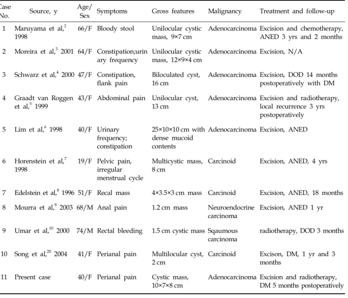
data for the nine previously reported cases are summarized in the Table 1. In Korea, one case of carcinoid tumor11 and one case of
adenocarci-noma12 derived from TGCs has been reported, and
there has been only one case of a serum CEA-producing adenocarcinoma within a TGC re-ported worldwide. To the best of our knowledge, our case is the first one that clearly showed an increase in serum CEA as well as serum CA19-9. An elevated level of serum CEA per se is not specific enough to permit a diagnosis of TGC adenocarcinoma to be made, but once a TGC malignancy has been diagnosed and it is shown to be associated with elevated level of CEA, it
seems logical that CEA measurements can be used as a simple measure to assess the tumor's re-sponse to treatment. In our patient, normalization of CEA after radical resection was related to the total excision of tumor mass, and the rebound of the CEA serum level could be considered as an index to judge tumor recurrence. There were two incidences of recurrence in our patient. The first tumor recurrence could be attributed to an in-complete surgical resection in the year of 2001. Because there is always a high probability of tumor spillage in the process of needle biopsy, our patient underwent postoperative adjuvant
radio-therapy. The high preoperative CEA serum level in our patient may serve to indicate the tumor's aggressive behavior, a poor prognosis and an early recurrence, as is the case in colorectal neo-plasms.18 In addition, the poorly differentiated
histology and tumor adhesion to adjacent organs probably contributed to the second recurrence of the tumor. The treatment guidelines for a recurred TGC tumor have not been documented due to the rarity of this disease. However, TGC embryologi-cally originates from hindgut1 and there was an
immunohistochemical study stating that the de-velopment of TGC adenocarcinoma follows the
Table 1. Cases of Malignancy Arising in Tailgut Cysts Case
No. Source, y
Age/
Sex Symptoms Gross features Malignancy Treatment and follow-up 1 Maruyama et al,2
1998 66/F Bloody stool Unilocular cysticmass, 9×7 cm Adenocarcinoma Excision and chemotherapy,ANED 3 yrs and 2 months 2 Moreira et al,3 2001 64/F Constipation;urin
ary frequency
Unilocular cystic mass, 12×9×4 cm
Adenocarcinoma Excision, N/A
3 Schwarz et al,4 2000 47/F Constipation,
flank pain Biloculated cyst,16 cm Adenocarcinoma Excision, DOD 14 monthspostoperatively with DM 4 Graadt van Roggen
et al,5 1999 43/F Abdominal pain Unilocular cyst,13 cm Adenocarcinoma Excision and radiotherapy,local recurrence 3 yrs postoperatively
5 Lim et al,6 1998 40/F Urinary frequency; constipation
25×10×10 cm with dense mucoid contents
Adenocarcinoma Excision, ANED
6 Horenstein et al,7
1998 19/F Pelvic pain,irregular menstrual cycle
Multicystic mass,
8 cm Carcinoid Excision, ANED, 4 yrs
7 Edelstein et al,8 1996 51/F Recal mass 4×3.5×3 cm mass Carcinoid Excision, ANED, 18 months 8 Mourra et al,9 2003 68/M Anal pain 1.2 cm mass Neuroendocrine
carcinoma
Excision, ANED 1 yr
9 Umar et al,10 2000 74/M Rectal bleeding 1.5 cm cystic mass Sqaumous carcinoma
radiotherapy, DOD 3 months
10 Song et al,20 2004 41/F Perianal pain Multilocular cyst,
2 cm Carcinoid Excison, DM, 1 yr and 3months 11 Present case 40/F Perianal pain Cystic mass,
10×7×8 cm Adenocarcinoma Excision and radiotherapy,DM 5 months postoperatively
similar dysplasia-carcinoma sequence as observed for colorectal adenocarcinoma.3 Therefore, we
de-cided to manage our patient along the lines of a rectal carcinoma and we started treatment with 5-Fluorouracil-based chemotherapy. The prognosis for malignant presacral lesions depends on the surgical performance of complete resection and the tumor histology, and there is a much better prognosis for endocrine tumors when compared with adenocarcinomas.7-9,19 The poor prognosis of
adenocarcinoma arising from TGC due to local recurrence and metastasis after surgery has been reported in many cases.3-5 Yet in contrast, the
patient reported on by Maruyama et al.2 was
given adjuvant chemotherapy and had a good outcome, suggesting that adjuvant therapy is effective. Because a preoperative biopsy may fail to confirm the diagnosis of malignancy and the procedure may carry significant hazards such as spillage of malignant cells into the peritoneal cavity, the transrectal or presacral needle biopsy is only indicated for patients who are considered at high risk for surgery.1,4 Especially in the setting
of a presacral heterogenous mass with CEA serum elevation, a biopsy should not be performed. The treatment of choice is complete excision of the lesion, with resection of the coccyx and an ade-quate surrounding margin of grossly normal tissue.1-6Simple cyst excision or drainage will lead
to recurrence or infection. Because of the multilo-cular nature of TGCs, a posterior surgical ap-proach is recommended for the operative treat-ment of retrorectal masses or cysts that are located below the level of S4.1 Lesions that occur at a
higher level are better addressed through an an-terior laparotomy.20 If a malignant process is proven or even suspected, the combined abdomi-nal/sacral approach, as we performed in our patient, is then recommended.4,21 In summary, our
case would suggest that the TGC should be completely resected at the time of diagnosis, and also that serum CEA measurements can be used to assess the treatment response.
ACKNOWLEDGEMENT
We are very thankful to Dr. Yong Sun Park for his careful review of this journal.
REFERENCES
1. Hjermstad BM, Helwig EB. Tailgut cysts, report of 53 cases. Am J Clin Pathol 1998;89:139-47.
2. Maruyama A, Murabayashi K, Hayashi M, Nakano H, Isaji S, Uehara S, et al. Adenocarcinoma arising in a tailgut cyst: report of a case. Surg Today 1998;28:1319-22.
3. Moreira AL, Scholes JV, Boppana S, Melamed J. p53 Mutation in adenocarcinoma arising in retrorectal cyst hamartoma (tailgut cyst): report of 2 cases-an immuo-histochemistry/immunoperoxidase study. Arch Pathol Lab Med 2001;125:1361-4.
4. Schwarz RE, Lyda M, Lew M, Paz IB. A carcinoembryo-nic antigen-secreting adenocarcinoma arising within a retrorectal tailgut cyst: clinicopathologic considerations. Am J Gastroenterol 2000;95:1344-7.
5. Graadt van Roggen JF, Welvaart K, de Roos A, Offer-haus GJ, Hogendoorn PC. Adenocarcinoma arising within a tailgut cyst: clinicopathological description and follow up of an unusual case. J Clin Pathol 1999; 52:310-2.
6. Lim KE, Hsu WC, Wang CR. Tailgut cyst with malig-nancy: MR imaging findings. Am J Roentgenol 1998; 170:1488-90.
7. Horenstein MG, Erlandson RA, Gonzalez-Cueto DM, Rosai J. Presacral carcinoid tumors: report of three cases and review of the literature. Am J Surg Pathol 1998;22:251-5.
8. Edelstein PS, Wong WD, La Valleur J, Rothenberger DA. Carcinoid tumor: an extremely unusual presacral lesion. Report a case. Dis Colon Rectum 1996;39:928-42. 9. Mourra N, Caplin S, Parc R, Flejou JF. Presacral neuro-endocrine carcinoma developed in a tailgut cyst: report of a case. Dis Colon Rectum 2003;46:411-3.
10. Umar T, Mikel JJ, Poller DN. Carcinoma arising in a tailgut cyst diagnosed on fine needle aspiration (FNA) cytology. Cytopathology 2000;11:129-32.
11. Song DE, Park JK, Hur B, Ro JY. Carcinoid tumor arising in a tailgut cyst of the anorectal junction with distant metastasis: a case report and review of the literature. Arch Pathol Lab Med 2004 May;128:578-80. 12. Chung KJ, Lee YL. Adenocarcinoma arising from the tailgut cyst (in Korean). Korean Radiol Society 1995;33: 399-402.
13. Kang JW, Kim SH, Kim KW, Moon SK, Kim CJ, Chi JG. Unusual perirenal location of a tailgut cyst. Korean J Radiol 2002;3:267-70.
14. Ahn BY, Jeong CS, Lee DH, Yu CS, Lee HJ, Lee MK, et al. Tailgut cyst- A case report (in Korean). J Korean Soc Coloproctol 1998;14:617-20.
15. Lee H, Oh JH, Cho SY, Yang DM, Ha SY. Two cases of Tailgut cyst (in Korean). J Korean Soc Coloproctol 2001;17:209-12.
16. Lim MJ, Lee SN, Kim SS, Koo H, Kim OK. Tailgut cyst with Glomus Coccygeum (in Korean). Korean J Pathol 1996;30:643-5.
17. Johnson AR, Ros PR, Hjermstad BM. Tailgut cyst: Diagnosis with CT and Sonography. Am J Roentgenol 1986;147:1309-11.
18. Marchena J, Acosta MA, Garcia-Anguiano F, Simpson H, Cruz F. Use of the preoperative levels of CEA in patients with colorectal cancer. Hepatogastroenterology 2003;50:1017-20.
19. Jao SW, Beart RW Jr, Spencer RJ, Reiman HM, Ilstrup DM. Retrorectal tumors. Dis Colon Rectum
1985;28:644-52.
20. Bohm B, Milsom JW, Fazio VW, Lavery IC, Church JM, Oakley JR. Our approach to the management of congenital presacral tumors in adults. Int J Colorectal Dis 1993;8:134-8.
21. Wang JY, Hsu CH, Changchien CR, Chen JS, Hsu KC, You YT, et al. Presacral tumor: A review of forty-five cases. Am Surg 1995;61:310-5.