Surgical Treatment of Primary Spinal Tumors
in the Conus Medullaris
In-Ho Han, M.D., Sung-Uk Kuh, M.D., Ph.D., Dong-Kyu Chin, M.D., Keun-Su Kim, M.D., Byung-Ho Jin, M.D., Yong-Eun Cho, M.D. Department of Neurosurgery, The Spine and Spinal Cord Institute, Yonsei University College of Medicine, Seoul, Korea
INTRODUCTION
The intramedullary spinal tumors (IMSTs) mainly occur in the cervicothoracic or thoracic region and rarely in conus medullaris, occupying approximately 10% of IMSTs16,17).
Specific intramedullary lesions of conus medullaris, including tumor and infection, have been reported in case reports2,3,6-8,18). Even though conus medullaris tumors have
different characteristics compared to IMSTs located at other site, but there has been no analysis on primary tumors in conus medullaris.
The objective of this study was to analyze the characteristics of clinical manifestation, radiological findings, treatment methods, histopathologic types and outcomes of conus medullaris tumor.
MATERIALS AND METHODS
We retrospectively reviewed 26 consecutive patients who underwent surgery for conus medullaris tumor at our hospital from August 1986 to July 2007. Conus medullaris tumor was defined as the primary tumor which occurred purely at conus medullaris. We excluded tumors originated from nerve root or meninges such as schwannoma and meningioma and lipomas associated with spinal dysra-phism. Medical informations analyzed for each patient included clinical manifestation, preoperative MRI findings, extent of surgical resection, histopathologic type, adjuvant therapy, and outcomes including tumor recurrence. The average of follow-up period was 50.2 (4-165) months. Final follow-up was done by the interview at final outpatient visit and by telephone. To evaluate of neurological outcome, we checked the modified JOA score pre- and post-operatively (Table 1). The modified JOA score was defined by exclu-ding the category of upper motor and sensory function in preexisting JOA score and focused on the bladder function related with the conus medullaris tumor5). The visual
analogue scale (VAS) was used for low back pain and leg pain. Objective : The objective of this study was to evaluate the characteristics and surgical outcome of the conus medullaris tumors.
Methods : We retrospectively reviewed 26 patients who underwent surgery for conus medullaris tumor from August 1986 to July 2007. We analyzed clinical manifestation, preoperative MRI findings, extent of surgical resection, histopathologic type, adjuvant therapy, and outcomes. Results : There were ependymoma (13), hemangioblastoma (3), lipoma (3), astrocytoma (3), primitive neuroectodermal tumor (PNET) (2), mature teratoma (1), and capillary hemangioma (1) on histopathologic type. Leg pain was the most common symptom and was seen in 80.8% of patients. Pain or sensory change in the saddle area was seen in 50% of patients and 2 patients had severe pain in the perineum and genitalia. Gross total or complete tumor resection was obtained in 80.8% of patients. On surgical outcome, modified JOA score worsened in 26.9% of patients, improved in 34.6%, and remained stable in 38.5%. The mean VAS score was improved from 5.4 to 1.8 among 21 patients who had lower back pain and leg pain.
Conclusion : The surgical outcome of conus medullaris tumor mainly depends on preoperative neurological condition and pathological type. The surgical treatment of conus medullaris tumor needs understanding the anatomical and functional characteristics of conus meudllaris tumor for better outcome.
KEY WORDS : Conus medullaris·Primary tumor.
Received:June 5, 2008 Accepted:July 24, 2008 Address for reprints: Sung-Uk Kuh, M.D., Ph.D.
Department of Neurosurgery, The Spine and Spinal Cord Institute, Yonsei University College of Medicine, 146-92 Dogok-dong, Gangnam-gu, Seoul 135-720, Korea
Tel:+82-2-2019-3404, Fax:+82-2-3461-9229 E-mail : kuhsu@yuhs.ac
The gross total resection was defined as the removal of at least 95% of the tumor on the microscopic view at the end of the surgery and the postoperative MRI. The subtotal resection was defined when 80-95% of the tumor was resected13,16,17,20).
RESULTS
Clinical presentation
Twenty-six patients underwent resection of conus medu-llaris tumors (Table 2). The mean age of the patients was 39.5 years (13-65 years). Fifteen patients (61%) were men. The mean symptom duration was 6.7 months (1-20 months) and abrupt onset of symptom was seen in 2 patients without prodromal symptoms. Leg pain was the most common symptom and was seen in 80.8% of patients (n=21), lower back pain in 69.2% (n=18), sensory change in 69.2% (n=18), bladder and bowel dysfunction in 57.7% (n=15), and motor weakness in 46.2% (n=12). Pain or sensory change in saddle area was seen in 50.0% of patients (n=13). Two patients had severe pain in the perineum and a
woman with severe genitalia pain underwent hysterectomy 6 months prior to the conus medullaris tumor resection because she was diagnosed with non-symptomatic uterine
Table 2. Demographics of 26 conus medullaris tumors Symtom
Follow-up Preoperative symptom and sign Specific Case Age, sex Level duration
(months) LBP/LP Motor Sensory B&B MRI finding
(weeks) weakness change dysfunction
1 28, M L1-2 1 6 +/+ - - - Tumor bleeding
2 37, F T12-L2 56 108 -/- + + +
-3 46, M T11-12 4 35 +/+ - - - Cystic mass, Syringomyelia
4 44, M T12-L3 50 58 +/+ - + +
5 13, M T11-L3 4 56 -/- + + - Tumor bleeding, CSF seeding
6 32, F T11-L3 48 128 -/- + + + 7 36, M L1 90 26 +/+ - + + -8 65, M L1-2 16 105 +/+ - + - -9 30, M L1-2 18 25 +/+ - + + -10 37, F L1-3 8 18 +/+ - - - -11 17, M T12-L2 4 54 +/+ - - + -12 48, F L1-T12 1 140 -/- + + + 13 34, F L1 96 139 -/- + + -14 46, F L1-2 3 13 +/+ - + + -15 40, M T9-L2 35 19 -/+ + + + -16 48, F L2-3 55 4 -/+ - - - -17 51, M T11-L1 12 15 +/+ + - + -18 39, F L1-2 12 9 +/+ - + + -19 41, M T11-L1 24 12 +/+ + + -20 57, M L1-2 9 81 -/+ - - - -21 61, M T12-L1 1 23 +/+ + + - Syringomyelia 22 37, F T12-L1 38 13 +/+ - + + Cystic mass 23 17, M T11-L2 2 24 +/+ + + + -24 40, F T11-L1 38 8 +/+ + + + -25 35, F T12-L2 22 21 +/+ + + + Cystic mass 26 49, M T11-L1 47 165 +/+ - -
-LBP/LP : Low back pain/Leg pain, B & B : bladder and bowl, GTR : gross total resection, STR : subtotal resection, ME : myxopapillary ependymoma
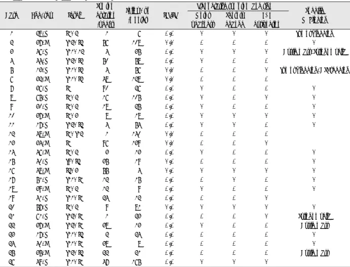
Table 1. Summary of modified JOA (Japanese Orthopaedic Association) score
Modified JOA score
I. Motor function of the lower extremity 0. Impossible to walk
1. Needs cane or aid on flat ground 2. Needs cane or aid only on stairs
3. Possible to walk without cane or aid but slow 4. Normal
II. Sensory function of the lower extremity 0. Apparent sensory loss
1. Minimal sensory loss 2. Normal
III. Bladder function 0. Complete retention
1. Severe disturbance (sense of retention, dribbling, incomplete continence)
2. Mild disturbance (urinary frequency, urinary hesitancy) 3. Normal
myoma on the gynecologic evaluation and was unaware that the genital pain comes from conus medullaris tumor not from uterine myoma (Fig. 1).
Preoperative MRI findings
Regarding the tumor extension on preoperative MRI, the involved masses less than 3 segments were 76.9% (n=20) and those more than 3 segments beyond conus medullaris were 23.1% (n=6). The mean involved segments were 2.8 segments. Three patients (11.5%) had cystic tumor mass, 2 patients (7.7%) had combined syringomyelia. Tumor bleeding was seen in 2 patients (7.7%).
Surgical Result
Twenty-four patients underwent laminectomy and 2 younger patients with tumors involving long segments underwent laminoplasty for prevention of postlaminectomy kyphosis. Gross total or complete tumor resection was performed in 80.8% of patients (n=21) and subtotal resection was performed in 19.2% (n=5) because of unclear demarcation between the tumor and normal neural tissue.
Pathology
The myxopapillary ependymoma, 13 cases (50.0%), was most common in conus medullaris tumors. Other histopathologic types were hemangioblastoma, 3 cases (11.5%); lipoma, 3 cases (11.5%); astrocytoma, 3 cases (11.5%); primitive neuroectodermal tumor (PNET), 2 cases (7.7%); capillary hemanigoma, 1 case (3.8%), and mature teratoma, 1 case (3.8%).
Adjuvant therapy
Postoperative radiotherapy was done in 6 patients (ependy-moma, 2; PNET, 2; astrocytoma, 1; hemangioblastoma, 1). Postoperative chemotherapy was done in 2 patients with
PNET, which is highly malignant.
Surgical outcomes
The surgical outcomes were summarized in Table 3. There was 1 case of CSF leakage as a postoperative complica-tion, which was controlled with dura repair and compression dressing. Tumor recurrence, including leptomeningeal seeding, occurred in 26.9% of patients (n=7)(myxopapillary ependymoma, 3; PNET, 2; astrocytoma, 3). Two patients with recurred myxopapillary ependymoma and a patient with pilocytic astrocytoma underwent radiotherapy and one patient with myxopapillary ependyomoma underwent radiotherapy and chemotherapy. One patient with recurred astrocytoma underwent reoperation. Two patients with astrocytoma and 1 patient with PNET died of tumor prog-ression at 12, 15, and 24 months after surgery. Another PNET tumor has been progressing in spite of postoperative radiotherapy and chemotherapy.
On the neurological outcome, modified JOA score wors-ened in 26.9% of patients (n=7), improved in 34.6% (n=9), and remained stable in 38.5% (n=18) compared to preoperative modified JOA score. Bladder and bowel function improved completely in 15.4% of patients (n=4), worsened in 15.4% (n=4), remained unchanged or minimally improved in 85.7% (n=18). The mean visual analogue scale (VAS) was improved from 5.4 to 1.8 among 21 patients who had low back pain and leg pain.
DISCUSSION
Conus medullaris tumor occupy approximately 10% of IMSTs and most of tumor are ependymoma, especially myxopapillary type18,19). Generally, IMSTs lead to myelop-athy such as sensory change or motor weakness16,19). However, in our study, the presenting symptoms of conus medullaris tumor were different from IMSTs at other site. The most com-mon symptoms were sciatica (90.5%) and low back pain (76.2%). In addi-tion, bladder and bowel dysfunction (61.9%) were also common. Uchiy-ama et al. reported that 58% of patients with epiconus/conus medull-aris tumors had voiding symptoms and especially, decreased urge to void, detrusor-sphinter dyssynergia and detrusor areflexia on voiding were the main symptom on urodynamic study21). Urinary symptoms may be the sole initial symptoms, but urinary Fig. 1. Pre- and Post-operative magnetic resonance image of lipoma at the conus medullaris
(case 16). A : The lipoma at the conus medullaris was revealed with high signal density on T1 weighted MRI. B : The lipoma suppressed typically on the Fat suppression MRI. C : The tumor was removed gross totally on the post operative MRI.
dysfunction tends to appear later. There may be no significant relationship between lower motor neuron signs and urinary symptoms. Two patients had severe perineal pain, and genitalia pain seemed to originate from sacral nerve root compression. These symptoms were misunderstood as a gynecological problem, and the patient underwent hysterectomy for the concordant uterine myoma before conus medullaris tumor detection. These symptomatic characteristics are due to the anatomical and functional characteristics of the conus medullaris as sacral segments and compression of lumbar nerve roots passing the conus medullaris, and should be considered for differential diagnosis of cauda equina syndrome or sciatica due to degenerative lumbar diseases.
In our series, laminectomy was performed in 21 patients and osteoplastic laminoplasty in 2 patients. There was no deformity requiring surgical correction in the patients who underwent only laminectomy at the final follow-up. Sandalcioglu et al. demonstrated osteoplastic laminoplasty did not reduce the risk of spinal deformities compared to patients who underwent laminectomy, and their current
surgical strategy consisted of reconstruction of the posterior spinal column and refixation of the laminotomy block with microplates16).
Recently, advanced microsurgical techniques and intraope-rative neurophysiological monitorings have made more aggressive approach for total or near-total resection of intra-medullary tumor10,15). In most reports, total or gross total resection of IMSTs has been reported as 65-90%11,18-20). However, there are the limitations of total resection due to unclear demarcation between the tumor and normal neural tissue and the resulting risk of injury to neural tissue. We achieved 80.8% of gross total resection and there may be no meaningful difference compared with IMSTs at other site. Several authors reported this variation of the resec-tability depends on the surgical technique and the surgeon’s attitude1,16). Others reported a difference of tumor resectability according to the histopathological type of tumors4,5,16,20). Generally, the rate of complete removal is approximately 90% for ependymoma and hemangioblastoma and 50%-76% in low grade astrocytoma. In our cases, total resection of ependymoma was achieved in 89%, which is similar to Table 3. Outcome of 26 conus medullaris tumors
Case Surgery Histology Adjuvant Recurrence Additional therapy Preop JOA score Postop JOA score Final therapy after recurrence (bladder function) (bladder function) outcome
1 GTR ME - - - 9 (3) 9 (3) 2 GTR ME - - - 5 (2) 5 (2) 3 GTR ME RT - - 9 (3) 9 (3) 4 GTR ME - - - 6 (2) 9 (3) 5 GTR ME - + RT, CT 5 (2) 2 (0) 6 GTR ME - - - 6 (2) 5 (1) 7 GTR ME - - - 7 (2) 8 (2) 8 GTR ME - - - 8 (3) 9 (3) 9 GTR ME RT - - 7 (2) 7 (2) 10 STR ME - + RT 9 (3) 9 (3) 11 GTR ME - + RT 7 (2) 7 (2) 12 GTR ME - - - 4 (2) 3 (2) 13 GTR ME - - - 7 (2) 4 (0) 14 GTR Lipoma - - - 8 (2) 9 (3) 15 STR Lipoma - - - 6 (2) 6 (2) 16 GTR Lipoma - - - 8 (3) 7 (2) 17 GTR Astrocytoma - + RT 1 (0) 0 (0) Death 18 GTR Astrocytoma - - - 8 (2) 9 (3) 19 STR Astrocytoma RT + - 6 (2) 0 (0) Death 20 GTR Hemangioblastoma - - - 9 (3) 9 (3) 21 STR Hemangioblastoma RT - RT 7 (3) 7 (3) 22 GTR Hemangioblastoma - - - 7 (2) 9 (3) 23 STR PNET RT, CT + 2 (0) 3 (0) Death 24 GTR PNET RT, CT + - 0 (0) 0 (0) Progress 25 GTR Mature teratoma - - - 6 (2) 7 (2) 26 GTR Capillary hemangioma - - - 7 (2) 8 (2)
reported rates. Tumor resectability of other histopatho-logical types varied due to intraoperative decisions to avoid injuring the normal neural tissue because the demarcation between the tumor and normal neural tissue was not clear. From our surgical experience of 26 patients, we noted that the important point in the surgical resection of conus tumor is the early detection and resection of filum terminale attached with the tumor. Sometimes, the discrimination between the conus medullaris and nerve roots is difficult to determine because the nerve roots around the conus meullaris are relatively thick compared to the distal portion of lumbosacral nerve roots. In the pathological type of spinal cord tumors, ependymoma was the most common, comprising of 33-83% of IMSTs5,16,17). Myxopapillary
subtype was common. Other common pathological types were lipoma, astrocytoma, and hemangioblastoma. In our cases, myxopapillary ependymoma was found in 50% of the total 26 cases and astrocytoma, lipoma, and hamangiob-lastoma were the second common, found in 14.3% cases. Rare tumor as PNET and mature teratoma occupied individually 7.7% and 3.8%. In particular, PNET is highly malignant as the average survival time is 20 months8). Intramedullary mature teratoma of the conus medullaris is also rare and 30 cases have been reported in our review of the literature7).
On functional outcome, modified JOA score worsened in 7 patients (26.9%), improved in 9 patients (34.6%), and remained stable in 10 patients (38.5%). The patient with worsened modified JOA score was aggravated mainly in urinary function, not motor function. The cause of aggra-vation was due to aggressive total resection in spite of large tumor size and unclear demarcation between tumor and normal neural tissue. Though complete resection brings relatively good tumor control, total resection should be carefully considered. Five patients who had lower modified JOA score below 5 points postoperatively also have poor preoperative neurological condition. Therefore, the func-tional outcome depended on the preoperative neurological condition as in other reports of IMSTs14,18). The postoperative bladder function may be the main factor on postoperative neurological function and play an important role in patient’s satisfaction in conus meudllaris tumor. In 3 patients with cystic component, gross total resection could be easily per-formed. All of the tumors were benign and the postoper-ative neurological function was improved in three patients. The cystic tumor lesion may be one predictor of good func-tional outcome.
Postoperative adjuvant radiotherapy is still controversial, especially for patients with low-grade tumor such as myxo-papillary ependymoma and pilocytic astrocytoma. In our
series, postoperative adjuvant radiotherapy was not routinely used in low-grade tumor and only used in the residual and highly malignant tumor. Lee et al.11)reported that postop-erative radiotherapy should only be considered as surgical adjunct where gross total resection is not achieved and Lin et al.12)reported postoperative local radiotherapy is recom-mend and can achieve excellent local control and survival for incomplete resection of ependymoma. We achieved good local control in ependymoma as well as in 3 ependy-momas with recurrence and leptomeningeal seeding after gross-total resection. Koh et al. reported that fractional external beam radiotherapy has a role in the management of residual hemangioblastoma, especially for von Hippel-Lindau disease9). We also used radiotherapy in one patient with residual hemangioblastoma and achieved good local control during follow-up period (23 months). Three patients with PNET and astrocytoma died of the systemic tumor progression at 12, 15, and 24 months after surgery. In 1 patient with PNET, tumor progressed in spite of the total tumor resection, postoperative adjuvant radiotherapy, and chemotherapy. The optimal treatment of PNET is unknown because of the rarity of the tumor. Literature review has shown that almost all spinal PNET cases were treated with surgery and high dose radiotherapy of the tumor region and entire neuraxis, and sometimes additional chemo-therapy8). It revealed the tendency of longer survival in
patient treated with additional chemotherapy than patients treated only surgical resection and radiotherapy8). Even though PNET is highly malignant, aggressive adjuvant therapy including chemotherapy may be considered.
CONCLUSION
The surgical outcome of conus medullaris tumor mainly depends on preoperative neurological condition and histopa-thological type. The surgical treatment of conus medullaris tumor needs understanding the anatomical and functional characteristics of conus medullaris tumor for better outcome.
Acknowledgement
This study was supported by a faculty research grant of Yonsei University College of Medicine for 2006 (No. 6-2006-164).
References
1. Brotchi J : Intrinsic spinal cord tumor resection. Neurosurgery 50 : 1059-1063, 2002
2. Chen CY, Chen PH, Yao MS, Chu JS, Chan WP : MRI of hemang-ioblastoma in the conus medullaris. Comput Med Imaging Graph 32 : 78-81, 2008
3. Choi SH, Kim SJ, Park SH, Cho YJ : Co-existence of lipoma and myxopapillary ependymoma in a filum terminale tumor. J Neurosurg Sci 39 : 378-381, 2006
4. Constantini S, Miller DC, Allen JC, Rorke LB, Freed D, Epstein FJ : Radical excision of intramedullary spinal cord tumors : Surgical morbidity and long-term follow-up evaluation in 164 children and young adults. J Neurosurg 93 : 183-193, 2000
5. Fukui M, Chiba K, Kawakami M, Kikuchi S, Konno S, Miyamoto M, et al : An outcome measure for patients with cervical myelopathy : Japanese orthopaedic association cervical myelopathy evaluation questionnaire (joacmeq) : Part 1. J Orthop Sci 12 : 227-240, 2007 6. Jaiswal AK, Jaiswal S, Gupta SK, Singh Gautam VK, Kumar S :
Intramedullary tuberculoma of the conus. J Clin Neurosci 13 : 870-872, 2006
7. Kahilogullari G, Erdem A, Heper AO, Erden E : Intramedullary mature cystic teratoma of the conus medullaris. A case report. J
Neurosurg Sci 50 : 55-58, 2006
8. Kim YW, Jin BH, Kim TS, Cho YE : Primary intraspinal primitive neuroectodermal tumor at conus medullaris. Yonsei Med J 45 : 533-538, 2004
9. Koh ES, Nichol A, Millar BA, Menard C, Pond G, Laperriere NJ : Role of fractionated external beam radiotherapy in hemangioblastoma of the central nervous system. Int J Radiat Oncol Biol Phys 69 : 1521-1526, 2007
10. Kothbauer KF : Intraoperative neurophysiologic monitoring for intramedullary spinal-cord tumor surgery. Neurophysiol Clin 37 : 407-414, 2007
11. Lee TT, Gromelski EB, Green BA : Surgical treatment of spinal ependymoma and post-operative radiotherapy. Acta Neurochir
(Wien) 140 : 309-313, 1998
12. Lin YH, Huang CI, Wong TT, Chen MH, Shiau CY, Wang LW, et al : Treatment of spinal cord ependymomas by surgery with or without postoperative radiotherapy. J Neurooncol 71 : 205-210,
2005
13. McGirt MJ, Chaichana KL, Atiba A, Attenello F, Woodworth GF, Jallo GI : Neurological outcome after resection of intramedullary spinal cord tumors in children. Childs Nerv Syst 24 : 93-97, 2008 14. Raco A, Esposito V, Lenzi J, Piccirilli M, Delfini R, Cantore G :
Long-term follow-up of intramedullary spinal cord tumors: A series of 202 cases. Neurosurgery 56 : 972-981; discussion 972-981, 2005 15. Sala F, Bricolo A, Faccioli F, Lanteri P, Gerosa M : Surgery for
intramedullary spinal cord tumors: The role of intraoperative (neurophysiological) monitoring. Eur Spine J 16 (Suppl 2) : S130-139, 2007
16. Sandalcioglu IE, Gasser T, Asgari S, Lazorisak A, Engelhorn T, Egelhof T, et al : Functional outcome after surgical treatment of intramedullary spinal cord tumors : experience with 78 patients. Spinal Cord 43 : 34-41, 2005
17. Shrivastava RK, Epstein FJ, Perin NI, Post KD, Jallo GI : Intra-medullary spinal cord tumors in patients older than 50 years of age : management and outcome analysis. J Neurosurg Spine 2 : 249-255, 2005
18. So WS, Lee WJ, Choi HY, Eun JP : Spinal intramedullary lipoma without dysraphism. J Korean Neurosurg Soc 42 : 42-45, 2007 19. Son YJ, Chung CK, Kim HJ : Surgical results of intramedullary
spinal cord ependymomas in adults : retrospective analysis of 51 cases. J Korean Neurosurg Soc 26 : 384-393, 1997
20. Tobias ME, McGirt MJ, Chaichana KL, Goldstein IM, Kothbauer KF, Epstein F, et al : Surgical management of long intramedullary spinal cord tumors. Childs Nerv Syst 24 : 219-223, 2008 21. Uchiyama T, Sakakibara R, Hattori T, Yamanishi T : Lower urinary
tract dysfunctions in patients with spinal cord tumors. Neurourol