Efficacy and Safety of Vancomycin versus Teicoplanin in Patients with
Health Care-Associated Methicillin-Resistant Staphylococcus aureus
Bacteremia
Young Kyung Yoon,aDae Won Park,aJang Wook Sohn,aHyo Youl Kim,bYeon-Sook Kim,cChang-Seop Lee,dMi Suk Lee,e
Seong-Yeol Ryu,fHee-Chang Jang,gYoung Ju Choi,hCheol-In Kang,iHee Jung Choi,jSeung Soon Lee,kShin Woo Kim,lSang Il Kim,m Eu Suk Kim,nJeong Yeon Kim,oKyung Sook Yang,pKyong Ran Peck,iMin Ja Kima
‹Division of Infectious Diseases, Department of Internal Medicine, Korea University, College of Medicine, Seoul, Republic of Koreaa
; Yonsei University Wonju College of Medicine, Wonju, Republic of Koreab
; Chungnam National University Hospital, Daejon, Republic of Koreac
; Chonbuk National University Medical School, Jeonju, Republic of Koread
; Kyung Hee University School of Medicine, Seoul, Republic of Koreae
; Keimyung University Dongsan Hospital, Daegu, Republic of Koreaf
; Chonnam National University Medical School, Gwangju, Republic of Koreag
; National Cancer Center, Seoul, Republic of Koreah
; Sungkyunkwan University School of Medicine, Seoul, Republic of Koreai
; Ewha Women’s University School of Medicine, Seoul, Republic of Koreaj
; Hallym University Sacred Heart Hospital, Anyang, Republic of Koreak
; Kyungpook National University Hospital, Daegu, Republic of Koreal
; Catholic University of Korea, College of Medicine, Seoul, Republic of Koream
; Seoul National University Bundang Hospital, Seoul, Republic of Korean
; Samyook Medical Center, Seoul, Republic of Koreao
; Department of Biostatistics, Korea University College of Medicine, Seoul, Republic of Koreap
The purpose of this study was to compare the clinical efficacy and safety of vancomycin to those of teicoplanin for the treatment
of adult patients with health care-associated methicillin-resistant Staphylococcus aureus (HA-MRSA) bacteremia. A multicenter
observational study was prospectively conducted in 15 teaching hospitals in Korea between February 2010 and July 2011. Adult
patients (>18 years old) with HA-MRSA bacteremia who were initially treated with vancomycin (VAN) (n ⴝ 134) or teicoplanin
(TEC) (n
ⴝ 56) were enrolled. Clinical and microbiological responses and drug-related adverse events were compared between
the two treatment groups using univariate and multivariate logistic regression analyses. The vancomycin and teicoplanin MICs
were determined by Etest. The MRSA-related mortality, duration of fever, and duration of MRSA bacteremia in the treatment
groups were not significantly different. There was no significant difference in the occurrence of drug-related adverse events.
Among the 190 MRSA isolates, the VAN MICs ranged from 0.5 to 2
g/ml (MIC
50and MIC
90, 1.5
g/ml), and the TEC MIC
ranged from 0.5 to 8
g/ml (MIC
50, 3
g/ml; MIC
90, 6
g/ml). In multivariate analyses, the antibiotic type (vancomycin or
teico-planin) was not associated with treatment outcomes. This study indicates that teicoplanin is an effective and safe alternative to
vancomycin for the treatment of HA-MRSA bacteremia.
N
osocomial bloodstream infections represent a major clinical
challenge in many health care institutions worldwide, despite
laborious and costly infection control efforts. Health
care-associ-ated methicillin-resistant Staphylococcus aureus (HA-MRSA)
bac-teremia has imposed a distinctly high burden on medical expenses
and has caused considerable morbidity and mortality (
1
,
2
).
Vancomycin (VAN) has widely been used for the treatment of
MRSA infection over the past decades. Increasingly, however,
therapeutic failures with VAN have been reported (
3
). There is
also growing evidence of bacteremia caused by MRSA isolates with
an increased VAN MIC (
4
,
5
). The VAN therapeutic monitoring
guidelines in 2009 recommended more aggressive VAN dosing
schemes, targeting VAN serum trough concentrations of 15 to 20
mg/liter for MRSA bacteremia (
6
). Similarly, optimizing the
phar-macokinetics of VAN to achieve an area under the curve (AUC)/
MIC ratio of
ⱖ211 has been shown to predict more favorable
treatment outcomes in cases of MRSA-associated complicated
bacteremia (
7
). However, if the MRSA strains’ MIC is
ⱖ2 g/ml,
conventional intermittent dosing might not achieve this ratio.
Rather, it may increase nephrotoxicity (
8
).
Teicoplanin (TEC) is a glycopeptide antibiotic with an
antibac-terial spectrum similar to that of VAN but is less toxic at daily
doses of less than 800 mg (
9
,
10
). It has a long half-life (45 to 70 h),
permitting once-daily dosing (
11
), and may enhance the
intracel-lular killing of bacteria by phagocytes (
12
). TEC is commonly used
for MRSA infections in Europe, while its use has not yet been
approved in the United States. TEC has been used as an alternative
agent for MRSA infections; however, there is a limited number of
studies that have evaluated the clinical efficacy of TEC in patients
with HA-MRSA bacteremia (
13
,
14
).
The purpose of this prospective observational study was to
compare the clinical efficacy and safety of VAN to those of TEC for
the treatment of adult patients with HA-MRSA bacteremia.
MATERIALS AND METHODS
Study design and patients. A prospective, multicenter observational study was conducted in 15 teaching hospitals in the Republic of Korea over an 18-month period from February 2010 to July 2011. The subjects comprised hospitalized adult patients (ⱖ18 years) with HA-MRSA
bac-Received 14 March 2013 Returned for modification 15 May 2013 Accepted 18 October 2013
Published ahead of print 28 October 2013
Address correspondence to Min Ja Kim, [email protected]. Copyright © 2014, American Society for Microbiology. All Rights Reserved. doi:10.1128/AAC.00520-13
on September 19, 2016 by Ewha Womans Univ
http://aac.asm.org/
teremia who were initially treated with VAN (n⫽ 134) or TEC (n ⫽ 56) and who were followed until death or hospital discharge. Only the first episode of HA-MRSA bacteremia and the first blood isolate of MRSA per patient that was susceptible to both VAN and TEC were included for analysis. Patients with polymicrobial bacteremia were excluded in order for this study to evaluate the impact of antibiotic therapy for MRSA bac-teremia specifically.
A loading dose of VAN (1 g every 12 h) or TEC (400 mg every 12 h) was administered for an initial 24 h or 36 h, respectively, and then followed by daily maintenance doses of each drug that were adjusted to the patient’s renal function, if needed (15). In the 11 participating hospitals (73.3%) that ran the therapeutic drug monitoring (TDM) practices for VAN, the TDM-guided VAN dosing was performed, targeting serum trough levels between 15 and 20g/ml. None of the participating hospitals ran the TDM for TEC. During the study period, there were no other standardized interventions for the management of MRSA bacteremia, and physicians treated the patients according to routine medical practice.
The study protocol was approved prior to study initiation by the in-stitutional review boards at each participating hospital. As this observa-tional study required no deviation from routine medical practice, the boards waived the need for informed consent.
Definitions. MRSA bacteremia was considered present if one or more blood cultures had positive results and if the clinical signs and course were consistent with MRSA infection (16).
The primary source of infection, based on the organs affected, was classified as one of the following: lower respiratory tract, intra-abdominal area, genitourinary tract, skin and soft tissue, bone and joint, central ner-vous system, and catheter. The origin of infection was considered un-known in cases of positive blood cultures without primary infection at another body site (16).
MRSA bacteremia was categorized epidemiologically as health care associated or nosocomial. Community-onset MRSA bacteremia within ⱕ48 h of hospital admission was considered health care associated if, during the preceding 12 months, the patient had any of the following: admission to other hospitals or health care facilities for more than 2 days, surgery, dialysis, specialized home care, care received at day hospitals, or permanent indwelling catheters. Patients defined as having community-acquired infections were excluded from this study. Infections occurring in patients after 48 h of hospital admission were considered nosocomial.
The duration of bacteremia after VAN or TEC treatment was calcu-lated as the number of days from the start of MRSA treatment to the day the first negative blood culture was drawn. Sepsis, severe sepsis, and septic shock were defined according to the standard criteria (17). The commu-nity-acquired phenotype for the MRSA isolates was defined as being sus-ceptible to clindamycin, erythromycin, and ciprofloxacin (18,19).
The primary endpoint was clinical failure, defined as a composite of mortality attributable to MRSA bacteremia, microbiological failure, and/or persistent fever, except drug fever. Mortality attributable to MRSA bacteremia was defined as positive blood cultures for MRSA, persistent fever, and no other definite causes of death. Microbiological failure was defined as positive blood cultures for MRSAⱖ7 days from the index culture under VAN or TEC therapy. Persistent fever was defined as ⱖ38.0°C for ⱖ7 days after the commencement of VAN or TEC treatment. Variables. Physicians or research coordinators of the participating hospitals entered the clinical data for each patient into a standardized web-based case report form. An infectious disease doctor at the coordi-nating center checked the entered data and supported the study sites by sending queries throughout the study period. The parameters collected for this analysis included demographic characteristics, comorbid medical conditions, including Charlson’s comorbidity index (20), factors predis-posing to infections, primary source of MRSA bacteremia, acute physiol-ogy and chronic health evaluation II (APACHE II) score (21) or Pitt’s bacteremia score (22) at the onset of MRSA bacteremia, diagnosis of se-vere sepsis or septic shock, hospital mortality, and microbiological data.
Microbiological tests. Bacterial identification and antibiotic suscep-tibility were performed at each study site using a Vitek II (bioMérieux, Hazelwood, MO) or MicroScan Pos Combo panel type 6 system (Baxter Diagnostics, West Sacramento, CA). All MRSA isolates from participating hospitals were sent to the coordinating center. All isolates received were immediately stored at⫺70°C until August 2012, when microbiologic tests were performed all at once. The VAN and TEC MICs for all 190 MRSA isolates were further determined by the Etest (bioMérieux, Marcy l’Etoile, France) at the coordinating center according to the manufacturer’s in-structions.
Statistical analysis. For comparisons between groups of continuous independent variables that were normally distributed, the two-sample Student’s t test was used. For comparisons of continuous independent variables that were not normally distributed, the Mann-Whitney U test was used. Summaries of the continuous variables were expressed as me-dians and interquartile ranges (IQR). Independent categorical variables were described using count (proportion), and comparisons between groups were made using the Pearson’s chi-squared test or Fisher’s exact test.
In the univariate analysis, the VAN and TEC MICs were evaluated as continuous variables as well as categorical variables. The cutoff values of the VAN MICs and the TEC MICs were determined with an analysis using the chi-squared automatic interaction detector (CHAID) decision tree algorithm, to predict treatment outcome in the respective treatment group. The cutoff values of the VAN MICs and the TEC MICs drawn from the CHAID algorithm were 1.5g/ml and 4.0 g/ml, respectively.
Multivariate logistic regression analyses using the backward stepwise variable selection based on the LR statistic were used to examine the im-pact of multiple independent predictors on the clinical failure as a depen-dent variable. Trauma, renal diseases, hepatic diseases, pneumonia, Pitt’s bacteremia score, C-reactive protein, acute renal injury, duration of fever or bacteremia after VAN or TEC treatment, and antibiotic type were eval-uated as independent variables for multivariable logistic regression anal-ysis if such independent variables were predictors of clinical failure at the 10% significance level. Hosmer-Lemeshow goodness-of-fit tests were per-formed to evaluate the models. Internal accuracy obtained by leave-one-out cross-validation was used to evaluate the performance of a predictive model. All tests were 2-tailed, and a P value⬍0.05 was considered statis-tically significant. All of the analyses were performed with IBM SPSS Sta-tistics version 20.0 (IBM Corporation, Armonk, NY), R 2.15.2 (The R Foundation for Statistical Computing, Vienna, Austria), and SAS 9.2 (SAS Institute Inc., Cary, NC).
RESULTS
Patients and clinical characteristics. During the study period,
426 patients with HA-MRSA bacteremia were enrolled from the
participating hospitals. Patients who were given antibiotics with
no activity against MRSA isolates (n
⫽ 81) and 49 patients from
whom MRSA isolates were not collected were excluded from the
analysis. Patients who initially received other antibiotics before
VAN or TEC (n
⫽ 96) and who received VAN or TEC for ⬍3 days
(n
⫽ 10) were also excluded. Eventually, 190 patients with
HA-MRSA bacteremia who were initially treated with VAN (n
⫽ 134)
or TEC (n
⫽ 56) for ⱖ3 days were included in this study.
The demographic and baseline characteristics of the 190
pa-tients are listed in
Table 1
. Of these, 158 patients (83.2%) had
nosocomial infections and 128 (67.4%) were male. The median
age was 66 years (IQR, 51 to 74 years). The univariate analyses
determined that there were no significant differences in sex, age,
and category of infection between the VAN and TEC treatment
groups (
Table 1
).
The most common source of MRSA bacteremia was
catheter-related infections (47.9%), followed by pneumonia (14.7%),
sur-gical wounds (10.0%), and bone and joint infections (5.8%). The
on September 19, 2016 by Ewha Womans Univ
http://aac.asm.org/
TABLE 1 Demographic and baseline characteristics of 190 patients with MRSA bacteremia according to treatment group and outcomea
Variable All (n⫽ 190)
Treatment group Treatment outcome Vancomycin (n⫽ 134) Teicoplanin (n⫽ 56) P Success (n⫽ 112, 58.9%) Failure (n⫽ 78, 41.1%) P
No. (%) receiving vancomycin 134 (70.5) 134 (100) 0 ⬍0.001 85 (75.9) 49 (62.8) 0.052 No. (%) of males 128 (67.4) 92 (68.7) 36 (64.3) 0.558 78 (69.6) 50 (64.1) 0.423 Median age, yrs (IQR) 66 (51–73) 64.5 (51–73) 68 (51.5–74) 0.521 65 (52–72) 67.5 (50–76) 0.508 No. (%) with time of bacteremia
ⱕ48 h 42 (25.6) 35 (26.1) 7 (23.3) 0.752 28 (26.9) 14 (23.3) 0.612
⬎48 h 122 (74.4) 99 (73.9) 23 (76.7) 76 (73.1) 46 (76.7)
No. (%) with category of infection
Health care associated 32 (16.8) 23 (17.2) 9 (16.1) 0.854 20 (17.9) 12 (15.4) 0.654 Nosocomial 158 (83.2) 111 (82.8) 47 (83.9) 92 (82.1) 66 (84.6)
Comorbid illness
No. (%) with cardiovascular disease 97 (51.1) 65 (48.5) 32 (57.1) 0.278 54 (48.2) 43 (55.1) 0.348 No. (%) with central nervous system disease 47 (24.7) 33 (24.6) 14 (25.0) 0.957 31 (27.7) 16 (20.5) 0.260 No. (%) with malignancy 57 (30.0) 47 (35.1) 10 (17.9) 0.018 36 (32.1) 21 (26.9) 0.440 No. (%) with trauma 18 (9.5) 12 (9.0) 6 (10.7) 0.706 5 (4.5) 13 (16.7) 0.005 No. (%) with renal disease 39 (20.5) 28 (20.9) 11 (19.6) 0.845 18 (16.1) 21 (26.9) 0.068 No. (%) with hepatic disease 20 (10.5) 17 (12.7) 3 (5.4) 0.133 16 (14.3) 4 (5.1) 0.043 No. (%) with respiratory disease 23 (12.1) 16 (11.9) 7 (12.5) 0.914 10 (8.9) 13 (16.7) 0.108 No. (%) with solid organ or bone marrow
transplant
4 (2.1) 3 (2.2) 1 (1.8) 1.000 3 (2.7) 1 (1.3) 0.645 No. (%) with metabolic disease 66 (34.7) 48 (35.8) 18 (32.1) 0.627 34 (30.4) 32 (41.0) 0.129 No. (%) with HIV infection 2 (1.1) 2 (1.5) 0 1.000 1 (0.9) 1 (1.3) 1.000 No. (%) with hematologic disease 30 (15.8) 26 (19.4) 4 (7.1) 0.035 19 (17.0) 11 (14.1) 0.595 No. (%) with gastrointestinal bleeding 8 (4.2) 5 (3.7) 3 (5.4) 0.695 3 (2.7) 5 (6.4) 0.276 Charlson’s comorbidity index, median (IQR) 2 (1–4) 3 (1–4) 1 (0–3) 0.008 2 (0–4) 2 (1–4) 0.693 No. (%) with primary source of bacteremia
Catheter-related infection 91 (47.9) 70 (52.2) 21 (37.5) 0.064 59 (52.7) 32 (41.0) 0.114 Pneumonia 28 (14.7) 19 (14.2) 9 (16.1) 0.737 10 (8.9) 18 (23.1) 0.007 Surgical wound infection 19 (10.0) 13 (10.4) 5 (8.9) 0.750 13 (11.6) 6 (7.7) 0.376 Bone and joint infection 11 (5.8) 10 (7.5) 1 (1.8) 0.179 5 (4.5) 2 (2.6) 0.702 Intra-abdominal infection 10 (5.3) 3 (2.2) 7 (12.5) 0.008 3 (2.7) 4 (5.1) 0.448 Urinary tract infection 7 (3.7) 3 (2.2) 4 (7.1) 0.198 7 (6.2) 4 (5.1) 1.000 Skin and soft tissue infection 7 (3.7) 5 (3.7) 2 (3.6) 1.000 1 (0.9) 3 (3.8) 0.307 Cardiovascular infection 4 (2.1) 2 (1.5) 2 (3.6) 0.583 5 (4.5) 5 (6.4) 0.743 Central nervous system infection 1 (0.5) 1 (0.7) 0 1.000 0 1 (1.3) 0.411
Head and neck infection 1 (0.5) 0 1 (1.8) 0.295 1 (0.9) 0 1.000
Unknown 11 (5.8) 7 (5.2) 4 (7.1) 0.734 8 (7.1) 3 (3.8) 0.530
Clinical severity at the onset of MRSA bacteremia
No. (%) with fever (ⱖ38.0°C) 142 (74.7) 104 (77.6) 38 (67.9) 0.158 90 (80.4) 52 (66.7) 0.033 No. (%) with SIRS 189 (99.5) 133 (99.3) 56 (100) 1.000 111 (99.1) 78 (100) 1.000 No. (%) with development of severe sepsis or
septic shock
66 (34.7) 43 (32.1) 23 (41.1) 0.236 31 (27.7) 35 (44.9) 0.014 Pitt’s bacteremia score [median (IQR)] 1 (0–3) 1 (0–3) 1 (0–4) 0.542 1 (0–3) 2 (0–3) 0.286 APACHE II score [median (IQR)] 17 (12–21) 15 (12–21) 19 (13–23) 0.211 17 (11–21) 17 (13–21) 0.667 No. (%) with APACHE II score ofⱖ20 42 (33.3) 23 (32.9) 19 (33.9) 0.899 22 (30.6) 20 (37.0) 0.445 No. (%) with complicated condition
Foreign body retention 8 (4.2) 7 (5.2) 1 (1.8) 0.440 7 (6.2) 1 (1.3) 0.144 Infective endocarditis 5 (2.6) 4 (3.0) 1 (1.8) 1.000 3 (2.7) 2 (2.6) 1.000 Metastatic infectionsb 11 (5.8) 9 (6.7) 2 (3.6) 0.512 8 (7.1) 3 (3.8) 0.530 Laboratory findings at the onset of MRSA
bacteremia
C-reactive protein (mg/liter) 10.8 (4.8–22.1) 11.1 (5.2–20.2) 9.9 (4.1–23.0) 0.914 9.2 (3.9–17.4) 14.3 (6.8–24.4) 0.039 No. (%) with hematocrit of⬍30% 98 (51.6) 66 (49.3) 32 (57.1) 0.321 53 (47.3) 45 (57.7) 0.159 No. (%) with platelet count of⬍100,000/l 41 (21.6) 30 (22.4) 11 (19.6) 0.675 22 (19.6) 19 (24.4) 0.437
(Continued on following page)
on September 19, 2016 by Ewha Womans Univ
http://aac.asm.org/
univariate analyses revealed no significant differences in the
pri-mary source of infection between the 2 treatment groups, except
for intra-abdominal infections (
Table 1
).
The median Charlson comorbidity index was 2 (IQR, 1 to 4),
and univariate analyses determined that the VAN group had a
significantly higher Charlson comorbidity index than the TEC
group. In particular, underlying malignancy and hematologic
dis-eases were significantly more common in the VAN group than the
TEC group (
Table 1
). Sixty-six patients (34.7%) had severe sepsis
or septic shock, and the median APACHE II score at the onset of
HA-MRSA bacteremia was 17 (IQR, 12 to 21). There was no
sig-nificant difference in the APACHE II score of HA-MRSA
bactere-mia between the two treatment groups (
Table 1
).
Microbiological characteristics. All 190 MRSA isolates
un-derwent microbiological analysis. The VAN MIC range was 0.5 to
2
g/ml, and the VAN MIC
50and MIC
90were both 1.5
g/ml. The
TEC MIC range was 0.5 to 8
g/ml, and the TEC MIC
50and
MIC
90were 3
g/ml and 6 g/ml, respectively. Distribution of the
VAN and TEC MICs and the antibiotic phenotype among the
MRSA isolates, categorized by treatment group and treatment
outcome, are shown in
Table 2
. In a total of 190 patients analyzed,
the VAN or TEC MICs were not associated with clinical failure.
When the influences of the VAN MICs on clinical outcomes in
the VAN-treated group were evaluated, a VAN MIC of
ⱖ1.5
g/ml was the significant risk factor for in-hospital mortality
(VAN MIC,
⬍1.5 g/ml versus ⱖ1.5 g/ml; 19.1% [9/47] versus
41.4% [36/87]; P
⫽ 0.009) but not for clinical failure (40.4% [19/
47] versus 34.5% [30/87]; P
⫽ 0.495). In the TEC-treated group, a
TEC MIC of
ⱖ4 g/ml in the TEC group was not significantly
associated with treatment failure (TEC MIC,
⬍4 g/ml versus ⱖ4
g/ml; 53.3% [24/45] versus 45.5% [5/11]; P ⫽ 0.639) or
in-hospital mortality (40.0% [18/45] versus 27.3% [3/11]; P
⫽
0.508).
The proportion of MRSA isolates with phenotypic expression
of community-acquired MRSA was 16.3% (31/190) and was not
significantly different between the 2 treatment groups (
Table 2
).
Treatment outcomes. The overall all-cause in-hospital
mor-tality and MRSA-related mormor-tality were 34.7% (66/190) and
14.7% (28/190), respectively. There were no significant
differ-ences in the all-cause in-hospital mortality and MRSA-related
mortality between the treatment groups. After the
commence-ment of VAN or TEC therapy, a significant difference was not
exhibited for the duration of fever and MRSA bacteremia between
the treatment groups (
Table 3
). The median durations of VAN
and TEC treatment showed no significant differences (median,
[IQR], 14 days [IQR, 9 to 23 days] versus 13 days [IQR, 8 to 21
days]; P
⫽ 0.239).
In total, 36 patients (18.9%) received alternative drugs due to
poor clinical response (n
⫽ 23), drug-related adverse events (n ⫽
12), or other reasons (n
⫽ 4). In the VAN group, VAN was
switched with alternative antibiotics in 20 patients (14.9%): TEC
(n
⫽ 11), linezolid (n ⫽ 6), tigecycline (n ⫽ 2), or levofloxacin
plus rifampin (n
⫽ 1). In the TEC group, 16 patients (28.6%)
received alternative antibiotics: VAN (n
⫽ 9), linezolid (n ⫽ 4), an
TABLE 1 (Continued)Variable All (n⫽ 190)
Treatment group Treatment outcome Vancomycin (n⫽ 134) Teicoplanin (n⫽ 56) P Success (n⫽ 112, 58.9%) Failure (n⫽ 78, 41.1%) P
No. (%) with albumin of⬍3.0 g/dl 84 (44.2) 56 (41.8) 28 (50.0) 0.299 49 (43.8) 35 (44.9) 0.878 No. (%) with total bilirubin ofⱖ2.0 mg/dl 37 (19.5) 27 (20.1) 10 (17.9) 0.716 20 (17.9) 17 (21.8) 0.500 No. (%) with creatinine ofⱖ2.0 mg/dl 52 (27.4) 37 (27.6) 15 (26.8) 0.907 33 (29.5) 19 (24.4) 0.272 No. (%) with serum sodium of⬍130.0
mmol/liter
17 (8.9) 15 (11.2) 2 (3.6) 0.093 13 (11.6) 4 (5.1) 0.124 aHIV, human immunodeficiency virus; SIRS, systemic inflammatory response syndrome; IQR, interquartile range; APACHE II, acute physiology and chronic health evaluation II;
SD, standard deviation; MRSA, methicillin-resistant Staphylococcus aureus.
bSites of metastatic infections include bones and joints, the epidural space, intervertebral disks, heart valves, and intra-abdominal organs.
TABLE 2 Microbiological characteristics of 190 patients with MRSA bacteremia according to treatment group and outcome
Variable All (n⫽ 190)
Treatment group Treatment outcome Vancomycin (n⫽ 134) Teicoplanin (n⫽ 56) P Success (n⫽ 112, 58.9%) Failure (n⫽ 78, 41.1%) P
MIC,g/ml [median (IQR)]
Vancomycin 1.5 (1.0–1.5) 1.5 (1.0–1.5) 1.0 (1.0–1.5) ⬍0.001 1.5 (1.0–1.5) 1.5 (1.0–1.5) 0.324 Teicoplanin 3.0 (2.0–4.0) 3.0 (2.0–4.0) 3.0 (2.0–3.0) 0.039 3.0 (2.0–4.0) 3.0 (2.0–4.0) 0.476 No. (%) with vancomycin MIC of
ⱖ1.5 g/ml
106 (55.8) 87 (64.9) 19 (33.9) 0.001 67 (59.8) 39 (50.0) 0.180
No. (%) with teicoplanin MIC of MICⱖ 4 g/ml
64 (33.7) 53 (39.6) 11 (19.6) 0.008 42 (37.5) 22 (28.2) 0.182
No. (%) with CA-MRSAa phenotype (18,19)
31 (16.3) 25 (18.7) 6 (10.7) 0.101 21 (18.8) 10 (12.8) 0.199 aCA-MRSA, community-acquired methicillin-resistant Staphylococcus aureus.
on September 19, 2016 by Ewha Womans Univ
http://aac.asm.org/
aminoglycoside (n
⫽ 1), clindamycin (n ⫽ 1), or
trimethoprim-sulfamethoxazole plus rifampin (n
⫽ 1).
The median duration of VAN in alternation from VAN to TEC
(n
⫽ 11) and vice versa (n ⫽ 9) was 9 days (IQR, 6 to 17 days) and
10 days (IQR, 7 to 13 days), respectively.
There was no significant difference in the occurrence of
drug-related adverse events between the 2 treatment groups (20.9%
[28/134] versus 14.3% [8/56], P
⫽ 0.289) (
Table 3
). Among the 8
patients who received alternative glycopeptides due to
drug-re-lated adverse events, cross-reactivity was not observed between
VAN and TEC. One patient with VAN-induced acute kidney
in-jury developed TEC-induced neutropenia.
In the multiple logistic regression modeling, the antibiotic type
(VAN or TEC) was not an independent risk factor for clinical
failure in the patients with HA-MRSA bacteremia, regardless of
variable selection (
Table 4
). The statistically significant factors
as-TABLE 4 Multivariable logistic regression analysis of risk factors associated with clinical failure in the 190 patients with MRSA bacteremiaa
Independent variable
Multivariate logistic regression analysis without variable selection
Multivariate logistic regression with backward variable selection based on LR
OR (95% CI for OR) P OR (95% CI for OR) P
Antibiotic type (vancomycin) 0.73 (0.18, 2.98) 0.666
Trauma (yes) 7.38 (0.85, 63.77) 0.069
Renal disease (yes) 1.62 (0.39, 6.76) 0.509 Hepatic disease (yes) 1.09 (0.12, 10.21) 0.939
Pneumonia (yes) 1.94 (0.43, 8.78) 0.391
Pitt’s bacteremia score 1.60 (1.13, 2.26) 0.008 1.51 (1.10, 2.06) 0.010 C-reactive protein 1.00 (0.99, 1.02) 0.740
Acute renal injury (yes) 18.41 (1.76, 192.26) 0.015 15.99 (1.81, 141.16) 0.013 Duration of fever (days) 1.78 (1.34, 2.37) ⬍0.001 1.77 (1.36, 2.32) ⬍0.001 Duration of bacteremia (days) 1.83 (1.34, 2.48) ⬍0.001 1.76 (1.34, 2.31) ⬍0.001 aLR, Logistic regression analysis; OR, odds ratio; 95% CI, 95% confidence interval.
TABLE 3 Antibiotic treatment outcomes and related adverse events for 190 patients with health care-associated MRSA bacteremiaa
Variable
Treatment group Treatment outcome
All (n⫽ 190) Vancomycin (n⫽ 134) Teicoplanin (n⫽ 56) P Success (n⫽ 112, 58.9%) Failure (n⫽ 78, 41.1%) P Antibiotic treatment
No. (%) with interval from culture to VAN or TEC treatment ofⱖ48 h
64 (41.8) 42 (39.3) 22 (47.8) 0.324 38 (40.9) 26 (43.3) 0.762 Duration of VAN or TEC treatment (days),
median (IQR)
14 (3–23) 14 (9–23) 13 (8–21) 0.239 14 (9–21) 13 (8–24) 0.995
Clinical response
Duration of bacteremia after VAN or TEC treatment (days), median (IQR)
1 (0–2) 1 (0–2) 0 (0–1) 0.254 0 (1–0) 1 (0–7) ⬍0.001 No. (%) with bacteremiaⱖ7 days after
VAN or TEC treatment
20 (10.9) 15 (11.7) 5 (9.1) 0.601 0 20 (25.6) ⬍0.001 Duration of fever after VAN or TEC
treatment (days), median (IQR)
4 (2–7) 4 (2–6) 5 (2–11) 0.084 3 (2–5) 8 (4–17) ⬍0.001 No. (%) with feverⱖ7 days after VAN or
TEC treatment
51 (29.5) 29 (24.0) 22 (42.3) 0.015 0 51 (67.1) ⬍0.001 No. (%) with drug-related adverse events
during treatment)
36 (18.9) 28 (20.9) 8 (14.3) 0.289 19 (17.0) 17 (21.8) 0.403 No. (%) with acute renal injury 17 (8.9) 14 (10.4) 3 (5.4) 0.262 6 (5.4) 11 (14.1) 0.038 No. (%) with hepatotoxicity 3 (1.6) 1 (0.7) 2 (3.6) 0.208 3 (2.7) 0 0.270 No. (%) with bone marrow toxicity 10 (5.3) 8 (6.0) 2 (3.6) 0.726 8 (7.1) 2 (2.6) 0.202 No. (%) with fever 8 (4.2) 6 (4.5) 2 (3.6) 1.000 5 (4.5) 3 (3.8) 1.000
No. (%) with rash 1 (0.5) 1 (0.7) 0 1.000 0 1 (1.3) 0.411
No. (%) with change of initial antibiotics 36 (18.9) 20 (14.9) 16 (28.6) 0.029 17 (15.2) 19 (24.4) 0.112 Outcome
No. of days of hospital stay after bacteremia, median (IQR)
23 (11–49) 23 (12–49) 25 (9–52) 0.706 23 (10–47) 24 (12–53) 0.867 No. (%) with in-hospital mortality 66 (34.7) 45 (33.6) 21 (37.5) 0.605 27 (24.1) 39 (50.0) ⬍0.001 No. (%) with mortality attributable to
MRSA
28 (14.7) 18 (13.4) 10 (17.4) 0.433 0 28 (35.9) ⬍0.001 aIQR, interquartile range; VAN, vancomycin; TEC, teicoplanin; MRSA, methicillin-resistant Staphylococcus aureus.
on September 19, 2016 by Ewha Womans Univ
http://aac.asm.org/
sociated with clinical failure included Pitt’s bacteremia score
(odds ratio [OR], 1.51; 95% confidence interval [CI], 1.10 to
2.06), acute renal injury (OR, 15.99; 95% CI, 1.81 to 141.16),
duration of fever (OR, 1.77; 95% CI, 1.36 to 2.32), and duration of
bacteremia (OR, 1.76; 95% CI, 1.34 to 2.31) (
Table 4
). The P
values for the Hosmer-Lemeshow goodness-of-fit test were
greater than 0.05. Hence, there is no significant evidence of lack of
fit for any of the final models.
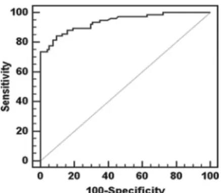
Leave-one-out cross-validation was performed to assess the
predictive accuracy of each final model. The AUCs for the clinical
failure model were greater than 0.90 for both the raw data set and
leave-one-out cross-validation. For this mode, the sensitivity,
specificity, positive predictive value, and negative predictive value
obtained with an optimal cutoff point were greater than 0.80 (
Fig.
1
;
Table 5
).
DISCUSSION
This multicenter prospective study compared the clinical efficacy
and safety of VAN versus TEC for the treatment of adult patients
with HA-MRSA bacteremia in hospital settings where MRSA
prevalence was about 70% (
23
). This study found that TEC has
efficacy and safety comparable to those of VAN for the treatment
of HA-MRSA bacteremia.
The in-hospital mortality rate of HA-MRSA bacteremia in the
VAN and TEC treatment groups of this study were 33.6% and
37.5%, respectively, which is comparable to the range of 14% to
60% reported previously in other studies (
3
,
24–26
). Based on the
Charlson comorbidity index or Pitt’s bacteremia score, the clinical
severity of the infections of the patients in our study was
compa-rable to that of health care-associated and community-acquired
MRSA infections (
14
,
26
). In addition, 4 risk factors for clinical
failure of HA-MRSA bacteremia, namely, Pitt’s bacteremia score,
acute renal injury, duration of fever, and bacteremia, were not
different from those reported previously (
26–29
).
In this study, the type of glycopeptide, i.e., VAN or TEC, was
not the risk factor associated with clinical failure. Meta-analysis
studies have reported that there were no differences in clinical
cure, microbiological cure, and mortality between VAN and TEC
treatments for Gram-positive infections, including bacteremia,
pneumonia, febrile neutropenia, and skin and soft tissue
infec-tions (
10
,
30
,
31
). However, studies on the comparative efficacy of
VAN versus TEC against MRSA bacteremia (
13
,
32
,
33
) are still
limited. Liu et al. (
33
) demonstrated that TEC was as efficacious as
VAN in terms of treatment success rate for MRSA bacteremia
(TEC group, 85% [17/20], versus VAN group, 75% [15/20]; P
⫽
0.69). On the other hand, Huang and Hsu (
32
) reported that there
was no statistically significant difference in the hospital mortality
rate (42% versus 47%) and microbiological failure rate (34%
ver-sus 40%) between the VAN group (n
⫽ 36) and the TEC group
(n
⫽ 15) among patients with MRSA infective endocarditis.
In this study, there was no significant difference in the
occur-rence of drug-related adverse events between the VAN and TEC
treatment groups. In meta-analysis studies, the incidence of total
drug-related adverse events, including nephrotoxicity and red
man syndrome, was lower with TEC (
10
,
30
,
31
). This discrepancy
might have resulted from the closed TDM of VAN in our study,
carried out according to recent clinical practice guidelines (
34
).
On the other hand, TEC was administered as directed in the
pack-age insert because TDM of TEC is not routinely available.
In this study, the use of an alternative agent, i.e., switching
from VAN to TEC or vice versa, was common in the patients with
HA-MRSA bacteremia due to the reimbursement system in
Ko-rea. VAN was replaced with TEC as an alternative agent, or vice
versa, in 20 (14.9%) and 16 patients (28.6%), respectively. Lin et
al. (
13
) reported no significant difference in 30-day mortality
among 3 treatment groups of elderly patients with persistent
MRSA bacteremia (VAN versus TEC versus VAN/TEC
alter-nately, 59.6% [65/109] versus 50.0% [7/14] versus 65.5% [19/
29]). They also reported that alternation between VAN and TEC
treatment was not more effective than either VAN or TEC
treat-ment alone (
13
). However, the appropriateness of this alternative
therapy needs to be evaluated in prospective randomized
con-trolled trials. Antibiotics such as linezolid or daptomycin as
prom-ising salvage agents or a novel strategy of combined antibiotic
treatment should be considered for better treatment outcomes of
HA-MRSA bacteremia (
27
,
35
).
In our study, the adverse cross-reactions between VAN and
TEC were not remarkable, although a limited number of cases
were evaluated. Previous studies have reported that the alternate
use of TEC in cases of VAN intolerance was associated with a high
incidence of drug-related adverse events, most notably
neutrope-nia (
36
,
37
). Therefore, the potential cross-reactivity between
these 2 glycopeptides remains to be clarified in future studies.
In recent meta-analysis studies, VAN MICs of
ⱖ1.5 g/ml or
ⱖ2.0 g/ml are associated with increased mortality as well as
clin-ical failure among patients with MRSA infections (
4
,
5
). In this
FIG 1 Receiver operating characteristic curve for clinical failure obtainedusing the predictive probability of multivariate logistic regression model and validation results.
TABLE 5 Validation results for the clinical failure variablea
Validation AUC Sensitivity (%) Specificity (%) PPV (%) NPV (%)
Validation for raw data set 0.939 (0.903–0.974) 84.2 (74.0–91.6) 90.4 (82.6–95.5) 87.7 (78.1–93.5) 87.6 (79.1–94.1) Leave-one-out cross-validation 0.926 (0.876–0.961) 80.3 (69.5–88.5) 91.5 (83.9–96.3) 88.4 (78.4–94.9) 85.1 (76.6–91.5) a
All values except AUC are optimal values in each final model. AUC, area under the curve; PPV, positive predictive value; NPV, negative predictive value; MRSA, methicillin-resistant Staphylococcus aureus.
on September 19, 2016 by Ewha Womans Univ
http://aac.asm.org/
study, the VAN MICs of
ⱖ1.5 g/ml from the VAN-treated group
and the TEC MICs of
ⱖ4.0 g/ml from TEC-treated group were
more common in the nonsurvivors than the survivors but were
not a significant factor for clinical failure. These findings indicate
that the threshold VAN or TEC MICs for clinical outcomes might
be different among the study populations.
This study has some limitations. First, this prospective study
was not designed to include the detailed complications
associ-ated with MRSA bacteremia. This may have resulted in a falsely
low complication rate. However, catheter-related infections
accounted for 47.9% of the HA-MRSA bacteremia cases in this
study, which were easily controlled with catheter removal and
antibiotic therapy. Thus, the related confounding factors might be
minimal. Second, this was not a randomized clinical trial: the
doc-tors chose VAN to initiate treatment due to the reimbursement
system in Korea. Therefore, the patients who received TEC
ther-apy may not be representative of the larger population with
HA-MRSA bacteremia. Third, individualized dosing regimens of VAN
or TEC relative to the MICs were not undertaken in this study.
Implementation of the individualized VAN dosing approach
tar-geting an AUC/MIC ratio of 400
g · h/ml or greater, rather than
a trough serum concentration, may lead to improved clinical
out-comes in critically ill patients (
38
). Lastly, other antibiotics
switched from VAN or TEC or concomitant antibiotics with VAN
or TEC might have influenced the treatment outcome. The
clini-cal effect of these antibiotics were not evaluated owing to the small
number of study cases.
In conclusion, this multicenter prospective study indicates that
TEC is an effective and safe alternative to VAN for the treatment of
HA-MRSA bacteremia. Further studies that take the AUC/MIC
ratio of VAN and TEC into account may be required for better
clinical outcomes in treating patients with HA-MRSA bacteremia.
ACKNOWLEDGMENTS
This work was supported by a grant (A102065) from the Korean Health 21 R&D project of the Ministry for Health, Welfare and Family Affairs, Re-public of Korea.
We have no conflicts of interest.
REFERENCES
1. Primo MG, Guilarde AO, Martelli CM, Batista LJ, Turchi MD. 2012. Healthcare-associated Staphylococcus aureus bloodstream infection: length of stay, attributable mortality, and additional direct costs. Braz. J. Infect. Dis. 16:503–509.http://dx.doi.org/10.1016/j.bjid.2012.10.001. 2. Park SY, Son JS, Oh IH, Choi JM, Lee MS. 2011. Clinical impact of
methicillin-resistant Staphylococcus aureus bacteremia based on propen-sity scores. Infection 39:141–147.http://dx.doi.org/10.1007/s15010-011 -0100-1.
3. Moise-Broder PA, Sakoulas G, Eliopoulos GM, Schentag JJ, Forrest A, Moellering RC, Jr. 2004. Accessory gene regulator group II polymor-phism in methicillin-resistant Staphylococcus aureus is predictive of fail-ure of vancomycin therapy. Clin. Infect. Dis. 38:1700 –1705.http://dx.doi .org/10.1086/421092.
4. Jacob JT, Diazgranados CA. 2013. High vancomycin minimum inhibi-tory concentration and clinical outcomes in adults with methicillin-resistant Staphylococcus aureus infections: a meta-analysis. Int. J. Infect. Dis. 17:e93– e100.http://dx.doi.org/10.1016/j.ijid.2012.08.005. 5. van Hal SJ, Lodise TP, Paterson DL. 2012. The clinical significance of
vancomycin minimum inhibitory concentration in Staphylococcus au-reus infections: a systematic review and meta-analysis. Clin. Infect. Dis. 54:755–771.http://dx.doi.org/10.1093/cid/cir935.
6. Rybak MJ, Lomaestro BM, Rotschafer JC, Moellering RC, Craig WA, Billeter M, Dalovisio JR, Levine DP. 2009. Vancomycin therapeutic guidelines: a summary of consensus recommendations from the
infec-tious diseases Society of America, the American Society of Health-System Pharmacists, and the Society of Infectious Diseases Pharmacists. Clin. Infect. Dis. 49:325–327.http://dx.doi.org/10.1086/600877.
7. Brown J, Brown K, Forrest A. 2012. Vancomycin AUC24/MIC ratio in patients with complicated bacteremia and infective endocarditis due to methicillin-resistant Staphylococcus aureus and its association with at-tributable mortality during hospitalization. Antimicrob. Agents Che-mother. 56:634 – 638.http://dx.doi.org/10.1128/AAC.05609-11. 8. Lodise TP, Patel N, Lomaestro BM, Rodvold KA, Drusano GL. 2009.
Relationship between initial vancomycin concentration-time profile and nephrotoxicity among hospitalized patients. Clin. Infect. Dis. 49:507–514.
http://dx.doi.org/10.1086/600884.
9. Verbist L, Tjandramaga B, Hendrickx B, Van Hecken A, Van Melle P, Verbesselt R, Verhaegen J, De Schepper PJ. 1984. In vitro activity and human pharmacokinetics of teicoplanin. Antimicrob. Agents Chemother. 26:881– 886.http://dx.doi.org/10.1128/AAC.26.6.881.
10. Svetitsky S, Leibovici L, Paul M. 2009. Comparative efficacy and safety of vancomycin versus teicoplanin: systematic review and meta-analysis. An-timicrob. Agents Chemother. 53:4069 – 4079.http://dx.doi.org/10.1128 /AAC.00341-09.
11. Williams AH, Gruneberg RN. 1988. Teicoplanin revisited. J. Antimicrob. Chemother. 22:397– 401.http://dx.doi.org/10.1093/jac/22.4.397. 12. Pedrera MI, Barriga C, Rodriguez AB. 1995. Intracellular activity of both
teicoplanin and vancomycin against Staphylococcus aureus in human neutrophils. Comp. Immunol. Microbiol. Infect. Dis. 18:123–128.http: //dx.doi.org/10.1016/0147-9571(95)98853-A.
13. Lin SH, Lai CC, Tan CK, Liao WH, Hsueh PR. 2011. Comparative efficacy of vancomycin and teicoplanin in the treatment of hospitalised elderly patients with persistent meticillin-resistant Staphylococcus aureus (MRSA) bacteraemia. Int. J. Antimicrob. Agents 37:179 –181.http://dx .doi.org/10.1016/j.ijantimicag.2010.10.018.
14. Chang HJ, Hsu PC, Yang CC, Siu LK, Kuo AJ, Chia JH, Wu TL, Huang CT, Lee MH. 2012. Influence of teicoplanin MICs on treatment outcomes among patients with teicoplanin-treated methicillin-resistant Staphylo-coccus aureus bacteraemia: a hospital-based retrospective study. J. Anti-microb. Chemother. 67:736 –741.http://dx.doi.org/10.1093/jac/dkr531. 15. Gilbert DN, Moellering RC, Jr, Eliopoulos GM (ed). 2010. The Sanford
guide to antimicrobial therapy 2010, 40th ed. Antimicrobial Therapy, Sperryville, VA.
16. Horan TC, Andrus M, Dudeck MA. 2008. CDC/NHSN surveillance definition of health care-associated infection and criteria for specific types of infections in the acute care setting. Am. J. Infect. Control 36:309 –332.
http://dx.doi.org/10.1016/j.ajic.2008.03.002.
17. Longo DL, Fauci A, Kasper D, Hauser S, Jameson J, Loscalzo J. 2012. Harrison’s principles of internal medicine, 18th ed. McGraw-Hill, New York, NY.
18. O’Brien FG, Lim TT, Chong FN, Coombs GW, Enright MC, Robinson DA, Monk A, Said-Salim B, Kreiswirth BN, Grubb WB. 2004. Diversity among community isolates of methicillin-resistant Staphylococcus aureus in Australia. J. Clin. Microbiol. 42:3185–3190.http://dx.doi.org/10.1128 /JCM.42.7.3185-3190.2004.
19. David MZ, Glikman D, Crawford SE, Peng J, King KJ, Hostetler MA, Boyle-Vavra S, Daum RS. 2008. What is community-associated methi-cillin-resistant Staphylococcus aureus? J. Infect. Dis. 197:1235–1243.http: //dx.doi.org/10.1086/533502.
20. Charlson M, Szatrowski TP, Peterson J, Gold J. 1994. Validation of a combined comorbidity index. J. Clin. Epidemiol. 47:1245–1251.http://dx .doi.org/10.1016/0895-4356(94)90129-5.
21. Knaus WA, Draper EA, Wagner DP, Zimmerman JE. 1985. APACHE II: a severity of disease classification system. Crit. Care Med. 13:818 – 829.
http://dx.doi.org/10.1097/00003246-198510000-00009.
22. Chow JW, Fine MJ, Shlaes DM, Quinn JP, Hooper DC, Johnson MP, Ramphal R, Wagener MM, Miyashiro DK, Yu VL. 1991. Enterobacter bacteremia: clinical features and emergence of antibiotic resistance during therapy. Ann. Intern. Med. 115:585–590.http://dx.doi.org/10.7326/0003 -4819-115-8-585.
23. Lee K, Kim MN, Kim JS, Hong HL, Kang JO, Shin JH, Park YJ, Yong D, Jeong SH, Chong Y. 2011. Further increases in carbapenem-, amika-cin-, and fluoroquinolone-resistant isolates of Acinetobacter spp. and P. aeruginosa in Korea: KONSAR study 2009. Yonsei Med. J. 52:793– 802.
http://dx.doi.org/10.3349/ymj.2011.52.5.793.
24. Cosgrove SE, Sakoulas G, Perencevich EN, Schwaber MJ, Karchmer AW, Carmeli Y. 2003. Comparison of mortality associated with
on September 19, 2016 by Ewha Womans Univ
http://aac.asm.org/
cillin-resistant and methicillin-susceptible Staphylococcus aureus bacte-remia: a meta-analysis. Clin. Infect. Dis. 36:53–59.http://dx.doi.org/10 .1086/345476.
25. Moore CL, Lu M, Cheema F, Osaki-Kiyan P, Perri MB, Donabedian S, Haque NZ, Zervos MJ. 2011. Prediction of failure in vancomycin-treated methicillin-resistant Staphylococcus aureus bloodstream infection: a clin-ically useful risk stratification tool. Antimicrob. Agents Chemother. 55: 4581– 4588.http://dx.doi.org/10.1128/AAC.00115-11.
26. Hall RG, II, Giuliano CA, Haase KK, Hazlewood KA, Frei CR, Forcade NA, Brouse SD, Bell T, Bedimo RJ, Alvarez CA. 2012. Empiric guide-line-recommended weight-based vancomycin dosing and mortality in methicillin-resistant Staphylococcus aureus bacteremia: a retrospective cohort study. BMC Infect. Dis. 12:104.http://dx.doi.org/10.1186/1471 -2334-12-104.
27. Lin SH, Liao WH, Lai CC, Liao CH, Tan CK, Wang CY, Huang YT, Hsueh PR. 2010. Risk factors for mortality in patients with persistent methicillin-resistant Staphylococcus aureus bacteraemia in a tertiary care hospital in Taiwan. J. Antimicrob. Chemother. 65:1792–1798.http://dx .doi.org/10.1093/jac/dkq188.
28. Liao CH, Chen SY, Huang YT, Hsueh PR. 2008. Outcome of patients with meticillin-resistant Staphylococcus aureus bacteraemia at an Emer-gency Department of a medical centre in Taiwan. Int. J. Antimicrob. Agents 32:326 –332.http://dx.doi.org/10.1016/j.ijantimicag.2008.04.011. 29. Wang JL, Wang JT, Sheng WH, Chen YC, Chang SC. 2010. Nosocomial
methicillin-resistant Staphylococcus aureus (MRSA) bacteremia in Tai-wan: mortality analyses and the impact of vancomycin, MIC⫽ 2 mg/L, by the broth microdilution method. BMC Infect. Dis. 10:159.http://dx.doi .org/10.1186/1471-2334-10-159.
30. Cavalcanti AB, Goncalves AR, Almeida CS, Bugano DD, Silva E. 2010. Teicoplanin versus vancomycin for proven or suspected infection. Co-chrane Database Syst. Rev. 2010:CD007022.http://dx.doi.org/10.1002 /14651858.CD007022.pub2.
31. Wood MJ. 1996. The comparative efficacy and safety of teicoplanin and vancomycin. J. Antimicrob. Chemother. 37:209 –222.http://dx.doi.org /10.1093/jac/37.2.209.
32. Huang JH, Hsu RB. 2008. Treatment of infective endocarditis caused by methicillin-resistant Staphylococcus aureus: teicoplanin versus vancomy-cin in a retrospective study. Scand. J. Infect. Dis. 40:462– 467.http://dx.doi .org/10.1080/00365540701837126.
33. Liu C-Y, Lee W-S, Fung C-P, Cheng N-C, Liu C-L, Yang S-P, Chen S-L. 1996. Comparative study of teicoplanin vs vancomycin for the treatment of methicillin-resistant Staphylococcus aureus bacteraemia. Clin. Drug Invest. 12:80 – 87.http://dx.doi.org/10.2165/00044011-199612020-00003. 34. Liu C, Bayer A, Cosgrove SE, Daum RS, Fridkin SK, Gorwitz RJ, Kaplan SL, Karchmer AW, Levine DP, Murray BE, M JR, Talan DA, Chambers HF. 2011. Clinical practice guidelines by the Infectious Diseases Society of America for the treatment of methicillin-resistant Staphylococcus aureus infections in adults and children: executive summary. Clin. Infect. Dis. 52:285–292.http://dx.doi.org/10.1093/cid/cir034.
35. Park HJ, Kim SH, Kim MJ, Lee YM, Park SY, Moon SM, Park KH, Chong YP, Lee SO, Choi SH, Woo JH, Kim YS. 2012. Efficacy of linezolid-based salvage therapy compared with glycopeptide-based ther-apy in patients with persistent methicillin-resistant Staphylococcus aureus bacteremia. J. Infect. 65:505–512.http://dx.doi.org/10.1016/j.jinf.2012.08 .007.
36. Hsiao SH, Chou CH, Lin WL, Lee EJ, Liao LH, Chang HJ, Yeh PY, Lin CY, Wu TJ. 2012. High risk of cross-reactivity between vancomycin and sequential teicoplanin therapy. J. Clin. Pharm. Ther. 37:296 –300.http: //dx.doi.org/10.1111/j.1365-2710.2011.01291.x.
37. Hung YP, Lee NY, Chang CM, Lee HC, Wu CJ, Chen PL, Lee CC, Chung CH, Ko WC. 2009. Tolerability of teicoplanin in 117 hospitalized adults with previous vancomycin-induced fever, rash, or neutropenia: a retrospective chart review. Clin. Ther. 31:1977–1986.http://dx.doi.org/10 .1016/j.clinthera.2009.09.010.
38. DeRyke CA, Alexander DP. 2009. Optimizing vancomycin dosing through pharmacodynamic assessment targeting area under the concen-tration-time curve/minimum inhibitory concentration. Hosp. Pharm. 44: 751–765.http://dx.doi.org/10.1310/hpj4409-751.