Sak Lee and Sang-Ho Cho
Huge Ascending Aortic Pseudoaneurysm Caused By a Penetrating Atherosclerotic Ulcer
Print ISSN: 1941-9651. Online ISSN: 1942-0080
Copyright © 2008 American Heart Association, Inc. All rights reserved. Dallas, TX 75231
is published by the American Heart Association, 7272 Greenville Avenue,
Circulation: Cardiovascular Imaging
doi: 10.1161/CIRCIMAGING.108.788133
2008;1:e19-e20
Circ Cardiovasc Imaging.
http://circimaging.ahajournals.org/content/1/3/e19
World Wide Web at:
The online version of this article, along with updated information and services, is located on the
http://circimaging.ahajournals.org//subscriptions/
is online at:
Circulation: Cardiovascular Imaging
Information about subscribing to Subscriptions:
http://www.lww.com/reprints
Information about reprints can be found online at: Reprints:
document.
Permissions and Rights Question and Answer
information about this process is available in the
requested is located, click Request Permissions in the middle column of the Web page under Services. Further Center, not the Editorial Office. Once the online version of the published article for which permission is being
can be obtained via RightsLink, a service of the Copyright Clearance
Circulation: Cardiovascular Imaging
in
Requests for permissions to reproduce figures, tables, or portions of articles originally published Permissions:
at CONS KESLI on October 15, 2014 http://circimaging.ahajournals.org/
Downloaded from http://circimaging.ahajournals.org/ at CONS KESLI on October 15, 2014 Downloaded from
Cardiovascular Images
Huge Ascending Aortic Pseudoaneurysm Caused By a
Penetrating Atherosclerotic Ulcer
Sak Lee, MD; Sang-Ho Cho, MD
A
47-year-old man was transferred to the emergency department for a huge ascending aortic pseudoaneurysm diagnosed at another hospital. The patient presented with chest discomfort for several weeks, which aggravated re-cently. He had a history of arterial hypertension and severe aortic regurgitation that were treated with aortic valve re-placement with a mechanical valve 5 years ago in the other hospital. He had also been taking captopril (angiotensin-converting enzyme inhibitor), furosemide (diuretics), and warfarin sodium. On physical examination, a mechanical valve click was heard without a murmur, and his heart rate was 77 bpm, with blood pressure of 105/70 mm Hg. Labo-ratory data revealed anemia (hemoglobin, 9.2 g/dL), pro-longed prothrombin time (International Normalization Ratio[INR], 5.16), and activated partial thromboplastin time (63.8 seconds). Initial chest x-ray showed mediastinal enlargement and a large left hilar shadow (Figure 1, arrow). Computed tomography of the chest and abdomen with iodinated contrast showed a huge (106 mm) ascending aortic aneurysm suspi-cious of impending rupture (Figures 2 and 3). Urgent aneu-rysmectomy, graft replacement of ascending aorta, and hemi-arch were performed. Operative findings showed a 6-cm pseudoaneurysm arising from the anterolateral side of the ascending aorta, with a 1-cm opening that was filled with organized thrombi (Figure 4). Pathological findings sug-gested that the cause of the pseudoaneurysm was a penetrat-ing atherosclerotic ulcer of the ascendpenetrat-ing aorta. The patient’s postoperative course was uneventful, and he was discharged from the hospital on postoperative day 7.
Disclosures
None.
Figure 1. Initial chest x-ray showed mediastinal enlargement
with hilar shadow (arrow).
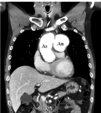
Figure 2. Chest computed tomography with contrast showed a
huge (106 mm) ascending aortic aneurysm. As indicates ascending aorta; An, aneurysm.
Received May 27, 2008; accepted July 11, 2008.
From the Department of Thoracic and Cardiovascular Surgery, Yonsei Cardiovascular Center, Yonsei University College of Medicine, Seoul, Korea. Correspondence to Sak Lee, MD, 134 Shinchon-dong, Seodaemoon-gu, Seoul, Korea, Division of Thoracic and Cardiovascular Surgery, Cardiovascular Center, Yonsei University College of Medicine, Seoul, Korea. E-mail [email protected]
(Circ Cardiovasc Imaging. 2008;1:e19-e20.)
© 2008 American Heart Association, Inc.
Circ Cardiovasc Imaging is available at http://circimaging.ahajournals.org DOI: 10.1161/CIRCIMAGING.108.788133
e19 at CONS KESLI on October 15, 2014 http://circimaging.ahajournals.org/
Figure 4. Operative findings showed a 6-cm pseudoaneurysm
arising from the anterolateral side of the ascending aorta, with a 1-cm opening (asterisk) that was filled with organized thrombi. H indicates head; F, feet.
Figure 3. Chest computed tomography with contrast showed a
huge (106 mm) ascending aortic aneurysm. As indicates ascending aorta; An, aneurysm.
e20 Circ Cardiovasc Imaging November 2008
at CONS KESLI on October 15, 2014 http://circimaging.ahajournals.org/