pISSN 1738-1843 © 2014 The Korean Society of Pathologists/The Korean Society for Cytopathology
Anaplastic carcinoma is an aggressive tumor that frequently develops in old age, and although rare cases have been reported to occur at a young age, it is extremely rare before age 30.1
Al-though anaplastic carcinoma is known to occur de novo, ana-plastic transformation by dedifferentiation from a
well-differen-tiated thyroid carcinoma (WDTC) has been reported.2 We
de-scribe an anaplastic carcinoma in a young adult. This study met criteria for exemption from review from the institutional review board.
A 31-year-old man with a relapsed tumor that was diagnosed as an anaplastic carcinoma of the thyroid died of respiratory failure 4 months after diagnosis. Eight years previously, he had a thyroid tumor diagnosed as a diffuse sclerosing papillary thy-roid carcinoma that was removed by total thythy-roidectomy with central compartment neck dissection (CCND) and right inter-nal jugular neck dissection. He also received postoperative ra-dioisotope treatment, but 5 years later the tumor relapsed in the left surgical site and was removed. The histologic features of the initial diagnosis, the first relapse, and the second anaplastic
carcinoma as well as the immunohistochemical staining and
BRAF mutation results were reviewed and a literature search
was performed to understand the mechanism of the anaplastic transformation of the WDTC.
CASE REPORT
Clinical manifestation
At 23 years of age, our patient presented with a palpable neck mass with no other underlying disease. Sonographic findings re-vealed an enlarged isoechoic mass (4×2.5×2.5 cm) with periph-eral rim-like calcification in the right lobe of the thyroid. A pre-operative diagnosis of papillary thyroid carcinoma was deter-mined and a bilateral total thyroidectomy with CCND and right internal jugular neck dissection was performed. The pathologic diagnosis determined that the mass was a diffuse sclerosing vari-ant of papillary thyroid carcinoma confined to the right thyroid parenchyma (Fig. 1A, B). The tumor metastasized to several lymph nodes on the right side of the neck and the following
re-Anaplastic Transformation of Papillary Thyroid Carcinoma in a Young
Man: A Case Study with Immunohistochemical and
BRAF Analysis
Ji Hye Park · Hyeong Ju Kwon Cheong Soo Park1· SoonWon Hong2
Department of Pathology, Severance Hospital, Departments of 1Surgery and 2Pathology,
Gangnam Severance Hospital, Yonsei University College of Medicine, Seoul, Korea
This study reports a case of anaplastic transformation from a well-differentiated thyroid carcinoma in a young patient. The first recurrent tissue contained poorly differentiated foci that revealed lower thyroglobulin, thyroid transcription factor 1 (TTF-1), and galectin-3 expression than the well-differ-entiated area. However there was no increased p53 or Ki-67 expression in the poorly differwell-differ-entiated foci, nor in the well-differentiated area. The tissue subsequently relapsed and revealed only ana-plastic features, complete loss of thyroglobulin, TTF-1, and galectin-3 expression and revealed an increase in p53 and Ki-67 expression. The BRAF V600E and BRAF V600V mutation were found in the initially diagnosed papillary thyroid carcinoma and the poorly differentiated foci of the recurring papillary thyroid carcinoma; however, only the BRAF V600V mutation was found in the anaplastic carcinoma. These results suggest that overexpression of p53 and Ki-67 contributed to the ana-plastic transformation. We also found that the BRAF type changed during the tumor relapse.
Key Words: Thyroid cancer, anaplastic; Young adult; Proto-oncogene proteins B-raf; Immunohisto-chemistry Received: November 12, 2013 Revised: December 9, 2013 Accepted: December 10, 2013 Corresponding Author SoonWon Hong, M.D.
Department of Pathology, Gangnam Severance Hospital, Yonsei University College of Medicine, 211 Eonju-ro, Gangnam-gu, Seoul 135-720, Korea Tel: +82-2-2019-3543
Fax: +82-2-3463-2103 E-mail: soonwonh@yuhs.ac
gional lymph nodes (8/30): level II (2/6), level III (2/13), level IV (3/10), and perithyroidal (1/1). Approximately 30 mCi of radioactive iodine (131I) was used for postoperative adjuvant
treatment.
After 5 years, a tumor was found at the previous left surgical site. Excision of the left surgical site with left modified radical neck dissection and right level II and III selective node dissec-tion were performed. Microscopic findings were similar to the previous primary thyroid carcinoma findings. The tumor had again metastasized to several lymph nodes on the left side of the neck and the following regional lymph nodes (8/33): right level II (0/3); left level II (1/9), level III (1/4), and level IV (4/15), and perithyroidal (2/2). Postoperatively, 200 mCi of radioactive io-dine (131I) was administered and a daily dose of 200 µg of
levo-thyroxine was prescribed for adjuvant treatment. Even though the patient continued with treatment, the tumor recurred at the left surgical site and the tumor size increased.
At 31 years of age, the patient identified another newly devel-oped, palpable small mass at the right postauricular area, which grew rapidly within a month. Radiologic examination revealed a pretracheal tumor and bilateral neck lymph node enlargement. Excision of the trachea with right modified radical neck node dissection and left level VI selective neck dissection was per-formed. Histologic diagnosis of the pretracheal lesion deter-mined that the lesion was an anaplastic thyroid carcinoma with a focal papillary carcinoma component, and the tumor metasta-sized to the following bilateral lymph nodes (8/18): right level II (5/14) and level V (0/1) and left level VI (3/3). After surgery, the patient experienced dysphagia and dyspnea, and despite inten-sive care for respiratory failure, he died 4 months later.
Histopathologic findings
The first surgical specimen was comprised of bilateral thy-roid glands and lymph nodes. The right thythy-roid gland con-tained a 4×2.5-cm ill-defined, yellow-tan mass. Histologically, the tumor showed typical papillary thyroid carcinoma features with lymphocytic thyroiditis, squamous morules, and abun-dant psammoma bodies in the outside of the tumor. These findings are consistent with diffuse sclerosing papillary thyroid carcinoma.
The histologic findings of the recurred tumor showed a pat-tern similar to the previous primary thyroid carcinoma. Our re-view of this tumor histology revealed a focal (<5%) poorly dif-ferentiated carcinoma component in the first recurrent papillary thyroid carcinoma of the left surgical site (Fig. 1C, D). We de-fined a poorly differentiated carcinoma component as a tumor
with 1) a focal solid pattern of growth, 2) absence of conven-tional nuclear features of papillary carcinoma, and 3) presence of convoluted nuclei.3
The anaplastic carcinoma of the second recurrent tumor re-vealed typical histologic findings: large pleomorphic, epitheli-oid, spindle, and multinucleated giant tumor cells, abundant eosinophilic cytoplasm, pleomorphic nuclei, more than two prominent nucleoli, frequent mitosis, and focal tumor necrosis (Fig. 1E, F). The periphery of this anaplastic carcinoma showed a remnant of papillary carcinoma.
Immunohistochemical staining and BRAF mutation analysis
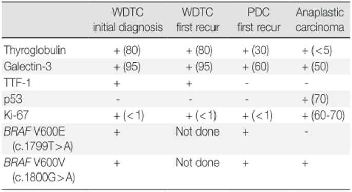
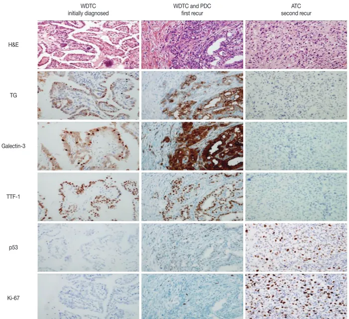
Immunohistochemical staining for thyroglobulin, galectin-3, thyroid transcription factor 1 (TTF-1), p53, and Ki-67 was per-formed. The antibodies used in this study are shown in Table 1. Table 2 and Fig. 2 summarize the immunohistochemical stain-ing results. Thyroglobulin showed diffuse, granular, cytoplasmic staining in the tumor cells of the initially diagnosed and re-curred papillary thyroid carcinoma. These tumor cells were posi-tive for galectin-3 and TTF-1 but negaposi-tive for p53. Ki-67 ex-pression was present in <1% of the initially diagnosed tumor cells and recurred papillary thyroid carcinoma. In the poorly dif-ferentiated tumor carcinoma component of the first recurrent tumor, thyroglobulin, TTF-1, and galectin-3 expression was lower than in the other part of the first recurrent tumor, but p53 and Ki-67 expression was the same. Thyroglobulin, TTF-1, and galectin-3 expression were decreased in the anaplastic carcinoma of the second recurrent tumor. However, nuclear p53 and Ki-67 expression levels were 70% and 60% to 70%, respectively, of the anaplastic carcinoma component.
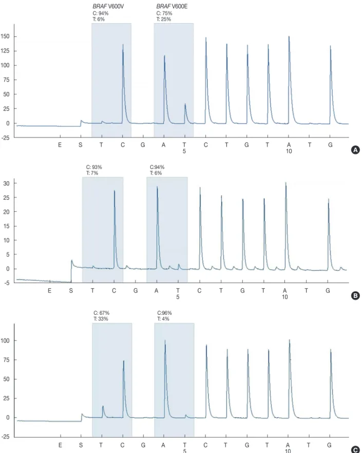
The BRAF gene mutation analysis of each tumor tissue per-formed by a pyrosequencing method revealed BRAF gene mu-tations in all tissues (Fig. 3). We defined the mutant allele peak
of 5% to 95% as the optimal cutoff value.4 The BRAF V600E
(c.1799T>A) and a low level of the BRAF V600V (c.1800G> A) mutation were found in the initially diagnosed papillary thy-roid carcinoma and poorly differentiated carcinoma component of the recurred papillary thyroid carcinoma, but only the promi-nent BRAF V600V (c.1800G>A) mutation was found in the anaplastic carcinoma component.
DISCUSSION
We report a rare case of anaplastic carcinoma in a young pa-tient, analyze the follow-up pathology data related to anaplastic transformation, and discuss the mechanism of anaplastic
trans-formation.
Anaplastic thyroid carcinoma occurs mainly in the elderly. The mean age of diagnosis is 55 to 65 years, with the peak inci-dence in the sixth to seventh decade of life. Only 25% of pa-tients are younger than 60 years at diagnosis. The age of most
reported cases was more than 30 years, and only a few reported cases were younger than 30 years.1 Most reported cases of
ana-plastic transformation by dedifferentiation from a WDTC were in the elderly.5 Therefore, thorough clinical history of previous
thyroid diseases should be determined and the possibility of an C
A
D B
E F
Fig. 1. (A, B) Papillary thyroid carcinoma at 23 years of age. (C, D) Recurrent papillary thyroid carcinoma after 5 years. Poorly differentiated cells are seen at focal areas of the papillary thyroid carcinoma. (E, F) Recurrent anaplastic thyroid carcinoma in the pretrachea adjacent thy-roid bed.
anaplastic transformation should be considered when a diagno-sis of anaplastic carcinoma is confirmed in an elderly patient.
Anaplastic thyroid carcinoma is a highly aggressive neoplasm with a poor prognosis. The mortality rate is >90%, with a mean survival of 6 months after diagnosis.6 Almost all patients
complain of a rapidly growing neck mass and symptoms associ-ated with a large mass such as hoarseness, dysphagia, vocal cord paralysis, cervical pain, and dyspnea are the most frequent and important. The overall 5-year survival ranges from 0% to 14% and the median survival is 4 to 12 months. The enlarged tumor mass often causes death by obstruction or invasion of a vital structure, similar to the occurrence in our patient.6
Anaplastic thyroid carcinoma may arise de novo or, more com-monly, through anaplastic transformation (or dedifferentiation) of a preexisting papillary or follicular thyroid carcinoma. The mechanism of this transformation, however, is not well under-stood.5
In this study, we performed immunohistochemical staining and BRAF mutation analysis to determine the mechanism of anaplastic transformation in the WDTC, and we found that the well-expressed markers in the WDTC showed loss of expression in the anaplastic carcinoma. We also confirmed that the mark-ers known to participate in transformation including p53, Ki-67, and others were not initially expressed but were expressed at the full anaplastic transformation. Todd and Wenig7 reported
that thyroglobulin and TTF-1 immunoreactivity was com-pletely reduced in recurrent or metastatic thyroid carcinoma and anaplastic thyroid carcinoma, which showed less differenti-ated histologic features, and these results were identical to the immunostaining results of our case. Ozaki et al.8 reported that
high Ki-67 expression was found in more malignant tumors, which was also similar to the findings in our case. Many studies have examined the role of the p53 gene in the thyroid as well as other organs, and most of these studies have shown that a loss in p53 gene expression is related to malignant transformation. Furthermore, studies of the thyroid have reported to be related
to anaplastic transformation in WDTC.9 The p53-positive
staining that appears in the full anaplastic transformation of our case is similar to other reports. However, additional research is necessary to determine the stage of anaplastic transformation in which the change in p53 and Ki-67 expression appears.
Mutation of the BRAF gene represents the most common ge-netic alteration in papillary thyroid cancer and is found in 45% of these tumors in Western countries and up to 90% of these tu-mors in Korea.10 The vast majority of BRAF alterations in
papil-lary thyroid cancer are BRAF V600E mutations,10 but BRAF
K601E mutations have been reported in both follicular and solid variants.11 In multifocal thyroid cancer, different tumor nodules
have distinct genetic alterations, such as different types of RET/
PTC rearrangement or variation in the presence of the BRAF
mutation.12 Recently, in Korea, Kim et al.11 reported BRAF
mu-tations that appeared in different forms according to the histo-logic subtype in a multifocal papillary carcinoma. Because the conventional papillary carcinoma component in their patient re-vealed the BRAF V600E mutation, but the follicular variant component revealed the BRAF K601E mutation, they deter-mined that the multifocal papillary carcinoma occurred de novo rather than as a result of intrathyroid metastasis, although no clear classification can be determined yet. In our case, the BRAF V600E and BRAF V600V mutations were found in the initially diagnosed papillary thyroid carcinoma and the poorly differenti-ated carcinoma component of the recurred papillary thyroid car-cinoma, but only the BRAF V600V mutation was found in the anaplastic carcinoma component. However, there has not been any report of a BRAF V600V (c.1800G>A) mutation in thy-roid carcinoma. We found two reports of a BRAF V600V muta-tion in malignant melanoma13 and colorectal cancer,14 but the
reports did not attempt to explain the relevance of the BRAF V600V mutation expression. We found that the BRAF
muta-Table 1. Panel of antibodies used in this study
Antibody Source Clone Dilution Thyroglobulin Dako (Carpinteria, CA, USA) A251 1:10,000 Ki-67 Dako (Carpinteria, CA, USA) M7240 1:150 TTF-1 Dako (Carpinteria, CA, USA) M3575 1:200 Galectin-3 NovoCastra
(Newcastle Upon Tyne, UK)
NCL-GAL3 1:50 p53 NovoCastra
(Newcastle Upon Tyne, UK) D02 1:300 TTF-1, thyroid transcription factor 1.
Table 2. Immunohistochemical staining and BRAF mutation results WDTC initial diagnosis WDTC first recur PDC first recur Anaplastic carcinoma Thyroglobulin + (80) + (80) + (30) + (<5) Galectin-3 + (95) + (95) + (60) + (50) TTF-1 + + - - p53 - - - + (70) Ki-67 + (<1) + (<1) + (<1) + (60-70) BRAF V600E (c.1799T>A) + Not done + - BRAF V600V (c.1800G>A) + Not done + +
Values in parentheses indicate percentage.
WDTC, well differentiated thyroid carcinoma; PDC, poorly differentiated carcinoma; TTF-1, thyroid transcription factor 1.
WDTC initially diagnosed H&E Galectin-3 p53 TG TTF-1 Ki-67 WDTC and PDC
first recur second recurATC
Fig. 2. Immunohistochemical staining for thyroglobulin, galectin-3, thyroid transcription factor 1 (TTF-1), p53, and Ki-67. Initially diagnosed and recurred well-differentiated thyroid carcinoma (WDTC) show diffuse positivity for thyroglobulin (TG), galectin-3, and TTF-1 but is negative for p53 and less than 1% of Ki-67 expression. In the poorly differentiated tumor carcinoma (PDC) component of the first recurrent tumor, thyroglobulin, TTF-1, and galectin-3 expression are lower than in the other part of the first recurrent tumor, but p53 and Ki-67 expression are the same. Thyroglobulin, TTF-1, and galectin-3 expression is decreased in the anaplastic carcinoma of the second recurrent tumor. Howev-er, nuclear p53 and Ki-67 expression are 70% and 60% to 70%, of the anaplastic carcinoma component, respectively. PDC, poorly differen-tiated carcinoma; ATC, anaplastic thyroid carcinoma; H&E, hematoxylin and eosin.
tion type changed during the tumor relapse process, but addi-tional research is necessary to determine whether the alteration of the BRAF mutation is just an incidental finding or a pheno-typic change relevant to the anaplastic transformation.
In conclusion, we evaluated the rare occurrence of anaplastic carcinoma in young patients and did not find p53 or Ki-67 pression or decreased thyroglobulin, TTF-1, and galectin-3 ex-pression in the poorly differentiated tumor cells.
Thyroglobu-lin, TTF-1, and galectin-3 were well expressed in the WDTC. Therefore, we considered the possibility that overexpression of p53 and Ki-67 contributed to the anaplastic transformation. Additional studies are necessary to ascertain the intermediate steps of anaplastic transformation. If the markers that are well manifested in WDTC are lost even though p53 and Ki-67 are not expressed, then we can consider those changes in expression as precursors to anaplastic transformation. Therefore, future
150 125 100 75 50 25 0 -25 E S T C G A T C T G T A T G 5 10 BRAF V600V C: 94% T: 6% BRAF V600E C: 75% T: 25% 30 25 20 15 10 5 0 -5 E S T C G A T C T G T A T G 5 10 C: 93% T: 7% C:94%T: 6% 100 75 50 25 0 -25 E S T C G A T C T G T A T G 5 10 C: 67% T: 33% C:96%T: 4%
Fig. 3. BRAF gene mutation analysis of each tumor tissue by pyrosequencing. The BRAF V600E (c.1799T>A) and a low level of the BRAF V600V (c.1800G>A) mutation are found in the initially diagnosed papillary thyroid carcinoma (A) and poorly differentiated carcinoma compo-nent of the recurred papillary thyroid carcinoma (B), but only a promicompo-nent BRAF V600V (c.1800G>A) mutation is found in the anaplastic car-cinoma component (C). The mutant allele peak of 5% to 95% is defined as the optimal cutoff value.
A
B
studies with thorough and careful histologic examination may allow us to predict the anaplastic transformation of the tumor.
Conflicts of Interest
No potential conflict of interest relevant to this article was reported.
Acknowledgments
We gratefully and sincerely thank H. J. Jeong for performing the immunohistochemical staining and BRAF mutation analy-sis in the Department of Pathology, Gangnam Severance Hos-pital.
REFERENCES
1. Tan RK, Finley RK 3rd, Driscoll D, Bakamjian V, Hicks WL Jr, Shedd DP. Anaplastic carcinoma of the thyroid: a 24-year experi-ence. Head Neck 1995; 17: 41-7.
2. Togashi S, Oka K, Kanayama R, et al. Thyroid anaplastic carcinoma transformed from papillary carcinoma in extrathyroid area. Auris Nasus Larynx 2004; 31: 287-92.
3. Tallini G. Poorly differentiated thyroid carcinoma. Are we there yet? Endocr Pathol 2011; 22: 190-4.
4. Yeo MK, Liang ZL, Oh T, et al. Pyrosequencing cut-off value identi-fying BRAFV600E mutation in fine needle aspiration samples of thyroid nodules. Clin Endocrinol (Oxf) 2011; 75: 555-60.
5. Al-Qsous W, Miller ID. Anaplastic transformation in lung metasta-ses of differentiated papillary thyroid carcinoma: an autopsy case report and review of the literature. Ann Diagn Pathol 2010; 14: 41-3.
6. Are C, Shaha AR. Anaplastic thyroid carcinoma: biology, patho-genesis, prognostic factors, and treatment approaches. Ann Surg Oncol 2006; 13: 453-64.
7. Todd WU 4th, Wenig BM. Thyroid follicular epithelial cell-derived carcinomas: an overview of the pathology of primary and recurrent disease. Otolaryngol Clin North Am 2008; 41: 1079-94.
8. Ozaki O, Ito K, Mimura T, Sugino K, Ito K. Anaplastic transforma-tion of papillary thyroid carcinoma in recurrent disease in regional lymph nodes: a histologic and immunohistochemical study. J Surg Oncol 1999; 70: 45-8.
9. Ito Y, Motoo Y, Yoshida H, et al. High level of tumour protein p53-induced nuclear protein 1 (TP53INP1) expression in anaplastic car-cinoma of the thyroid. Pathology 2006; 38: 545-7.
10. Park JY, Kim WY, Hwang TS, et al. BRAF and RAS mutations in fol-licular variants of papillary thyroid carcinoma. Endocr Pathol 2013; 24: 69-76.
11. Kim WY, Ko YS, Hwang TS, et al. A case of multifocal papillary thyroid carcinoma consisting of one encapsulated follicular variant with BRAF K601E mutation and three conventional types with BRAF V600E mutation. Korean J Pathol 2013; 47: 293-8.
12. Bansal M, Gandhi M, Ferris RL, et al. Molecular and histopathologic characteristics of multifocal papillary thyroid carcinoma. Am J Surg Pathol 2013; 37: 1586-91.
13. Bahadoran P, Allegra M, Le Duff F, et al. Major clinical response to a BRAF inhibitor in a patient with a BRAF L597R-mutated melano-ma. J Clin Oncol 2013; 31: e324-6.
14. Mao C, Zhou J, Yang Z, et al. KRAS, BRAF and PIK3CA mutations and the loss of PTEN expression in Chinese patients with colorectal cancer. PLoS One 2012; 7: e36653.