A Thesis
For the Degree of Master of Veterinary Medicine
Tetracycline resistance determinants
in
Staphylococcus intermedius
isolated from dogs in Jeju
Graduate School, Cheju National University
Department of Veterinary Medicine
Sung-up Moon
Tetracycline resistance determinants
in
Staphylococcus intermedius isolated
from dogs in Jeju
Sung-up Moon
(Supervised by professor Du-Sik Lee)
A thesis submitted in partial fulfillment of the requirement
for the degree of Master of Veterinary Medicine
2007. 8.
This thesis has been examined and approved.
Thesis director, Dusik Lee, Prof. of Veterinary Medicine
Thesis director, Wongeun Son, Prof. of Veterinary Medicine
Thesis director, Youngmin Yun, Prof. of Veterinary Medicine
ABSTRACT
Tetracycline resistance determinants in
Staphylococcus
intermedius isolated from dogs in Jeju
Sung-up Moon
(Supervised by professor Du-Sik Lee)
Department of Veterinary Medicine, Graduate School, Cheju National University
One hundred-two Staphylococcus intermedius were isolated from diseased dogs and healthy dogs to investigate the antimicrobial resistant rates on 15 commonly used drugs and tetracycline resistant gene (tet gene) was analysed on 78 tetracycline resistant isolates. Fifty-four S. intermedius isolates were recovered from oral cavity, nasal cavity and/or cranial hair coat cultures of 20 clinically healthy dogs. S. intermedius was colonized at more than one sites, including 18 (90.0%) cranial hair coat, 10 (50.0%) oral cavity, and 8(40.0%) nasal cavity of healthy dogs. Antibiograms of these commensal isolates were compared to antibiograms from 48 historical clinical isolates (2003-2006) obtained from cases of canine pyoderma (24), otitis externa (8), nasal discharge (12), pyometra (2) or cystitis (2). Antimicrobial resistant test were performed by disk diffusion test of CLSI and final resistance decisions on oxacillin and vancomycin were
made by minimum inhibitory concentration. All isolates from both healthy and diseased dog were susceptible to vancomycin and only those from the former were sensitive to amoxicilline/clavulanic acid, and cefazolin, while 8 % of those from the latter were resistant to both antibiotics. Among S. intermedius isolates recovered from diseased dogs, resistance was most often seen to penicillin (85%), ampicillin (81%) tetracycline (79%), erythromycin (52%), trimethoprim/sulfamethoxazole (48%), kanamycin (44%), norfloxacin and ciplofloxacin (38%), and gentamicin (35%). Resistance was also noted, but to a lesser degree, to neomycin (23%) and cloramphenicol (17%). Among the S. intermedius isolates recovered from healthy dogs, resistance was most often observed to penicillin (80%), ampicillin (80%), tetracycline (78%), kanamycin (65%), and erythromycin (44%), trimethoprim/ sulfamethoxazole (43%) and gentamicin (33%). Resistance was also noted, but to a lesser degree, to chloramphenicol (28%), norfloxacin (22%), ciprofloxacin (20%) and neomycin (24%). The commensal isolates were lesser resistant to most antimicrobials than those of diseased dogs, exception with chloramphenicol, kanamycin, and neomycin. The data from this study might serve as a guideline in selecting drugs to be used for treating dogs with staphylococcal infections.
S. intermedius harboring tet(M) and tet(K) were 62 (79.5%) and 3 (3.8%) strains, respectively and 3 (3.8%) and 4 (5.1%) isolates were harboring both tet(M) and tet(K), and tet(M) and tet(L), respectively.
CONTENTS
Abstract
Chapter I. Antimicrobial Resistance
of
Staphylococcus
intermedius Isolates
1. Introduction ……… 1
2. Materials and Methods ……… 3
3. Results ……… 6
4. Discussion ……… 12
5. Conclusion ……… 17
Chapter Ⅱ. Tetracycline resistance
determinants
1. Introduction ……… 25
2. Materials and Methods ……… 28
3. Results ……… 31
4. Discussion ……… 34
5. Conclusion ……… 38
Chapter I. Antimicrobial Resistance of
Staphylococcus intermedius
Isolates
Introduction
Staphylococcus intermedius is a coagulase-positive zoonotic pathogen found in pigeons, dogs, foxes, mink, and horses [16]. Initially, all coagulase-positive staphylococci were identified as S. aureus, until Hajek in 1976 established the unique identity of a group of organisms, originally identified as S. aureus biotypes E and F, as S. intermedius [16]. S. intermedius is a common commensal of oral, nasal, and skin flora in healthy dogs, where it can also be that the predominant pathogen isolated from dogs with cutaneous infections and is an important cause of ocular disease, otitis externa, cystitis, respiratory and wound infections [6, 16, 30, 35, 41, 45]. There are no vaccines to control these diseases [44]. This organism is increasingly reported to be resistant to many antibiotics [2, 19, 25, 27, 30, 34, 38, 39, 40, 46,47] and failures in treatment are a cause of problems in small animal practices. Moreover, S. intermedius can transfer occasionally from dogs to humans [12. 17] and the risk of owners being infected by resistant strains must be considered [14, 15]. However, little was known about the study of the antimicrobial susceptibility patterns of theses isolates from dogs in Korea. For these reasons, a survey of trends in the susceptibility of canine S. intermedius strains to antimicrobial drugs appeared to be great importance in the selective use of chemotherapeutics, the evaluation of new antimicrobial agents and the development of drug resistance
through continuous use of antimicrobial agents against field isolates. The present study was designed to evaluate the degree of in vitro activity of different antimicrobial agents against S. intermedius strains recently isolated in Jeju from clinically healthy and diseased dogs, such as pyoderma, otitis externa, respiratory disease, pyometra, and cystitis, and to provide a progress report on antimicrobial resistance patterns in this bacterial species.
MATERIALS AND METHODS
Bacterial isolation and identification
Fifty-four S. intermedius isolates were collected between April 2006 to July 2006 from oral cavity, nasal cavity and/or cranial hair coat cultures of 20 clinically healthy dogs visiting in private veterinary practice clinics in the Jeju-si, the south Korea for the vaccination. The specimens were collected by the method of the previous study [9]. A sterile BBL Culture Swab (Becton, Dickinson and CO., Franklin Lakes, NJ) was used to sample the tonsilar areas of the oral cavity. The cranial hair coat was sampled using a sterile BBL Culture-Swab (Becton, Dickinson and CO., Franklin Lakes, NJ) moistened in Stuart's medium and rubbed vigorously against the grain over the hair and skin of the shoulder area followed by the top of the head. Swabs were immediately placed in Stuart's transport medium (Becton, Dickinson and CO., Franklin Lakes, NJ) and kept at 4℃. Swabs were inoculated within 24 h by spread plating onto trypticase blood agar base supplemented with 5% sheep blood and Columbia CNA agar supplemented with 5% sheep blood. A single representative colony of each different morphological type resembling S. intermedius was isolated and identified from each culture using standard identification procedures [24]. A commercial identification system (ATB 32 Staph-system, BioMerieux, France) was used to further speciate the isolates. In addition, 48 S. intermedius isolates recovered between May 2003 and July 2006 from clinical cases of canine pyoderma(11) nasal discharge (n=12), otitis externa (n=6), cystitis (n=2), pyometra
recovered from veterinary schools in Cheju National University, Jeju-si, Jeju-do, South Korea. All isolates were stored at -80℃ until analysis.
Antimicrobial susceptibility test
Once samples were identified, the staphylococcal strains were tested for susceptibility to antibiotics by disc agar diffusion in accordance with NCCLS guidelines [31]. Discs of antibiotics commonly used in clinical veterinary medicine for suppurative diseases were tested: Ampicillin (AM), Amoxicillin/Clavulanic acid (AMC), Chlorampenicol (C), Ciprofloxacin (CIP), Cephazolin (CZ), Erythromycin (E), Gentamicin (GM), Kanamycin (KM), Neomycin (N), Norfloxacin (NOR), Oxacillin (OX), Penicillin (P), Trimethoprim/Sulfamethoxazole (SXT), Tetracycline (T), Vancomycin (Va). After measuring the zones of inhibition, the strains were classified as sensitive, intermediate or resistant to the drug according to the literature [21] and manufacturer [BD, Franklin Lakes, NJ, USA].
Oxacillin agar screening test
For the agar screening test, strains were plated on tryptic soy agar with 5% sheep blood and a 0.5 McFarland standard suspension was prepared for each sample. All isolates were plated on Mueller-Hinton agar supplemented with 4% (w/v) NaCl and Mueller-Hinton agar supplemented with 4% NaCl containing oxacillin at a concentration of
bacterial growth after 24 and 48 h of incubation at 35 ℃ on both plates. The minimum inhibitory concentration (MIC) for oxacillin was determined in the staphylococcal strains growing onto the plate containing 6 ㎍/ml of oxacillin.
Vancomycin agar screeing test
For the agar screening test, strains were prepared as oxacilline agar screening test. All isolates were plated on braine heart infusion agar (BHIA) and BHIA containing vancomycin at a concentration of 6 ㎍/ml according to Centers for Disease Control and Prevention guidelines [4]. Vancomycin resistance was conferred by bacterial growth after 24 h of incubation at 35 ℃ on plates. The minimum inhibitory concentration (MIC) for vancomycin was determined in the staphylococcal strains growing onto the plate containing 6 ㎍/ml of vancomycin.
Results
Bacterial strains and isolation
A total of 48 Staphylococcus intermedius were isolated from 24, 12, 8, 2 and 2 samples collected from pyoderma, nasal discharge, otitis externa, cystitis and pyometra, respectively (Table 1).
Table 1. Numbers of Staphylococcus intermedius isolated from 48 diseased dog.
Disease No. of Staphylococus intermedius isolated
Pyoderma 24 Nasal discharge 12 Otitis externa 8 Cystitis 2 Pyometra 2 Total 48
Among 20 healthy dogs, the S. intermedius were recovered from one or more sites of 19 dogs (95%). A total of 54 strains of S. intermedius were recovered from 10 (50%, 12 strains), 8 (40%, 12 strains) and 18 (90%, 30 strains) of each 20 samples taken from the oral and nasal cavities, and cranial hair coats, respectively (Table 2).
Table 2. Isolation rates of Staphylococcus intermedius from nasal cavity, oral cavity and skin of 20 healthy dog.
Sampling sites No. of dogs positive (%) No. of Staphylococcus intermedius isolated Oral cavity 10 (50) 12
Nasal cavity 8 (40) 12 Cranial hair coat 18 (90)a) 30
Total 19 (95) 54
a)
one sample was not able to isolate any bacteria for the overgrowth of Proteus sp.
Amtimicrobial resistance
All 102 S. intermedius isolates from both healthy (54 strains) and diseased dog (48 strains) were susceptible to vancomycin and only those from the former were sensitive to amoxicilline/clavulanic acid, and cefazolin, while 8 % of those from the latter were resistant to both antibiotics. Among S. intermedius isolates recovered from diseased dogs, resistance was most often seen to penicillin (85%), ampicillin (81%) tetracycline (79%), erythromycin (52%), trimethoprim/sulfamethoxazole (48%), kanamycin (44%), norfloxacin and ciplofloxacin (38%), and gentamicin (35%). Resistance was also noted, but to a lesser degree, to neomycin (23%) and cloramphenicol (17%). Among the S. intermedius isolates recovered from healthy dogs, resistance was most often observed to penicillin (80%), ampicillin (80%), tetracycline (78%), kanamycin (65%), and erythromycin (44%), trimethoprim/sulfamethoxazole (43%) and gentamicin (33%). Resistance was also noted, but to a lesser degree, to chloramphenicol (28%), norfloxacin (22%), ciprofloxacin (20%) and
neomycin (24%). The commensal isolates were lesser resistant to most antimicrobials than those of diseased dogs, exception with chloramphenicol, kanamycin, and neomycin (Fig. 1).
0 10 20 30 40 50 60 70 80 90
AM AMC C CIP CZ E GM KM N NOR OX P SXT T VA Antimicrobial agents R e s is ta n t ra te s ( % )
Diseased dog Healthy dog
Fig. 1. Antimicrobial resistant rates of Staphylococcus intermedius isolated from healthy and diseased dogs. Ampicillin(AM), Amoxicillin/Clavulanic acid (AMC), Chlorampenicol (C), Ciprofloxacin (CIP), Cephazolin (CZ), Erythromycin (E), Gentamicin (GM), Kanamycin (KM), Neomycin (N), Norfloxacin (NOR), Oxacillin (OX), Penicillin (P), Trimethoprim/Sulfamethoxazole (SXT), Tetracycline (T), Vancomycin (Va).
Antimicrobial resistance patterns
A total of 31 resistance patterns were observed in 48 S. intermedius strains from diseased dogs (Table 3). Fourteen antimicrobial agents, except for vancomycin were affected by resistance. Overall 47 of the 48 strains (98.0%) were resistant to two or more antimicrobial drugs and 43 strains (90%) were resistant to three or more antimicrobial drugs (multiresistance). Five oxacillin resistant strains were resistant to six to fourteen antibiotic agents and AM, P, T-mutiresistant strains were most prominent as 7 strains (14.6%).
A total of 37 resistance patterns were observed in 54 S. intermedius strains from healthy dogs (Table 4). As like as the results of diseased dogs, fourteen antimicrobial agents, except for vancomycin were affected by resistance. Overall, all 54 strains were resistant to one or more antimicrobial drugs and 45 strains (83.3%) were resistant to three or more antimicrobial drugs (multiresistance). Two oxacillin resistant strains were resistant to AM,CIP,E,GM,KM,NOR or AM,AMC,C,CIP,CZ,E,GM,KM,N,NOR,P,SXT,T.
S. intermedius with same antibiograms in mouth, nose and/or cranial hair coat were isolated from only 4 healthy dogs and other isolates showed totally different antibiograms, indicating that S. intermedius colonized in the dogs as transient, intermittent, or persistent might be different strains in same time.
Table 3. Antimicrobial resistance patterns of Staphylococcus intermedius strains isolated from diseased dogs
Antimicrobial resistance patterns Total, n (%) No resistance 1 (2.1) Resistance to SXT,T 1 (2.1) AM,P 3 (6.3) AM,C,T 1 (2.1) AM,P,SXT 1 (2.1) AM,P,T 7 (14.6) C,E,KM 1 (2.1) P,SXT,T 1 (2.1) AM,AMC,E,P 1 (2.1) AM,E,P,T 3 (6.3) C,E,GM,KM 1 (2.1) E,GM,KM,T 1 (2.1) AM,CIP,NOR,P,T 2 (4.2) AM,C,KM,P,T 1 (2.1) AM,KM,P,SXT,T 2 (4.2) AM,AMC,CZ,E,N,P 1 (2.1) AM,CIP,CZ,NOR,P,T 1 (2.1) AM,GM,KM,P,SXT,T 1 (2.1) AM,E,GM,P,SXT,T 1 (2.1) CIP,NOR,OX,P,SXT,T 1 (2.1) AM,E,GM,KM,P,SXT,T 1 (2.1) C,CIP,E,GM,N,NOR,R,T 1 (2.1) CIP,E,GM,KM,NOR,P,SXT,T 1 (2.1) AM,C,CIP,E,GM,KM,N,NOR,P 1 (2.1) AM,CIP,E,GM,KM,NOR,P,SXT,T 1 (2.1) AM,CIP,E,KM,N,NOR,P,SXT,T 2 (4.2) AM,CIP,E,KM,NOR,OX,P,SXT,T 1 (2.1) AM,CIP,E,GM,KM,N,NOR,P,SXT,T 2 (4.2) AM,AMC,C,CZ,E,GM,KM,P,SXT,T 1 (2.1) AM,CIP,E,GM,KM,NOR,OX,P,SXT,T 1 (2.1) AM,CIP,E,GM,KM,N,NOR,OX,P,SXT,T 3 (6.3) AM,AMC,C,CIP,CZ,E,GM,KM,N,NOR,OX,P,SXT,T 1 (2.1) Total 48 (100)
Ampicillin(AM), Amoxicillin/ Clabulanic acid (AMC), Chlorampenicol (C), Ciprofloxacin (CIP), Cephazolin (CZ), Erythromycin (E), Gentamicin (GM), Kanamycin (KM), Neomycin (N), Norfloxacin (NOR), Oxacillin (OX), Penicillin (P), Sulfamethoxazole/Trimethoprim (SXT), Tetracycline (T), Vancomycin (Va).
Table 4. Antimicrobial resistance patterns of Staphylococcus intermedius strains isolated from healthy dogs
Antimicrobial resistance patterns Total, n (%) Resistance to AM 1 (1.9) E 1 (1.9) T 5 (9.3) GM,KM 1 (1.9) N,T 1 (1.9) AM,P,T 4 (7.4) AM,NOR,P,SXT 1 (1.9) AM,KM,P,T 1 (1.9) AM,KM,P,SXT 1 (1.9) AM,P,SXT,T 2 (3.7) AM,C,E,KM,P 1 (1.9) AM,C,GM,P,T 1 (1.9) AM,C,KM,P,T 1 (1.9) AM,C,N,P,T 1 (1.9) AM,GM,KM,P,SXT 1 (1.9) AM,GM,KM,P,T 1 (1.9) AM,KM,P,SXT,T 1 (1.9) CIP,KM,NOR,P,T 1 (1.9) CIP,E,NOR,SXT,T 1 (1.9) AM,C,E,KM,N,P 5 (9.3) AM,C,GM,KM,P,T 1 (1.9) AM,CIP,KM,NOR,P,T 1 (1.9) AM,E,GM,P,SXT,T 1 (1.9) AM,E,KM,P,SXT,T 3 (5.6) AM,GM,KM,P,SXT,T 1 (1.9) AM,C,E,GM,KM,P,T 1 (1.9) AM,C,E,KM,N,P,SXT 1 (1.9) AM,CIP,E,GM,KM,NOR,OX 1 (1.9) AM,CIP,GM,KM,NOR,P,T 2 (3.7) AM,E,KM,N,P,SXT,T 2 (3.7) AM,C,E,GM,KM,N,PT 2 (3.7) C,CIP,E,GM,KM,NOR,SXT,T 1 (1.9) AM,C,E,KM,N,P,SXT,T 1 (1.9) AM,CIP,GM,KM,NOR,P,SXT,T 1 (1.9) AM,CIP,E,GM,KM,NOR,P,SXT,T 1 (1.9) AM,C,CIP,E,GM,KM,NOR,P,SXT,T 1 (1.9) AM,CIP,E,GM,KM,NOR,OX,P,SXT,T 1 (1.9) Total 54 (100)
Ampicillin(AM), Amoxicillin/ Clabulanic acid (AMC), Chlorampenicol (C), Ciprofloxacin (CIP), Cephazolin (CZ), Erythromycin (E), Gentamicin (GM), Kanamycin (KM), Neomycin (N), Norfloxacin (NOR), Oxacillin (OX), Penicillin (P),
Discussion
The present study confirms the occurrence of Staphylococcus intermedius strains in various sites of healthy dogs as well as variety diseases of dogs, due to the isolation of 54 strains of S. intermedius from oral and nasal cavities and cranial hair coats of 20 healthy dogs and 48 isolates from dogs with pyoderma, otitis externa, raspiratory disease, cystitis, and pyometra of 48 dogs. This is expected results, since privious study usually reports S. intermedius is a normal inhabitant of canine skin; however, it becomes the primary or secondary agents in certain diseases, such as the bacterial dermatitis, otitis externa [1, 5, 29, 43].
S. intermedius may function as a super-antigen and regulate the immune system [7, 18]. S. intermedius can produce numerous toxins and readily demonstrates antibiotic resistance [7]. In our study, of 102 S. intermedius isolates from both healthy and diseased dog were susceptible to vacomycin but 101 strains (99%) were resistant to one or more antimicrobial agents.
The antimicrobial resistance for S. intermedius is well documented in the literature outside of Korea [13, 33, 40, 43]. Most importantly the penicillins and tetracyclines are described virtually useless, as most strains are resistant to these compounds [7, 43]. Amoxicillin with clavulanic acid, cephalosporins, potentiated sulfa drugs, erythromycin and fluoroquinolones are usually effective [33, 36, 43].
Since the introduction of penicillin, methicillin and oxacillin into clinical use, staphylococci have obtained resistance to β-lactam antibiotics [38]. Resistant to penicillin in Staphylococcus spp. is a well-known problem in human and veterinary medicine. The secretion of β-lactamase is very frequent in S. intermedius strains from dogs and leads to the non-prescription of penicillin, ampicillin and
dogs: 50 to 74%. According to Lloyd et al. [27] the resistance to penicillin increased from 69% in 1980 to 89% in 1996. According to Reedy [42], the resistance to ampicillin increased from 41% to 67% in the United States, between 1982 and 1994. In this study, data revealed high both ampicillin and penicillin-resistant S. intermedius; around 80%, comparing with the previous reports of other countries [25, 26, 37, 38]. An increase over time of resistance to penicillin and ampicillin in staphylococci may be explained by the selective pressure exerted by considerable use of penicillin and ampicillin in treating many diseases in dogs.
The methicillin-resistant S. aureus strains are usually resistant to all the group M; penicillins, including oxacillin and cloxacillin. Methicillin has never been used in animal antibiotherapy in Korea, but the other two drugs are widely used in S. aureus cow mastitis treatment and in S. intermedius dog pyodermitis treatment [38]. Very different from the hospital methicillin-resistant S. aureus strains, is the situation in S. intermedius strains. Only few group M-penicillin resistance strains were found in dog S. intermedius strains. In this study, 3.7% (2/54) of healthy dog origins and 14.6% (7/48) of diseased dog origins were resistant to oxacillin. Investigations conducted in several other countries also revealed oxacillin resistant S. intermedius, but usually low occurrence (0 to 3.3%) were reported [25, 26, 37, 38].
Only 0 to 2.5% of the strains were resistant to cefalexine in the UK [27], in the United States [26], in Norway [25], in France [38], and in Sweden [37]. We found the relatively higher cefazolin resistant S. intermedius (8%) isolated from the diseased dogs, whereas did not from the healthy dogs. We also found the same situation on amoxycillin with clavulanic acid (8% resistant from the diseased dogs and 100% sensitive from the heathy dogs). This indicates that the dog continue administrated with antibiotics might have more resistant strains because our bacteria tested were isolated from dog failed to treat in the local animal hospital. However, oxacillin, amoxycillin/calvulanic acid or cefazolin is one of the best
qualities for the treatment of dog staphylococcal disease.
According to Pellerin et al. [38], the resistance to Kanamycin increased from 18.9% in 1987-88 to 22% in 1992, and Kunkle[26] has been reported 50% of the strains isolated in dogs with prior antibiotic therapy were resistant to kanamycin. According to Pellerin et al. [38], kanamycin should be replaced by gentamicin, but 44% of the strains from the diseased dogs and 65% from the healthy dogs were resistant to kanamycin, and both originated S. intermedius were moderately resistant (33 and 35%) to gentamicin. We do not have any explanation for this difference between them, but it should be replaced by other aminoglycosides, such as amikacin, tobramycin because both antibiotics, kanamycin and gentamicin have been used for long time without prescription by veterinarian in Korea.
Although chloramphenicol has rarely been used as therapy for S. intermedius infections, it has been used for the treatment of many bacterial disease and S. intermedius is commonly sensitive to the antibiotics (80 to 90%). According to Pellerin et al. [38] chloramphenicol is no longer of interest for the treatment of dog pyodermas, as the frequency of chloramphenicol resistance is increased with time. However, the resistant rates were around 25% in our results, indicating that the antimicrobial resistant rates may depend on the regions and countries.
The frequency of tetracycline resistance bacteria is well-known. As previously reported by others [25, 26, 30, 33]. Tetracycline shows a high percentage (35-60%) of resistant strains. According to Reedy, the resistance to tetracycline was, in the United States, at the level of 49% of the stains in 1982, and according to Pellerin et al. [38], the resistance to tetracycline increased from 19% to 40% of the strains between 1988 and 1993, and then was stable. However, our results showed much higher resistance rates to tetracycline in accordance to 90% resistant to the drugs of the previous report in
periods was the most significant finding by Kruse et al. [25] in 1996. Accoding to the Pellerin et al. [38], these drugs in canine dermatology has increased steadily over the last decade and a stable and relatively high frequency of resistance for 23% to 30% of the strains tested. However, 44% and 52% of the strains isolated from the healthy and the diseased dogs were respectively found in this study. This finding is in accordance to the study of Kunkle [25] with some difference, where nearly all the strains of S. intermedius isolated in dogs without prior antibiotic therapy are susceptible to macrolides, as compared to nearly half of the strains isolated in dogs with prior antibiotic therapy being resistant to macrolides.
Trimethoprim-sulphonamides combinations have been used extensively in most cases of first-time pyoderma in dogs [3, 8]. An interesting observation was to increase number of resistant strains to trimethoprim-sulphonamides with time, from 5% in 1987-88, to 20% in 1992 and to 36% in 1995-96 [38]. Some studies have revealed relatively low frequencies of resistance to trimethoprim or trimethoprim-sulphonamides [19, 25, 27, 30], but others have reported resistance rates reaching 70% [2]. In this study, 43% of healthy dog origins and 48% of diseased dog origins were resistant to trimethoprim-sulphonamides combinations.
Fluoroquinolones are recommended principally for antimicrobial therapy in mixed infection, in recurrent pyoderma, in chronic, deep pyoderma with extensive scar tissue, or when canine pyoderma has proved to be refractory to 'first-line' antibiotics [20]. The increasing use of fluoroquinolones over the past years has given rise to an increase in resistance to fluoroquinolones [10, 20, 28, 40, 46]. Ciprofloxacin and norfloxacin have been little used for the treatment of dog diseases in Korea, but in this study, 25% of healthy dog origins and 35% of diseased dog origins were resistant to ciprofloxacin and/or norfloxacin.
The many different resistance patterns observed confirm other studies, reporting a great variety of resistance patterns in staphylococci. However, any comparison between the proportions of
multidrug-resistant organisms and those referred to in other veterinary reports in confounded by inconsistencies in defining multiple drug resistance, and different reporting techniques. During this study, most of strains were resistant to three or more antimicrobial drugs. Multiple antimicrobial-resistant S. intermedius isolates from canine pyodermas [11] have been reportedly transmitted from infected dogs to their owners [15, 16]. Also, there was a report of a mastoid cavity infection in the owner caused by S. intermedius transmitted from his dog’s saliva [22]. Therefore, high occurrence of multi-antimicrobial resistant strains in dogs could increase the implications of zoonotic transmission of multi -antimicrobial resistant S. intermedius.
Conclusion
Staphylococcus intermedius strains isolated form dogs, were most highly resistant to penicillin, ampicillin, and tetracycline, and also high resistance were observed on erythromycin, kanamycin, fluoroqiunolons, trimethoprim/sulfamethoxazole, and gentamicin. Resistance is very rare to amoxicillin-clavulanic acid, cefazolin, and oxacillin. This must provide guidelines for the dog veterinary practitioner's choice of antibacterial compound. These results also show that there are many multidrug-resistance S. intermedius in dogs, including oxacillin-resistant strains, human potential pathogen.
References
1. Allaker RP, Lloyd DH, Bailey RM. Population sizes and frequency of staphylococci at mucocutaneous sites on healthy dogs. Vet Rec 1992, 130, 303-304.
2. Barrs VR, Malik R, Love DN. Antimicrobial susceptibility of staphylococci isolated from various disease conditions in dogs: a further survery. Aust Vet Pract 1995, 25, 37-42
3. Carlotti DN, Guaguere E, Pin D, Jasmin P, Thomas E, Guiral V. Therapy of difficult cases of canine pyoderma with marbofloxacin: a report of 39 dogs. J Small Anim Pract 1999, 40, 265-70.
4. Centers for Disease Control and Prevention. Investigation and control of vancomycin-intermediate and resistant Staphylococcus aureus: A Guide for Health Departments and Infection Control Personnel. Atlanta, GA, 2003.
5. Cox HU, Hoskins JD, Newman SS, Foil CS, Turnwald GH, Roy AF. Temporal study of staphylococcal species on healthy dogs. Am J Vet Res 1988, 49, 747-751.
6. Cox HU, Newman SS, Roy AF, Hoskins JD. Species of Staphylococcus isolated from animal infections. Cornell Vet 1984, 74, 124-135.
7. Crossley KB, Archer GL. The Staphylococci in Human Disease (K.B. Crossley, G.L. Archer eds.). Churchill Livingston, New York, NY, 1997.
by healthy dogs and comparison of antimicrobial susceptibility patterns to isolates from dogs with pyoderma. Vet Microbiol 2005, 108, 119-131
10. Ganiere JP, Medaille C, Limet A, Ruvoen N, Fontaine GA. Antimicrobial activity of enrofloxacin against Staphylococcus intermedius strains isolated from canine pyodermas. Vet Dermatol 2001, 12, 171-175.
11. Ganiere JP, Medaille C, Mangion C. Antimicrobial Drug Susceptibility of Staphylococcus intermedius Clinical Isolates from Canine Pyoderma. J Vet Med B Infect Dis Vet Public Health 2005, 52, 25-31
12. Goodacre R, Harvey R, Howell SA, Greenham LW, Noble WC. An epidemiological study of Staphylococcus intermedius strains isolated from dogs, their owners and veterinary surgeons. J Anal Appl Pyrol 1997, 44, 49-64.
13. Gortel K. Methicillin resistance among staphylococci isolated form dogs. Am J Vet Res 1999, 60, 1526-1530.
14. Guardabassi L, Loeber ME, Jacobson A. Transmission of multiple antimicrobial-resistant Staphylococcus intermedius between dogs affected by deep pyoderma and their owners. Vet Microbiol 2004, 98, 23-27.
15. Guardabassi L, Schwarz S, Lloyd DH. Pet animals as reservoirs of antimicrobial-resistant bacteria. J Antimicrob Chemother 2004, 54, 321-332.
16. Hajek V. Staphylococcus intermedius, a New Species Isolated from Animals. International J Syst Bacteriol 1976, 26, 401-408. 17. Harvey RG, Marples RR, Noble WC. Nasal carriage of
Staphylococcus intermedius in humans in contact with dogs. Microbiol Ecol Health Dis 1994, 7, 225-227.
18. Hendricks A, Schuberth HJ, Schueler K, Lloyd DH. Frequency of superantigen-producing Staphylococcus intermedius isolates from canine pyoderma and proliferation-inducing potential of superantigens in dogs. Vet Science 2002, 73, 273-277.
19. Holm BR, Petersson U, Morner A, Bergstrom K, Franklin A, Greko C. Antimicrobial resistance in staphylococci from canine pyoderma: a prospective study of first-time and recurrent cases in Sweden. Vet Rec 2002, 151, 600-605.
20. Ihrke PJ, Papich G, Demanuelle TC. The use of fluoroquinolones in veterinary dermatology. Vet Dermatol 1999, 10, 193-204.
21. Jorgensen JH, Tunridge JD, Washington JA. Antibacterial Susceptibility Tests: Dilution and Disk Diffusion Methods. In: Murray PR, Baron EJ, Pfaller MA, Tenover FC, Yolken RH. Manual of Clinical Microbiology. 7th edition ed. American Society for Microbiology, Washington, DC, 1999.
22. Kikuchi K, Karasawa T, Piao C, Itoda I, Hidai H, Yamaura H, Totsuka K, Morikawa T, Takayama M. Molecular confirmation of transmission route of Staphylococcus intermedius in mastoid cavity infection from dog saliva. J Infect Chemother 2004, 10, 46-48.
23. Kim TJ, Na YR, Lee JI. Investigations into the Basis of Chloramphenicol and Tetracycline Resistance in Staphylococcus intermedius Isolates from Cases of Pyoderma in Dogs. J Vet Med
Murray PR, Barron EJ, Phaller MA, Tenover FC, Yolken RH. (Eds.), Manual of Clinical Microbiology. American. 1999.
25. Kruse H, Hofshagen M, Thoresen SI, Bredal WP, Vollset I, Soli NE. The antimicrobial susceptibility of Staphylococcus species isolated from canine dermatitis. Vet Res Commun 1996, 20, 205-214. 26. Kunkel GA. Mew considerations for rational antibiotic therapy of
cutaneous Staphlococcal infection in the dog. Seminars in Veterinary Medicine and Surgery (Small animal) 1987, 2, 212-220. 27. Lloyd DH, Lamport AI, Feeney C. Sensitivity to antibiotics
amongest cutaneous and mucosal isolates of canine pathogenic staphylococci in the U.K. 1980-96. Vet Dermatol 1996, 7, 171-175. 28. Lloyd D, Lamport AI, Noble WC, Howell SA. Fluoroquinolone
resistance in Staphylococcus intermedius. Vet Dermatol 1999, 10, 193-204.
29. McEwan NA. Adherence by Staphylococcus intermedius to canine keratinocytes in atopic dermatitis. Res Vet Science 2000, 68, 279-283.
30. Medleau L, Long RE, Brown J, Miller WH. Frequency and antimicrobial susceptibility of Staphylococcus species isolated from canine pyodermas. Am J Vet Res 1986, 47, 229-231.
31. National Committee for Clinical Laboratory Standards. Performance standards for antimicrobial disk susceptibility tests. Approved standards. NCCLS document M2-A3. National Committee for Clinical Laboratory Standards, Wayne, Pa, 1997. 32. National Committee for Clinical Laboratory Standards. Methods for
dilution antimicrobial susceptibility tests for bacteria that grow aerobically, 5th ed. Approved standard M7-A8. NCCLS, Wayne, PA, 2000.
33. Noble WC, Kent LE. Antibiotic Resistance in Staphylococcus intermedius Isolated from Cases of Pyoderma in the Dog. Vet Dermatol 1992, 3, 71-74.
34. Normand EH, Gibson NR, Taylor DJ, Carmichael S, Reid SWJ. 2000. Trends of antimicrobial resistance in bacterial isolates from a small animal referral hospital. Vet. Rec. 146:151-155.
35. Oluoch AO, Weisiger R, Siegel AM, Campbell KL, Krawiec DR, McKiernan BC, Kakoma I. Trends of bacterial infections in dogs: characterization of Staphylococcus intermedius isolates (1990-1992). Canine Pract 1996, 21, 12-19.
36. Paradis M, Lemay S, Scott DW, Miller WH, Wellington J. Efficacy of Enrofloxacin in the Treatment of Canine Bacterial Pyoderma. Vet Dermatol 1990, 1, 123-127.
37. Pedersen-Morner A, Bergstrom K, Greko C, Franklin A. Antibiotic sensitivity of staphyloccocci isolated from cases of canine pyoderma. Supp Compend Contin Educ Pract Vet 1996, 18, 48. 38. Pellerin JL, Bourdeau P, Sebbag H, Person JM.
Epidemiosurveillance of antimicrobial compound resistance of Staphylococcus intermedium clinical isolates from canine pyodermas. Comp Immunol Microbiol Infect Dis 1998, 21, 115-133. 39. Petersen AD, Walker RD, Bowman MM, Rosser EJ. Frequency of
isolation and antimicrobial susceptibility patterns of Staphylococcus intermedius and Pseudomonas aeruginosa isolates from canine skin and ear samples over a 6-year period (1992-1997). J Am Anim Hosp Assoc 2002, 38, 407-413.
41. Raus J, Love DN. Characterization of coagulase-positive Staphylococcus intermedius and Staphylococcus aureus isolated from veterinary clinical specimens. J Clin Microbiol 1983, 18, 789-792.
42. Reedy LM. Comparative results of 100 antibiotic sensitivity tests on Staphylococcus intermedius (1982-1994). 11th Proceedings. of A.A.V.D./A.C.V.D. meeting. 1995.
43. Scott DW, Miller WH, Griffin CE. Muller and Kirk's Small Animal Dermatology, 6th ed. WB Saunders, Philadelphia, PA, 2001.
44. Shimizu A, Wakita Y, Nagase S, Okabe M, Koji T, Hayashi T, Nagase N, Sasaki A, Kawano J, Yamashita K, Takagi M. Antimicrobial susceptibility of Staphylococcus intermedius isolated from healthy and diseased dogs. J Vet Med Sci 2001, 63, 357-360. 45. Talan DA, Staatz D, Staatz A, Goldstein EJC, Singer K, Overturf
GD. Staphylococcus intermedius in canine gingival and canine inflicted human wound infections: laboratory characterization of a newly recognized zoonotic pathogen. J Clin Microbiol 1989, 27, 78-81.
46. Walker RD, Thornsberry C. Decrease in antibiotic susceptibility or increase in resistance. J Antimicrob Chemother 1998, 41, 1-4. 47. Werckenthin C, Cardoso M, Martel JL, Schwarz S. Antimicrobial
resistance in staphylococci from animals with particular reference to bovine Staphylococcus aureus, porcine Staphylococcus hyicus, and canine Staphylococcus intermedius. Vet Res 2001, 32, 341-362.
Chapter Ⅱ. Tetracycline resistance determinants
Introduction
Tetracyclines were the first major group of antibiotics to which the term 'broad-spectrum' was used [16]. Because of the spectrum of activity, the relative safety and low cost, tetracyclines have been widely used throughout the world and are second after penicillins in total tons used each year [4]. During many years, the therapeutic use of tetracyclines in human medicine has been reduced as bacterial resistance has become more widespread [3, 16, 24]. Oxytetracycline has been used to treat certain bacterial diseases which effect field crops and fruit trees and in subtherapeutic levels as food additives for growth promotion in billions of animals raised for food each year. Use of oxytetracycline in food production ranges from chickens, cows, honeybees, salmon, and catfish [16]. Tetracyclines are also used for therapy in food animals and in pets such as dogs, cats, and horses [16]. The bacteria which cause disease in animals and plants can be of the same species as those found in man, may belong to related species found within the same or related genera as those found in man, or more distantly related. Unique plasmids and antibiotic resistance genes first described in animal specific bacteria have made their way into human bacteria and vice versa [16, 24]. The reason this happens is due to the fact that bacteria exchange
and genera and ultimately influence the antibiotic resistance carriage of strictly human bacterial species [16, 24]. The result is that antibiotic use anywhere in the world, regardless of its original purpose, can effect the antibiotic resistance genes in bacteria in other ecosystems including those that are pathogenic for man. Thus, tetracycline resistance determinants are wide-spread among bacterial species and have been identified in as many as 32 Gram negative and 22 Gram positive organisms and are often found in multi-drug resistant bacteria [3,14, 23].
Staphylococcal infections are frequently treated with antimicrobial agents, but treatment is problematic because of the limited number of effective antimicrobial agents available. The frequent occurrence of antimicrobial resistance has previously been reported among S. intermedius in different countries [20, 32, 33, 35]. Strains used for these previous studies were multi-resistant to antimicrobials; mostly to ampicillin, chloramphenicol, gentamicin, erythromycin, penicillins, sulphonamides and tetracyclines.
Resistance to tetracycline occurs by three mechanisms: the use of an energy-dependent efflux of tetracycline, altering the ribosome to prevent effective binding of the tetracycline, and producing tetracycline-inactivating enzymes [23]. The tetracycline resistant genes associated with an efflux mechanism are tet(A), (B), (C), (D), (E), (G), (I), (M) and (K). The tetracycline resistance genes associated with a ribosomal protection mechanism and/or efflux mechanism are tet(K), (L), (M), (O), (S), (P), (Q), (B), (D), (H) and (C). The only example of a tetracycline resistance gene causing the enzymatic alteration of tetracycline is tet(X) [23]. Among those tet genes, tet(A)
to tet(G) genes are usually found in Gram negative bacterial species and are associated with efflux mechanism. The tet(L), tet(M) and tet(Q) genes, associated with a ribosomal protection mechanism and/or efflux mechanism, are also carried by Gram negative bacteria. Gram positive bacteria carry commonly tet(M) gene associated with efflux mechanism and also take tet(K), tet(L), tet(M), tet(O), tet(S) and tet(Q) genes associated with a ribosomal protection mechanism and/or efflux mechanism.
Several studies have determined the occurrence of different genes encoding antimicrobial resistance in S. intermedius of canine origin [5, 12, 26] Four genes, such as tet(K), tet(L), tet(M) and tet(O) encoding tetracycline resistance have been identified in Staphylococcus species [26, 32]. Nevertheless, there is limited information about the distribution of the tet genes in S. intermedius of canine origin in Jeju, Korea. This study describes the distribution of tetracycline resistance genes in S. intermedius of canine origin in Jeju, Korea.
Materials and Methods
Bacterial strains and growth conditions
A total 78 tetracycline resistant Stapylococcus intermedius were used for this study and included 40 and 38 strains originated from healthy and diseased dogs. The organisms were subcultured from the frozen stock (-80℃) onto Trypticase blood agar base containing 5% sheep blood.
DNA extraction
Bacterial DNA was extracted by the modification of the method of Richard et al [21]. S. intermedius isolates were grown onto Colombia blood agar base containing 5% sheep blood at 37℃ for 18-24 hr and one loopful bacterial cells were mixed with 1.0 ml of PBS (pH 7.2) in 1.7 ml microfuge tube and 0.25 g of Chelex-100 (Bio-Rad, Hercules, Calif.) was suspended into 0.5 ml of 10 mM TE buffer (pH 8.4). in another microfuge tube. The bacterial and 0.6 ml of Chelex-100 suspension were added to 2 ml screw-caped tube containing 1 g of 0.1-mm-diameter glass beads (Biospec Products, Inc., Bartlesville, Okla.). The samples were mixed and processed in the bead beater (Biospec Products; Inc.) at three-quarters speed for 5 min and then boiled for 5 min. The samples were then centrifuged for 5 min at 13,000g, and the supernatants were removed to clean 1.7-ml Eppendorf tubes. All DNA samples were measured for concentration using a DNA/RNA calculator (Pharmacia Biotech, Piscataway, N.J.).
Table 1. Tetracycline-resistant PCR primers Tetracycline
resistance gene PCR primer sequence 5'-3'
Amplicon size(bp) Tet(A) GCT ACA TCC TGC TTG CCT TC 210
CAT AGA TCG CCG TGA AGA GG
Tet(B) TTG GTT AGG GGC AAG TTT TG 659 GTA ATG GGC CAA TAA CAC CG
Tet(C) CTT GAG AGC CTT CAA CCC AG 418 ATG GTC GTC ATC TAC CTG CC
Tet(D) AAA CCA TTA CGG CAT TCT GC 787 GAC CGG ATA CAC CAT CCA TC
Tet(E) AAA CCA CAT CCT CCA TAC GC 278 AAA TAG GCC ACA ACC GTC AG
Tet(Ga) GCT CGG TGG TAT CTC TGC TC 468 AGC AAC AGA ATC GGG AAC AC
Tet(Gb) CAG CTT TCG GAT TCT TAC GG 844 GAT TGG TGA GGC TCG TTA GC
Tet(K) TCG ATA GGA ACA GCA GTA 169 CAG CAG ATC CTA CTC CTT
Tet(L) TCG TTA GCG TGC TGT CAT TC 267 GTA TCC CAC CAA TGT AGC CG
Tet(M) GTG GAC AAA GGT ACA ACG AG 406 CGG TAA AGT TCG TCA CAC AC
Tet(O) AAC TTA GGC ATT CTG GCT CAC 515 TCC CAC TGT TCC ATA TCG TCA
Tet(S) CAT AGA CAA GCC GTT GAC C 667 ATG TTT TTG GAA CGC CAG AG
Tet(P) CTT GGA TTG CGG AAG AAG AG 676 ATA TGC CCA TTT AAC CAC GC
Tet(Q) TTA TAC TTC CTC CGG CAT CG 904 ATC GGT TCG AGA ATG TCC AC
Tet(X) CAA TAA TTG GTG GTG GAC CC 468 TTC TTA CCT TGG ACA TCC CG
Primers
of amplicon are listed in Table 1.
Multiplex PCR conditions
Multiplex PCR was performed by the modification of the method of Ng et al. [18]. following the determination of DNA concentration using a ultraviolet spectrophotometer at A260. The PCR reaction mix
(total 50 ㎕) included 0.5 ㎍ template DNA, 1×PCR buffer, 2.5 U DNA Taq polymerase (Intron ), 2.5 mM of each of the deoxynucleotides dNTP (Gibco-BRL, Burlington, Ontario, Canada) and ddH2O. Group I contained primers for tet(B) (0.25 μM), tet(C) (0.25 μM) and tet(D) (2.0 μM) each (4.0mM MgCl2). Group II contained primers for tet(A) (1.0 μM), tet(E) (1.0 μM) and tet(G) (1.0 μM) each (3.0 mM MgCl2). Group III contained primers for tet(K) (1.25 μM), tet(L) (1.0 μM), tet(M) (0.5 μM), tet(O) (1.25 μM) and tet(S) (0.5 μM) each 3.0 mM MgCl2). Group IV contained primers for tetA(P) (1.25 μM), tet(Q) (1.25 μM) and tet(X) (1.25 μM) each (4.0 mM MgCl2). DNA amplification was carried out in a PCR Thermal cycler DICE Gradient Model TP600 (TaKaRa, Tokyo, Japan) using the following conditions: a 5 min initial denature at 94°C followed by 35 cycles of 94°C for 1 min, 55°C for 1 min and 72°C for 1.5 min. PCR products were analysed by gel electrophoresis (1% (w/v) agarose in 1×TAE buffer). DNA bands were stained with ethidium bromide and then visualized by u.v. transillumination. The sizes of the PCR products were determined by comparing them with the migration of 100-bp plus DNA ladder (Intron).
Results
Of the 102 S. intermedius isolated from dogs 78 (76.5%) was resistant to tetracycline. Among the 78 tetracycline resistance bacteria, 65 (83%) were also resistant to penicillin 62 (80%) to ampicillin, 42 (54%) to kanamycin, 38 (49%) to trimethoprim/sulfamethoxazole, 35 (45%) to erythromycin, 29 (37%) to gentamicin, 27 (35%) to both ciprofloxacin and norfloxacin, and 16 (21%) to neomycin. Of the 24 tetracycline sensitive bacteria, 19 (79%) were also resistant to ampicillin, 17 (71%) to penicillin, 15 (63%) to kanamycin, 14 (58%) to erythromycin, 12 (50%) to chloramphenicol, 7 (29%) to neomycin, 6 (25%) to gentamicin, and 5 (21%) to trimethoprim/sulfamethoxazole (Data not shown).
Table 1. Tetracycline resistant gene types of Staphylococcus intermedius isolated from dogs.
Origins
Tet gene type with
K M KM LM NT Total Diseased dog 0 (0.0) 33 (86.8) 1 (2.6) 2 (5.3) 2 ( 5.3) 38
Healthy dog 3 (7.5) 29 (72.5) 2 (5.0) 2 (5.0) 4 (10.0) 40 Total 3 (3.9) 62 (79.5) 3 (3.9) 4 (5.1) 6 (7.7) 78 * NT, non-typed
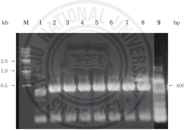
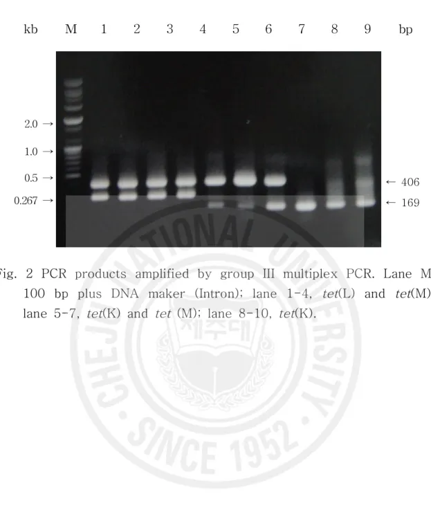
containing 38 strains from the diseased dogs and 40 strains from the healthy dogs (Table 1). Group III multiplex PCR was initially tested individually, and the template DNA of samples with ambiquous PCR products were re-tested using another 3 multiplex PCR groups. Overall, 62 (79.5%) were found to carry only tet(M), 3 (3.9%) carried only tet(K), 3 (3.9%) carried both tet(K) and tet(M), and 4 (5.1%) carried both tet(L) and tet(M). Seven strains (7.7%) did not carry any tetracycline resistance genes (Fig 1 and 2).
kb M 1 2 3 4 5 6 7 8 9 bp
2.0 → 1.0 →
0.5 → ← 406
Fig. 1 PCR products representing tet(M) gene amplified by group III multiplex PCR. Lane M, 100 bp plus DNA maker (Intron); lane 1, tetS S. intermedius; lane 2-8, tetR S. intermedius; lane 9, non-typed S. intermedius.
kb M 1 2 3 4 5 6 7 8 9 bp
2.0 → 1.0 →
0.5 → ← 406
0.267 → ← 169
Fig. 2 PCR products amplified by group III multiplex PCR. Lane M, 100 bp plus DNA maker (Intron); lane 1-4, tet(L) and tet(M); lane 5-7, tet(K) and tet (M); lane 8-10, tet(K).
Discussion
Tetracyclines are broad-spectrum antimicrobial agents with activity against a wide range of Gram positive and Gram-negative anaerobic and aerobic bacteria, cell-wall free mycoplasmas, chlamydiae, mycobacterium, rickettsia, Helicobacter, Listeria and protozoan parasites such as Entamoeba histolytica, Giardia lamblia, and Plasmodium falciparum [3, 7, 13, 15, 16, 24]. They have been used extensively for therapy in man for bacterial respiratory and urogenital tract diseases, periodontal, Lyme, and rickettsial diseases. In addition, Tetracyclines have non-antibacterial properties which include antiinflammatory and immunosuppressive properties [11]. Studies have linked tetracycline with suppression of antibody production in lymphocytes, reduction in phagocytic function of polymorphonuclear leukocytes, reduction of leukocyte and neutrophil chemotaxis, as a sclerosising agent, as an inhibitor of lipase and collagenase activity and as an enhancer of gingival fibroblast cell attachment [11, 34]. These additional properties have encouraged the use of tetracycline in non-infectious conditions such as resistant rheumatoid arthritis, rosacea, pyoderma gangrenosum, prurigo pigmentosa, pleural effusions, recurrent pneumothorax, recurrent thyroid cysts, and biliary-cutaneous fistula. These uses unfortunately expose the patient's normal flora to active tetracycline which can select for tetracycline-resistant bacteria as does tetracycline used for treatment of an infectious disease.
In 1953, the first tetracycline-resistant bacterium isolated was S. dysenteriae, which causes bacterial dysentery [6]. The first
multi-resistant Shigella was isolated in 1955 and was resistant to tetracycline, streptomycin, and chloramphenicol [1, 6, 31] and came from a total of 5327 (0.02%) isolates tested. By 1960, multi-resistant Shigella represented almost 10% of the strains tested in Japan [1, 6, 31], a dramatic increase in five years. The increase in multi drug resistant Shigella has continued to the present as illustrated by a recent study [17] where over 60% of the S. flexneri isolated between 1988-1993 were resistant to tetracycline, streptomycin, and chloramphenicol. This is the same combination of antibiotic resistance determinants found in the 1953 S. dysenteriae isolate: Subsequently, it was demonstrated that these antibiotic-resistant bacteria could transfer their antibiotic-resistant phenotypes to susceptible isolates by cocultivation. This transfer was dependent upon direct contact of the bacteria [31]. We now know that the Japanese studies were the first reports of tetracycline resistance genes carried on conjugative R-plasmids. These tetracycline genes coded for the efflux of tetracycline out of the cell and were the first of the three different tetracycline resistance mechanisms found in bacteria [24]. Multi-drug resistance, which includes tetracycline resistance, has also been seen in Gram-positive species. A recent 1994 study [8] indicated that approximately 90% of methicillin-resistant Staphylococcus aureus, 70% of Streptococcus agalactiae, 70% of multi-drug resistant Enterococcus faeca!is and: 60% of multi-drug resistant S. pneumoniae now are tetracycline-resistant. This suggests that tetracycline resistance has become widespread in pathogenic Gram-positive
extended use of tetracycline may select for both tetracycline and multi-drug resistant bacteria [16]. This situation applies equally to animals given low dose tetracycline for growth promotion and patients given long term tetracycline for control of acne [16]. Since 1960, investigators have found that bacterial resistance to tetracyclines is primarily due to acquisition of tetracycline resistance determinants rather than by mutation of existing chromosomal genes [22, 24]. There have been 16 different tetracycline resistant determinants characterized.
Of the 102 canine S. intermedius investigated in this study 78 (76.5%) were resistant to tetracycline and the resistant rate was much higher than that reported in studies conducted in the UN [19], Canada [10] and USA [26], but slightly lower than that reported in the previous study in Korea [12].
The tet(M) genes have been most commonly detected in tetracycline-resistant S. intermedius [26]. In this study, 69 strains (88.5%) of 78 tetracycline resistance isolates carried also tet(M) gene with or without other genes. The tet(K) gene was present in 3.9%, there are no strains with only tet(L) gene, and the tet(O) gene, including other tet genes were not found in any of these isolates.
All isolates carrying tet(M) and tet(O) genes are tetracycline- resistant by a ribosome protecting protein [30], and also exhibit resistance to minocycline, whereas tet(K) and tet(L) bearing isolates are sensitive to minocycline [26]. Tetracycline resistance in these latter isolates is based on a membrane-associated efflux system. S. intermedius strains carry both efflux and ribosomal protection genes in combination [23]. The tet(K) genes, which were previously known
Staphylococcus spp., but not in S. intermedius [26].
The tet(L) genes, which are commonly located on small plasmids, have rarely been detected in Staphylococcus spp. from animals [25] and S. intermedius harboured tet(L) gene independendly have been reported in 24% or the isolates investigated [12]. However, in this study, there were no strains harboured tet(L) in accordance with the report of Schwarz et al. (1998). The tet(K) and tet(L) genes can be found in single isolates of streptococci [2], though not in isolates in this study.
The observation that tet(M) genes are detected in most tetracycline-resistant S. intermedius isolates independently of their animal origin suggests that they are most readily acquired by these bacteria [26]. Most tet(M) genes are located on conjugative transposons [29], and are usually found in the chromosomal DNA rather than on plasmids [5]. The reason for the preferential acceptance of transposon-encoded resistance genes in S. intermedius is still unknown [26]. However, S. intermedius isolates differ from other staphylococcal species by their high number of chromosomally located insertion elements [9]. These may play a role in the development of chromosomal multi-resistance in this species. In the majority of Gram-positive species, the tet(M) determinant is found in the chromosome, most often on conjugative elements [26]. It has been reported that the presence of tet(O) genes in chromosomal DNA are uncommon in staphylococci [26] in accordance with the result of this study while there are some reports the tet(O) genes occur
Conclusion
Of the 102 S. intermedius isolated from dogs 78 (76.5%) was resistant to tetracycline. Among the 78 tetracycline resistance bacteria, 65 (83%) were also resistant to penicillin 62 (80%) to ampicillin, 42 (54%) to kanamycin, 38 (49%) to trimethoprim/ sulfamethoxazole, 35 (45%) to erythromycin, 29 (37%) to gentamicin, 27 (35%) to both ciprofloxacin and norfloxacin, and 16 (21%) to neomycin. S. intermedius harboring tet(M) and tet(K) independently were 62 (79.5%) and 3 (3.8%) strains, respectively and 3 (3.8%) and 4 (5.1%) isolates were harboring both tet(M) and tet(K), and both tet(M) and tet(L), respectively.
References
1. Akiba T, Koyama K, Ishiki Y, Kimura S, Fukushima T. On the mechanism of the development of multiple-drug-resistant clones of Shigella. Japan J Microbiol 1960, 4, 219.
2. Brown MB, Roberts MC. Tetracycline resistance determinants in streptococcal species isolated from the bovine mammary gland. Vet Microbiol 1991, 29, 173-180.
3. Chopra I, Hawkey PM, Hinton M. Tetracyclines, molecular and clinical aspects. J Antimicrob Chemother 1992, 29, 245-277.
4. Col NF, O'Connor RW. Estimating worldwide current antibiotic usage: Report of task force 1. Rev Infect Dis 1987, 9, 232-243. 5. Eady EA, Ross JI, Tipper JL, Walters CE, Cove JH, Noble WC.
Distribution of genes encoding erythromycin ribosomal methylases and an erythromycin efflux pump in epidemiologically distinct groups of staphylococci. J Antimicrob Chemother 1993, 31, 211-217.
6. Falkow S. Infectious Multiple Drug Resistance. Pion Limited, London, 1975.
7. Freeman CD, Nightingale CH, Quintiliani R. Minocycline: old and new therapeutic uses. Int J Antimicrob Agents 1994, 4, 325-335. 8. Goldstein FW, Kitzis MD, Acar JF. N,Ndimethylglycyl-amido
derivative of minocycline and 6-demethyl-6-desoxytetracycline, two new glycylcyclines highly effective against tetracycline-resistant
elements in Staphylococcus intermedius. Lett Appl Microbiol. 1995, 20, 180-183.
10. Hoekstra KA, Paulton RJ. Clinical prevalence and antimicrobial susceptibility of Staphylococcus aureus and Staph. intermedius in dogs. J Appl Microbiol 2002, 93, 406-413.
11. Humbert P, Treffel P, Chapuis JF, Buchet S, Derancourt C, Agache P. The tetracyclines in dermatology. J Am Acad Dermat. 1991, 25, 691-697.
12. Jaffe RI, Lane JD, Albury ST, Niemeyern DM. Rapid Extraction from and Direct Identification in Clinical Samples of Methicillin-Resistant Staphylococci Using the PCR. Society for Microbiology 2000, 38, 264-282.
13. Kim TJ, Na YR, Lee JI, Investigations into the Basis of Chloramphenicol and Tetracycline Resistance in Staphylococcus intermedius Isolates from Cases of Pyoderma in Dogs. J Vet Med B Infect Dis Vet Public Health 2005, 52, 119-124.
14. Klein NC, Cunha BA. Tetracyclines. Medical Clinics N America 1995, 79, 789-801.
15. Levy SB, McMurray LM, Barbosa TM, Burdett V, Courvalin P, Hillen W. Nomenclature for new tetracycline resistance determinants. Antimicrob Agents Chemother 1999, 43, 1523-1524. 16. Levy SB. Tetracycline resistance determinants are widespread.
ASM News 1988, 54, 418-421.
17. Levy SB. The Antibiotic Paradox: How Miracle Drugs are Destroying the Miracle. Plenum Press, New York, 1992.
18. Lima AAM, Lima NL, Pinho MC, Barros Jr, E A, Teixeria MJ, Marins MCV, Guerrant RL. High frequency of strains multiply
resistant to ampicillin, trimethoprim-sulfamethoxazole, streptomycin, chl0ramphenicol, and tetracycline isolated from patients with shigellosis in northeastern Brazil during the period 1988 to 1993. Antimicrob Agents Chemother 1995, 39, 256-259.
19. Ng LK, Martin I, Alfa M, Mulvey M. Multiplex PCR for the detection of tetracycline resistant genes. Mol Cell Probes 2001, 15, 209-215
20. Noble WC, Kent LE. Antibiotic resistance in Staphylococcus intermedius isolated from cases of pyoderma in the dog. Vet Dermatol 1992, 3, 71-74
21. Prescott JF, Hanna WJ, Reid-Smith R, Drost K. Antimicrobial drug use and resistance in dogs. Can Vet J 2002, 43, 107-116.
22. Roberts MC. Epidemiology of tetracycline resistance determinants. Trends Microbiol 1994, 2, 353-357.
23. Roberts MC. Tetracycline resistance determinants: mechanisms of action, regulation of expression, genetic mobility, and distribution. FEMS Microbiol Rev 1996, 19, 1-24.
24. Roberts MC. Gene transfer in the urogenital and respiratory tract. In: Gene Transfer in the Environment (Levy, S. and Miller, R. V., Eds.), pp. 347-375. McGraw-Hill Publishing Co, New York, 1989. 25. Schwarz S, Noble WC. Structure and putative origin of a plasmid
from Staphylococcus hyicus that mediates chloramphenicol and streptomycin resistance. Lett Appl Microbiol 1994, 18, 281-284. 26. Schwarz S, Roberts MC, Werckenthin C, Pang Y, Lange C.
conferring combined resistance to tetracycline and minocycline among group B streptococcal isolates from humans and various animals. Zentralbl Bakteriol 1994, 281, 526-533.
28. Soedermanto I, Schwarz S, Liebisch B, Lammler C. Tetracycline resistance determinants among streptococci of serological group G and L. Vet Microbiol 1995, 45, 331-337.
29. Speer BS, Shoemaker NB, Salyers AA. Bacterial resistance to tetracycline: mechanism, transfer, and clinical significance. Clin Microbiol Rev 1992, 5, 387-399.
30. Taylor DE, Chau A. Tetracycline resistance mediated by ribosomal protection. Antimicrob Agents Chemother 1996, 40, 15. 31. Watanabe T. Infectious heredity of multiple drug resistance in
bacteria. Bacteriol Rev 1963, 27, 87-115.
32. Werckenthin C, Cardoso M, Martel JL, Schwarz S. Antimicrobial resistance in staphylococci from animals with particular reference to bovine Staphylococcus aureus, porcine Staphylococcus hyicus, and canine Staphylococcus intermedius. Vet Res 2001, 32, 341, 362.
33. Wissing A, Nicolet J, Boerlin P. The current antimicrobial resistance situation in Swiss veterinary medicine. Schweiz Arch Tierheilkd 2001, 143, 503-510.
34. Yanagimura M, Koike F, Hara K. Collagenase activity in gingival crevicular fluid and inhibition by tetracyclines. J Dental Res 1989, 68, 1691-1693.
35. Yoo H, Park SW, Hwang CY, Youn HY, Han HR. Aerobic antimicrobial susceptibility patterns of bacteria isolated from dogs. J Vet Clin 2002, 19, 303-311.