저작자표시-비영리-변경금지 2.0 대한민국 이용자는 아래의 조건을 따르는 경우에 한하여 자유롭게 l 이 저작물을 복제, 배포, 전송, 전시, 공연 및 방송할 수 있습니다. 다음과 같은 조건을 따라야 합니다: l 귀하는, 이 저작물의 재이용이나 배포의 경우, 이 저작물에 적용된 이용허락조건 을 명확하게 나타내어야 합니다. l 저작권자로부터 별도의 허가를 받으면 이러한 조건들은 적용되지 않습니다. 저작권법에 따른 이용자의 권리는 위의 내용에 의하여 영향을 받지 않습니다. 이것은 이용허락규약(Legal Code)을 이해하기 쉽게 요약한 것입니다. Disclaimer 저작자표시. 귀하는 원저작자를 표시하여야 합니다. 비영리. 귀하는 이 저작물을 영리 목적으로 이용할 수 없습니다. 변경금지. 귀하는 이 저작물을 개작, 변형 또는 가공할 수 없습니다.
Master’s Thesis
PARP in glycolysis and
mitochondrial respiration
Hana Song
Department of Biomedicine & Drug Development
Graduate School
PARP 에 의한 해당과정과
미토콘드리아 호흡 연구
지도교수 김 진 우
송 하 나
이 논문을 이학 석사학위 논문으로 제출함
2016년 2월
송하나의 이학 석사학위 논문을 인준함
심사위원장 ________________________________
위 원 ________________________________
위 원 ________________________________
제주대학교 대학원
2016년 2월
PARP in glycolysis and
mitochondrial respiration
Hana Song
(Supervised by Professor Jinu Kim)
A thesis submitted in partial fulfillment of the requirement for
degree of Master of Science in Biomedical and New Drug
Development
2016.02
This thesis has been examined and approved.
...
...
...
...
Department of Biomedical & New Drug Development
Graduate School
CONTENTS
LIST OF ABBREVIATIONS ...1
LIST OF FIGURES ...2
I. ABSTRACT...3
II. INTRODUCTION...4
III. MATERIALS AND METHODS...6
1. Cell culture ...6
2. Enzyme activity ...6
3. Extracellular acidification rate ...7
4. Oxygen consumption rate ...7
IV. RESULTS...9
1. Glucose increases PARP activation in kidney proximal tubule epithelial cells. ..9
2. PARP inactivation increases glycolytic activity in kidney proximal tubule epithelial cells...11
3. Mitochondrial function is independent of PARP activation in kidney proximal tubule epithelial cells. ...14
4. PARP inactivation augments glycolytic enzyme activity induced by glucose in kidney proximal tubule epithelial cells...17
V. DISCUSSION ...19
LIST OF ABBREVIATIONS
PARP Poly(ADP-ribose) polymerase
3-AB 3-Aminobenzamide
PGI Phosphoglucose isomerase
PFK1 Phosphofructokinase-1
GAPDH Glyceraldehyde-3-phosphate dehydrogenase
ECAR Extracellular acidification rate
BG Basal glycolysis
GA Glycolytic activity
GC Glycolytic capacity
2-DG 2-deoxyglucose
OCR Oxygen consumption rate
FCCP Carbonyl cyanide-4-(trifluoromethoxy)phenylhydrazone
R&A Rotenone plus antimycin A
BR Basal respiration
MA Mitochondrial ATP
LIST OF FIGURES
Figure 1 : Treatment with 3-AB attenuates PARP activation increased by glucose in
kidney proximal tubule epithelial cells ………. 10
Figure 2 : PARP1 inactivation augments glycolytic activity in kidney proximal tubule
epithelial cells ………12
Figure 3 : PARP1 is not involved in mitochondrial function during treatment with
glucose in kidney proximal tubule epithelial cells ………15
Figure 4 : PARP1 inactivation augments activities of glycolytic enzymes increased by
I. ABSTRACT
After renal injury, selective damage occurs in the proximal tubules as a result of inhibition of glycolysis; but the molecular mechanism is not known. Poly(ADP-ribose) polymerase (PARP) activation plays a critical role of proximal tubular cell death in several renal disorders. Here, we studied the role of PARP on glycolytic flux in pig kidney proximal tubule epithelial LLC-PK1 cells using XFp extracellular flux analyzer. Poly(ADP-ribosyl)ation by PARP activation was increased by 10 mM glucose in LLC-PK1 cells, but treatment with 3-aminobenzamide as a PARP inhibitor does-dependently prevented the PARP activation induced by glucose. Treatment with 1 mM 3-aminobenzamide significantly enhanced extracellular acidification rate increased by glucose, but not oligomycin; indicating that PARP inactivation increases only glycolytic activity during glycolytic flux including basal glycolysis, glycolytic activity, and glycolytic capacity in kidney proximal tubule epithelial cells. Glucose increased the activities of glycolytic enzymes including hexokinase, phosphoglucose isomerase, phosphofructokinase-1, glyceraldehyde-3-phosphate dehydrogenase, enolase, and pyruvate kinase in LLC-PK1 cells. Furthermore, PARP inactivation selectively augmented the activities of hexokinase, phosphofructokinase-1, and glyceraldehyde-3-phosphate dehydrogenase. In conclusion, these data suggest that PARP activation regulates glycolytic activity through poly(ADP-ribosyl)ation of hexokinase, phosphofructokinase-1, and glyceraldehyde-3-phosphate dehydrogenase in kidney proximal tubule epithelial cells.
Key Word: Poly (ADP-ribose) polymerase, glycolysis, kidney proximal tubule,
II. INTRODUCTION
Poly(ADP-ribose) polymerase (PARP) is a nuclear protein that regulates gene transactivation as a transcription coactivator and protein function via poly(ADP-ribosyl)ation (16). Alternatively, PARP activation is important for DNA repair, but its excessive activation plays a prominent role in necrotic cell death (12). The necrotic cell death via PARP activation is caused by NAD+-dependent poly(ADP-ribosyl)ation that
leads to ATP depletion and metabolic collapse (5, 12). Our previous reports and those by other researchers have demonstrated that either pharmacological or genetic inhibition of PARP is renoprotective against ischemia reperfusion injury (20, 25), cisplatin nephrotoxicity (14, 23), and obstructive nephropathy (15). The kidney proximal tubule among renal tubules is most sensitive to lethal injury as a result of difference in their capacity to generate energy by glycolysis (18). The proximal tubule has low capacity for glycolysis, as demonstrated by their failure to produce lactate under either control conditions or loss of oxidative phosphorylation using antimycin A (3). Furthermore, the activity of hexokinase as a glycolytic enzyme is less in the proximal tubule than in the other tubules (24).
Glycolysis is the sequence of reactions that metabolizes one molecule of glucose to two molecules of pyruvate. During glycolytic flux, two molecules of ATP and two molecule of NADH are produced under an anaerobic condition. Phosphofructokinase-1 (PFK1) is one of the most important regulatory enzymes in the mammalian glycolytic pathway (6). The phosphorylation of fructose 6-phosphate to fructose 1,6-biphosphate by PFK1 is the first point of commitment of glucose to the glycolytic pathway (22). Glyceraldehyde-3-phosphate dehydrogenase (GAPDH) is also one of the most important regulatory enzymes in glycolysis and gluconeogenesis by reversibly catalyzing the oxidation and
phosphorylation of glyceraldehyde-3-phosphate to 1,3-diphosphoglycerate (7). Recent reports indicate that poly(ADP-ribosyl)ation induced by PARP activation inhibits PFK1 and GAPDH activities in brain-derived and endothelial cells, respectively (9, 10). However, the role of PARP in glycolysis in kidney proximal tubule epithelial cells remained unclear. Therefore, we investigated the effect of treatment with a PARP inhibitor during glycolytic flux in kidney proximal tubule epithelial cells.
III. MATERIALS AND METHODS
1. Cell culture
The kidney proximal tubule epithelial cell line derived from pig (LLC-PK1) was obtained from American Type Culture Collection (Rockville, MD, USA). The LLC-PK1 cells were maintained in Dulbecco’s modified Eagle’s medium (DMEM)/high-glucose medium containing 10% fetal bovine serum (FBS) at 37°C with 5% CO2. The cells were grown
until 70 % confluence on culture plates, and then changed to glucose- and serum-free DMEM medium. After treatment with 0.01, 0.1, or 1 mM 3-aminobenzamide (3-AB; R&D Systems, Minneapolis, MN) in glucose- and serum-free DMEM medium (vehicle) for 30 minutes, the cells were incubated with 10 mM glucose in XF base medium (Seahorse Bioscience, Billerica, MA) with 4 mM glutamine for 30 minutes.
2. Enzyme activity
PARP activity in the pig kidney proximal tubule epithelial cells was measured using a universal PARP assay kit according to manufacturer’s instructions (Trevigen, Gaithersburg, MD, USA). Activities of Hexokinase, phosphoglucose isomerase (PGI), GAPDH, enolase, and pyruvate kinase in the cells were measured using respective colorimetric assay kits purchased from BioVision Inc. (Mountain View, CA, USA) according to manufacturer’s instructions. PFK1 activity in the cells was measured as previously described. Briefly, the cells were homogenized in cold sucrose buffer (0.32 M sucrose and 10 mM Tris-HCl, pH 7.4). Homogenates were centrifuged at 13,000 rpm for 20 minutes. The supernatants were incubated in 50 mM Tris-HCl buffer (pH 8.0) including 2.6 mM DTT, 2 mM MgCl2, 5 mM (NH4)2SO4, 1 mM EDTA, 40 units aldose,
250 units triosephosphate isomerase, 40 units a-glycerophosphate dehydrogenase, 100 mM fructose-6-phosphate, 100 mM ATP, and 16 mM NADH. The decrease in optical density at 340 nm due to the oxidation of NADH was the measured for 60 seconds.
3. Extracellular acidification rate
Extracellular acidification rate (ECAR) in the pig kidney proximal tubule epithelial cells was measured using the XFp extracellular flux analyzer (Seahorse Bioscience). The cells were seeded in XFp cell culture miniplates (Seahorse Bioscience) at 4 x 10⁵per well
in DMEM/high-glucose medium containing 10% FBS and incubated overnight. The following day, the cells were treated with 1 mM 3-AB in glucose- and serum-free DMEM medium (vehicle) for 30 minutes, and then incubated at 37°C with XF base medium containing 4 mM glutamine in a CO2-free incubator for 60 minutes. Glycolytic flux
(basal glycolysis, glycolytic activity, and glycolytic capacity) as assessed by extracellular acidification rate (ECAR) was analyzed by the sequential injection of 10 mM glucose, 1 mM oligomycin, and 50 mM 2-deoxyglucose. ECAR was measured at 37°C with a 3-minutes mix, 0-minute wait, and 3-3-minutes measurement protocol. The levels of ECAR were made three times in respective phases, and expressed as units of milli-pH (mpH) per minute.
4. Oxygen consumption rate
The pig kidney proximal tubule epithelial cells seeded in XFp cell culture miniplates (Seahorse Bioscience) at 4 x 10⁵per well in DMEM/high-glucose medium containing 10%
FBS were incubated overnight. After that, the cells were treated with 1 mM 3-AB in glucose- and serum-free DMEM medium (vehicle) for 30 minutes; changed to XF base medium containing 4 mM glutamine, 1 mM pyruvate, and 25 mM glucose; and incubated at 37°C in a CO2-free incubator for 60 minutes. Mitochondrial function (basal
respiration, mitochondrial ATP production, and maximal respiration) as assessed by oxygen consumption rate (OCR) was analyzed by the sequential injection of 1 M oligomycin, 2 M carbonyl cyanide 4 -(trifluoromethoxy)phenylhydrazone (FCCP), and 0.5 M rotenone plus antimycin A in the XFp extracellular flux analyzer (Seahorse
Bioscience). OCR was measured at 37°C with a minutes mix, 0-minute wait, and 3-minutes measurement protocol. The levels of OCR were made three times in respective phases, and expressed as units of picomoles (pmol) per minute.
5. Statistical analyses
Analysis of variance was used to compare data among groups using Systat SigmaPlot (Systat Software Inc., San Jose, CA, USA). Differences between two groups were assessed by two-tailed unpaired Student’s t-tests. P values of <0.05 were considered statistically significant.
IV. RESULTS
1. Glucose increases PARP activation in kidney proximal tubule epithelial cells.
To study the role of PARP on glycolysis in kidney proximal tubule epithelial cells, PARP activity after incubation with glucose was measured in LLC-PK1 cells. The cells incubated with glucose for 30 minutes showed the significant increase in the PARP activity, compared to that in glucose-starved control cells (Figure 1). We also tested whether treatment with 3-AB, a PARP inhibitor, reduced the PARP activity increased by glucose. Treatment with 3-AB at 30 minutes prior to incubation with glucose dose-dependently diminished the increment in the PARP activity after 30 minutes of incubation with glucose (Figure 1). However, treatment with 3-AB in glucose-starved control cells did not significantly alter the PARP activity (Figure 1). These data indicate that PARP inhibition is efficacious against PARP activation induced by glucose in kidney proximal tubule epithelial cells.
Fig. 1 Treatment with 3-AB attenuates PARP activation increased by glucose in kidney proximal tubule epithelial cells.
LLC-PK1 cells were maintained in DMEM/high-glucose medium containing 10% FBS at 37°C with 5% CO2. The cells were grown until 70 % confluence on culture plates; and
then treated with 0.01, 0.1, or 1 mM 3-AB in glucose- and serum-free DMEM medium (vehicle) for 30 minutes. After that, the cells were incubated with 10 mM glucose in XF base medium with 4 mM glutamine (control) for 30 minutes. PARP activity in the cells measured using the universal PARP assay kit was expressed as units (U) per mg protein. Error bars represent SD (n=4 experiments). *P<0.05 versus control; #P<0.05 versus
vehicle. Control Glucose P A R P a ct iv ity (U /m g pr ot ei n) 0 20 40 60 80 100
*
#*
*
Vehicle 0.01 mM 3-AB 0.1 mM 3-AB 1 mM 3-AB2. PARP inactivation increases glycolytic activity in kidney proximal tubule epithelial cells.
To analyze the effect of glucose-induced PARP activation on glycolysis in kidney proximal tubule epithelial cells, we investigated a comprehensive real-time analysis of glycolytic flux using XFp extracellular flux analyzer in LLC-PK1 cells treated with vehicle and 3-AB. No significant difference in basal glycolysis as a basal ECAR rate reached by the cells during glucose starvation was found in cells treated with vehicle and 3-AB (Figure 2, A and C). To measure glycolytic activity, glucose was injected into culture wells. The ECAR level was increased by glucose in cells treated with vehicle, and the level was more increased in cells treated with 3-AB (Figure 2A). The results indicates that glycolytic activity in cells treated with 3-AB is greater than that in cells treated with vehicle (Figure 2D). To measure glycolytic capacity as a maximum ECAR rate reached by the cells, then oligomycin was injected into culture wells. Oligomycin increased ECAR levels in both cell groups treated with vehicle and 3-AB, resulting in no significant difference of glycolytic capacity in cells treated with vehicle and 3-AB (Figure 2, A and E). These data indicate that PARP inactivation increases glycolytic activity in kidney proximal tubule epithelial cells.
Fig. 2 PARP1 inactivation augments glycolytic activity in kidney proximal tubule epithelial cells.
LLC-PK1 cells were treated with 1 mM 3-AB in glucose- and serum-free DMEM medium (vehicle) for 30 minutes, and then incubated at 37°C with XF base medium containing 4 mM glutamine in a CO2-free incubator for 60 minutes. After that, ECAR
in the cells was measured using the XFp extracellular flux analyzer. Glucose (G), oligomycin (O), and 2-deoxyglucose (2-DG) were sequentially injected into the miniplate at a final concentration of 10 mM, 1 mM, and 50 mM, respectively. The levels of ECAR were made three times in respective phases, and expressed as milli-pH (mpH) per minute. (A) ECAR analysis in 3-AB-treated cells. (B) Profile of ECAR analysis. BG, basal glycolysis; GA, glycolytic activity; GC, glycolytic capacity. (C) Basal glycolysis indicates a basal ECAR rate reached by the cells during glucose starvation. It was calculated by the average of three ECAR baselines before glucose injection minus the average of three non-glycolytic ECAR levels after 2-DG injection. (D) Glycolytic activity indicates an ECAR rate reached by the cells after the injection of saturating
GC Time E C A R Time (min) 0 20 40 60 80 E C A R ( m pH /m in ) 0 50 100 150 200 250 Vehicle 3-AB
B
# # # Basal Glycolysis Vehicle 3-AB E C A R ( m p H /m in ) 0 1 2 3 4 5 6 7 Glycolytic Activity Vehicle 3-AB E C A R ( m p H /m in ) 0 20 40 60 80 100 120 140 Glycolytic Capacity Vehicle 3-AB E C A R ( m p H /m in ) 0 20 40 60 80 100 120 140 160 180A
E
C
D
# G O 2-DG G O 2-DG BG GAamounts of glucose. It was calculated by the average of three ECAR levels after glucose injection minus the average of three ECAR baselines. (E) Glycolytic capacity indicates a maximum ECAR rate reached by the cells. It was calculated by the average of three ECAR levels after oligomycin injection minus the average of three non-glycolytic ECAR levels after 2-DG injection. Error bars represent SD (n=4 experiments). #P<0.05
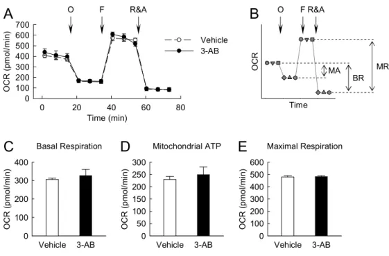
3. Mitochondrial function is independent of PARP activation in kidney proximal tubule epithelial cells.
To determine whether PARP activation causes mitochondrial dysfunction in kidney proximal tubule epithelial cells, the OCR was monitored using XFp extracellular flux analyzer in LLC-PK1 cells. No significant difference in basal respiration as an energetic demand of the cells under the baseline condition, mitochondrial ATP production as a FCCP-sensitive OCR rate, and maximal respiration as a maximum OCR rate of respiration that the cells can achieve was found in cells treated with vehicle and 3-AB (Figure 3, A to E). These data suggest that PARP activation is not attributable to mitochondrial function in kidney proximal tubule epithelial cells.
Fig. 3 PARP1 is not involved in mitochondrial function during treatment with glucose in kidney proximal tubule epithelial cells.
LLC-PK1 cells on an XFp cell culture miniplate were treated with 1 mM 3-AB in glucose- and serum-free DMEM medium (vehicle) for 30 minutes; and then incubated with XF base medium containing 25 mM glucose, 1 mM pyruvate, and 4 mM glutamine in a 37°C CO2-free incubator for 60 minutes. After that, OCR was measured using the
XFp extracellular flux analyzer. Oligomycin (O), FCCP (F), and rotenone plus antimycin A (R&A) were sequentially injected into the miniplate at a final concentration of 1 M, 2 M, and 0.5 M, respectively. The levels of OCR were made three times in respective phases, and expressed as picomoles (pmol) per minute. (A) OCR analysis in 3-AB-treated LLC-PK1 cells. (B) Profile of OCR analysis. BR, basal respiration; MA, mitochondrial ATP; MR, maximal respiration. (C) Basal respiration indicates an energetic demand of the cells under the baseline condition. It was calculated by the average of three OCR baselines before oligomycin injection minus the average of three
Basal Respiration Vehicle 3-AB O C R ( p m o l/m in ) 0 100 200 300 400 Mitochondrial ATP Vehicle 3-AB O C R ( p m o l/m in ) 0 50 100 150 200 250 300 Time (min) 0 20 40 60 80 O C R ( pm ol /m in ) 0 100 200 300 400 500 600 700 Time O C R Vehicle 3-AB O FR&A
B
MR BR MAA
Maximal Respiration Vehicle 3-AB O C R ( p m o l/m in ) 0 100 200 300 400 500 600E
C
D
O F R&Aproduced by mitochondria. It was calculated by the average of three OCR baselines before oligomycin injection minus the average of three ECAR levels after oligomycin injection. (E) Maximal respiration indicates a maximum OCR rate of respiration that the cells can achieve. It was calculated by the average of three OCR levels after FCCP injection minus the average of three OCR levels after R&A injection. Error bars represent SD (n=4 experiments).
4. PARP inactivation augments glycolytic enzyme activity induced by glucose in kidney proximal tubule epithelial cells.
To clarify the effect of PARP inactivation on glycolysis in kidney proximal tubule epithelial cells, we measured activities of glycolytic enzymes in LLC-PK1 cells. Glucose increased the activities of glycolytic enzymes including hexokinase, PGI, PFK1, GAPDH, enolase, and pyruvate kinase, compared to that in glucose-starved control cells (Figure 4, A to F). Treatment with 3-AB in cells incubated with glucose markedly increased further the levels of activities in hexokinase, PFK1, and GAPDH (Figure 4; A, C, and D). However, the enzyme activities were not significantly changed in glucose-starved control cells after 30 minutes of treatment with 3-AB (Figure 4, A to F). These data suggest that PARP activation regulates hexokinase, PFK1, and GAPDH activities increased by glucose in kidney proximal tubule epithelial cells.
Fig. 4 PARP1 inactivation augments activities of glycolytic enzymes increased by glucose in kidney proximal tubule epithelial cells.
LLC-PK1 cells were treated with 1 mM 3-AB in glucose- and serum-free DMEM medium (vehicle) for 30 minutes; and then incubated with 10 mM glucose in XF base medium with 4 mM glutamine (control) for 30 minutes. (A, B, D, E, F) Activities of hexokinase, PGI, GAPDH, enolase, and pyruvate kinase in the cells were measured by hexokinase, phosphoglucose isomerase, GAPDH, enolase, and pyruvate kinase colorimetric assay kits, respectively. (C) PFK1 activity in the cells was measured as previously described (13). All results were expressed as units (U) per mg protein per minutes. Error bars represent SD (n=4 experiments). *P<0.05 versus control; #P<0.05
versus vehicle. Vehicle 3-AB Control Glucose G A P D H a ct iv ity (m U /m g/ m in ) 0 20 40 60 80 100 120 Control Glucose E n ol as e ac tiv ity (m U /m g/ m in ) 0 2 4 6 8 10 12 14 Control Glucose P yr u va te k in as e a ct iv ity (m U /m g/ m in ) 0 2 4 6 8 10 12 14 Control Glucose H ex o ki n as e a ct iv ity (m U /m g/ m in ) 0 5 10 15 20 Control Glucose P G I a ct iv ity (m U /m g/ m in ) 0 2 4 6 8 10 12 Control Glucose P F K 1 a ct iv ity (m U /m g/ m in ) 0 2 4 6 8
E
F
D
B
C
A
*
*
* *
*
*
*
*
*
*
*
*
# # #V. DISCUSSION
The present study shows that PARP activation induced by glucose does not affect mitochondrial function in kidney proximal tubule epithelial cells. Instead, PARP activation leads to inhibition of glycolytic activity as determined by the significant increment induced by the injection of glucose during glycolytic flux. Furthermore, the inhibition of glycolytic activity is caused by the significant decrement of activity in glycolytic enzymes including hexokinase, PFK1, and GAPDH.
The activation of PARP is displayed by poly(ADP-ribose) polymerization because PARP builds up homopolymers of ADP-ribose units on various nuclear proteins as well as PARP itself (16). The poly(ADP-ribosylated) proteins lose their affinity for DNA following genotoxic injury and then the proteins are inactivated (1). In the metabolic pathway of glycolysis; hexokinase, PFK1, and GAPDH contain a poly(ADP-ribose)-binding domain such as poly(ADP-ribose)-binding motif, poly(ADP-ribose)-binding zinc finger domain, macro domain, and domain with conservative multiple sequence alignment of two tryptophan and a glutamate residues (10, 17). Hexokinase is the first regulatory enzyme to initiate glycolysis by converting glucose to glucose-6-phosphate (19), and its activity is inhibited by PARP activation in primary mouse cortical neurons (2). In support of this notion, hexokinase has been shown to contain a poly(ADP-ribose)-binding motif, and coimmunoprecipitate with poly(ADP-ribose) after PARP activation, indicating that it is a poly(ADP-ribose)-binding protein (2). PFK1, one of the most important regulatory enzymes of glycolysis, contains a poly(ADP-ribose)-binding domain; and its activity increases when the ratio of ATP to AMP is lowered (6). PFK1 activity is inhibited by poly(ADP-ribosyl)ation induced by PARP activation in brain-derived cells (10).
oxidative modifications of thiols that inhibits its activity (21). It has been reported that GAPDH activity is also inhibited by poly(ADP-ribosyl)ation in kidney proximal tubule epithelial cells after ischemia reperfusion injury (8). Poly(ADP-ribose) is detected in GAPDH, and then its activity is subsequently decreased (10). The previous findings are consistent with our present results, suggesting that PARP activation induced by injecting glucose into kidney proximal tubule epithelial cells generates poly(ADP-ribose) on its binding site in hexokinase, PFK1, and GAPDH; and reduces their activities. The poly(ADP-ribose)-binding domain in other glycolytic enzymes including PGI, aldoase, triose phosphate isomerase, phosphoglycerate kinase, phosphoglyceromutase, enolase, and pyruvate kinase has not been reported. In our present data, the activity of PGI, enolase, and pyruvate kinase has not been consistently altered by PARP inhibition, indicating that their enzymes may not contain the poly(ADP-ribose)-binding domain.
Glycolysis of glucose metabolism processes is the metabolic pathway that converts glucose into pyruvate in cytoplasm, which produces ATP (26). Exogenous glucose, the most important energy-producing molecule of organisms, strictly induces activities of glycolytic enzymes in the whole pathway of glycolysis, containing 10 steps of chemical reactions with each catalyzed by a specific enzyme (11). A recent report has demonstrated that PARP inhibits glycolysis in kidneys after ischemia reperfusion injury, as demonstrated by lactate production increased by PARP deficiency in injured tissues (8). Our present data using XFp extracellular flux analyzer have showed that glucose increases glycolytic activity during glycolytic flux in kidney proximal tubule epithelial cells. Furthermore, treatment with the PARP inhibitor 3-AB in those cells has markedly lifted up glycolytic activity induced by glucose. Intriguingly, the mitochondrial function containing basal respiration, mitochondrial ATP production, and maximal respiration has not been significantly different between treatment with 3-AB and vehicle in those cells.
This result contrasts with the previous data demonstrating increased mitochondrial function through SIRT1 in PARP-deficient myoblasts (4). This result suggests that the alteration of mitochondrial function by PARP activation may be dependent on cell type.
Taken together, the results of our present study demonstrate that exogenous glucose increases PARP activation in kidney proximal tubule epithelial cells, and further, the PARP activation regulates glycolytic activity through poly(ADP-ribosyl)ation of hexokinase, PFK1, and GAPDH. PARP may be a pivotal molecule involved in regulation of glucose metabolism.
VI. REFERENCE
1. Althaus FR, and Richter C. ADP-ribosylation of proteins. Enzymology and biological
significance. Molecular biology, biochemistry, and biophysics 37: 1-237, 1987.
2. Andrabi SA, Umanah GK, Chang C, Stevens DA, Karuppagounder SS, Gagne JP, Poirier GG, Dawson VL, and Dawson TM. Poly(ADP-ribose) polymerase-dependent energy
depletion occurs through inhibition of glycolysis. Proceedings of the National Academy of
Sciences of the United States of America 111: 10209-10214, 2014.
3. Bagnasco S, Good D, Balaban R, and Burg M. Lactate production in isolated
segments of the rat nephron. The American journal of physiology 248: F522-526, 1985.
4. Bai P, Canto C, Oudart H, Brunyanszki A, Cen Y, Thomas C, Yamamoto H, Huber A, Kiss B, Houtkooper RH, Schoonjans K, Schreiber V, Sauve AA, Menissier-de Murcia J, and Auwerx J. PARP-1 inhibition increases mitochondrial metabolism through SIRT1 activation. Cell metabolism 13: 461-468, 2011.
5. Berger NA, and Berger SJ. Metabolic consequences of DNA damage: the role of poly
(ADP-ribose) polymerase as mediator of the suicide response. Basic life sciences 38: 357-363, 1986.
6. Chesney J. 6-phosphofructo-2-kinase/fructose-2,6-bisphosphatase and tumor cell
glycolysis. Current opinion in clinical nutrition and metabolic care 9: 535-539, 2006.
7. Chuang DM, Hough C, and Senatorov VV. Glyceraldehyde-3-phosphate
dehydrogenase, apoptosis, and neurodegenerative diseases. Annual review of pharmacology and
toxicology 45: 269-290, 2005.
8. Devalaraja-Narashimha K, and Padanilam BJ. PARP-1 inhibits glycolysis in
ischemic kidneys. Journal of the American Society of Nephrology : JASN 20: 95-103, 2009. 9. Du X, Matsumura T, Edelstein D, Rossetti L, Zsengeller Z, Szabo C, and Brownlee M. Inhibition of GAPDH activity by poly(ADP-ribose) polymerase activates three major pathways
of hyperglycemic damage in endothelial cells. The Journal of clinical investigation 112: 1049-1057, 2003.
10. Gagne JP, Isabelle M, Lo KS, Bourassa S, Hendzel MJ, Dawson VL, Dawson TM, and Poirier GG. Proteome-wide identification of poly(ADP-ribose) binding proteins and
poly(ADP-ribose)-associated protein complexes. Nucleic acids research 36: 6959-6976, 2008. 11. Greiner EF, Guppy M, and Brand K. Glucose is essential for proliferation and the
glycolytic enzyme induction that provokes a transition to glycolytic energy production. The
Journal of biological chemistry 269: 31484-31490, 1994.
12. Ha HC, and Snyder SH. Poly(ADP-ribose) polymerase is a mediator of necrotic cell
death by ATP depletion. Proceedings of the National Academy of Sciences of the United States of
America 96: 13978-13982, 1999.
13. Kim J, Devalaraja-Narashimha K, and Padanilam BJ. TIGAR regulates glycolysis in
F298-308, 2015.
14. Kim J, Long KE, Tang K, and Padanilam BJ. Poly(ADP-ribose) polymerase 1
activation is required for cisplatin nephrotoxicity. Kidney international 82: 193-203, 2012.
15. Kim J, and Padanilam BJ. Loss of poly(ADP-ribose) polymerase 1 attenuates renal
fibrosis and inflammation during unilateral ureteral obstruction. American journal of physiology
Renal physiology 301: F450-459, 2011.
16. Kraus WL, and Lis JT. PARP goes transcription. Cell 113: 677-683, 2003.
17. Krietsch J, Rouleau M, Pic E, Ethier C, Dawson TM, Dawson VL, Masson JY, Poirier GG, and Gagne JP. Reprogramming cellular events by poly(ADP-ribose)-binding
proteins. Molecular aspects of medicine 34: 1066-1087, 2013.
18. Lieberthal W, and Nigam SK. Acute renal failure. I. Relative importance of proximal
vs. distal tubular injury. The American journal of physiology 275: F623-631, 1998.
19. Lunt SY, and Vander Heiden MG. Aerobic glycolysis: meeting the metabolic
requirements of cell proliferation. Annual review of cell and developmental biology 27: 441-464, 2011.
20. Martin DR, Lewington AJ, Hammerman MR, and Padanilam BJ. Inhibition of
poly(ADP-ribose) polymerase attenuates ischemic renal injury in rats. American journal of
physiology Regulatory, integrative and comparative physiology 279: R1834-1840, 2000.
21. Mohr S, Stamler JS, and Brune B. Mechanism of covalent modification of
glyceraldehyde-3-phosphate dehydrogenase at its active site thiol by nitric oxide, peroxynitrite and related nitrosating agents. FEBS letters 348: 223-227, 1994.
22. Mor I, Cheung EC, and Vousden KH. Control of glycolysis through regulation of
PFK1: old friends and recent additions. Cold Spring Harbor symposia on quantitative biology 76: 211-216, 2011.
23. Park S, Yoon SP, and Kim J. Cisplatin induces primary necrosis through
poly(ADP-ribose) polymerase 1 activation in kidney proximal tubular cells. Anatomy & cell biology 48: 66-74, 2015.
24. Vandewalle A, Wirthensohn G, Heidrich HG, and Guder WG. Distribution of
hexokinase and phosphoenolpyruvate carboxykinase along the rabbit nephron. The American
journal of physiology 240: F492-500, 1981.
25. Yoon SP, and Kim J. Poly(ADP-ribose) polymerase 1 activation links ischemic acute
kidney injury to interstitial fibrosis. The journal of physiological sciences : JPS 65: 105-111, 2015. 26. Zwerschke W, Mazurek S, Stockl P, Hutter E, Eigenbrodt E, and Jansen-Durr P.
Metabolic analysis of senescent human fibroblasts reveals a role for AMP in cellular senescence.
ACKNOWLEDGEMENT
길다면 길고 짧다면 짧은 2년이 흘러 이렇게 석사 졸업 논문을 쓰게 되었습 니다. 짧지만 이 글로나마 감사한 분들께 감사인사를 전하고자 합니다. 먼저 아낌없는 지도와 조언을 해주신 김진우 교수님께 먼저 감사의 인사를 전 하고 싶습니다. 많은 것을 배울 수 있는 기회와 경험을 할 수 있게 언제나 신 경을 써주시고 가르쳐주셔서 감사합니다. 또한, 교수님께서 저를 위해 해주셨 던 말들은 제가 석사를 밟는 2년 동안 저를 바꿀 수 있는 조언이 되었습니다. 요즘 몸이 많이 안 좋으신데 얼른 건강 되찾으셔서 항상 건강하셨으면 좋겠습 니다. 그리고 논문 심사를 맡아 주시고 조언을 해주신 김영석 교수님과 박수 제 교수님께 진심으로 감사 드립니다. 제가 학위를 하는 동안 많은 관심과 격려를 아껴주시지 않았던 조직학교실의 이영기 교수님과 박덕배 교수님, 약리학교실의 강희경 교수님과 유은숙 교수 님, 생리학교실의 은수용 교수님과 정성철 교수님께 감사 드립니다. 그리고 해부학교실 연구조교를 학위를 편안히 밟도록 신경을 써주셨던 윤상필 교수님 과 조사선 교수님께도 감사 드립니다. 학위 과정 동안 연구조교를 병행하면서 많은 도움과 조언을 주셨던 강원석 선 생님, 이정희 선생님께도 감사 인사를 전합니다. 함께 연구조교 생활을 하면 서 힘들 때 언제나 의지할 수 있었던 보람언니랑 지현이에게도 고맙다고 전하 고 싶습니다. 언제나 나를 더 신경 써주고 생각해줘서 고마워요. 예전처럼 함 께 어울릴 시간이 적어 아쉽지만 지금 서로가 있는 자리에서 더 열심히 해서 각자가 꾸는 꿈이 이루어지길 바랍니다. 함께 연구조교 생활을 하고 있는 승헌오빠, 해경이, 수은이, 현지, 수연이, 주 연이, 현애, 지민이, 민철이, 정문오빠, 그리고 다예주. 언제나 나를 걱정해주 고 많이 신경 써줘서 고맙다고 전하고 싶습니다. 그리고 바쁜데도 의생명∙신 약개발학과 조교까지 하면서 많이 신경 써주시고 항상 웃는 얼굴로 반겨주는 우리 희란 언니에게도 감사 인사를 전합니다. 막바지 힘든 시기에 친해져 아쉽고 고마운 생리학실험실에 언제나 많은 이야 기를 들어주고 조언을 해주는 지형언니, 너무 뒤늦게야 친해져 아쉬운 승혜언 니, 항상 기분 좋게 해주고 힘들 때 옆에서 힘이 되어준 홀란, 언제나 많은 것을 아끼지 않고 내준 상찬쌤, 깊게 와닿는 조언을 아낌없이 해준 윤실언니,대학원 과정 동안 잘 챙겨준 지연이와 가영이 그리고 항상 우리의 엔돌핀인 상희에게도 고맙다는 말을 전합니다. 제대로 된 실험을 알게 해주시고 이끌어주신 생물학과 분자생물학 실험실에 김세재 교수님, 희철오빠, 성일오빠, 혜선언니, 선아언니, 승우오빠, 정환오빠, 지연이에게도 감사의 인사 드립니다. 그리고 학사를 밟던 4년 동안 언제나 많 은 조언과 힘이 되어주셨던 생물학과 교수님들, 선배님들, 동기들, 후배들에게 도 감사합니다. 그리고 민지, 수연이, 문호, 지현이, 문석이. 지금은 연락하기 도 힘들고 다들 바빠서 만나기도 힘들지만 너희랑 함께여서 내 대학생활이 재 미있었고 기억에 남는거 같아. 고마워. 마지막으로 세상 그 어떤 좋은 것과도 바꿀 수 없는 나의 가족들에게 감사인 사를 전합니다. 옆에서 힘이 되어주기 위해 노력해주시고 응원해주셔서 힘들 었던 2년 동안 잘 버틸 수 있었고 그래서 이렇게 좋은 결과물을 얻을 수 있 었던 거 같습니다. 사랑하는 내 하나뿐인 엄마. 엄마가 있어서 내가 이만큼 할 수 있었던 것 같 아요. 지금까지 믿어줬던 만큼 앞으로도 더 믿음을 줄 수 있는 딸이 될 테니 깐 너무 걱정 말고 항상 노력하고 열심히 하는 딸이 될게요. 아프지 말고 더 욱 건강해지면 좋겠어요. 이제 제 앞에 놓인 상황이 막연하고 힘들겠지만 저를 아껴주시고 응원해주신 많은 분들이 있어 더욱 힘차게 제가 앞날을 나아갈 수 있을 것 같습니다. 제 가 더 멋있는 앞날을 만드는 모습 끝까지 지켜봐 주시고 언제나 건강하시길 바랍니다. 감사합니다.