Rapid Analysis for the Isolation of Novel Genes Encoding
Putative Effectors to the Position-specific Regulatory
Element of Murine
Hoxa-7
Myungsun Cho, Chuog Shin, Wongi Min and Myoung Hee Kim
*
tGenome Center, Korea Research Institute of Bioscience and Biotechnology, KIST, Taejon 305-600, Korea
(Received on December 15, 1996) Hox
genes are known to
play
a critical role in pattern formation during vertebrate
development by being expressed at
the
specific time and
in
the specific position along the
antero-posterior body axis. In order to
understand
the regulatory mechanism for the
position-specific expression of murine
Hoxa-7,yeast one-hybrid system was applied. DNA
fragment conferring a position specificity to the
Hoxa-7gene was placed ju
s
t
up
s
tream
from the
y
east
Cye1promoter
and
lacZgene
in
a reporter. Selection of LacZ positive
clones after cotransformation of the reporter and mouse embryonic cDNA
library
as an
effector, which was
designed
to be expressed as fusion proteins to the GAlA activation
domain, allowed us to isolate
putative
factors
interacting
with the position-
s
pecific
regulatory element of murine
Hoxa-7.A total of 28 positive clone
s
were
s
creened from 5
x
105yeast transformants. About 70
%
of the clones turned out to be novel and most of the
can-didate clones selected in this study showed a temporally restricted expression pattern
dur-ing embryonic development, suggestdur-ing that this method could provide an efficient way for
isolating
novel genes who
s
e expressions are temporally regulated during embryogenesis.
Hox genes, homeobox contammg genes in ve r-tebrates, play critical roles in pattern formation during embryonic development and their functions are pe r-formed in a coordinated fashion temporally as well as spatially (Krumlauf, 1994). So far, 39 Hox genes be -ing clustered at 4 different loci (HoxA, HoxB, HoxC, and HoxD) of the chromosomes have been identified in both mice and humans. Their organization and nu -cleotide sequence showed strong conservation through an evolutionary process. Proteins encoded by these Hox genes have been identified as transcriptional fac -tors controlling the expression of specific target genes during development (Kim and Kessel, 1993).
The Hoxa-7 gene located at the middle of a HoxA cluster is expresssed in the restricted area along the antero-posterior axis during gastrulation; the anterior limit of expression at C5 in the ectoderm derived neural tube and spinal ganglia, and T3 to T4 in the mesoderm derived prevertebrae around day 12.5 p.c. Temporally, the expression started from the beg in-ning of gastrulation around day 7.5 p.c., peaked at 12.5 p.c. and decreased since then (Mahon et aI.,
1988;
Puschel et aI., 1990). The upstream spatio-temporal regulatory regions have beendoned from both mice*
To whom correspondence should be addressed.t Present address: Department of Anatomy, Yonsei U ni-versity College of Medicine, P.O. Box 8044, Seoul, Korea.
(Puschel et aZ., 1990; 1991) and humans (Knittel et aZ., 1995; Min et aZ., 1996), and their roles have been analyzed in transgenic mice. This region sho w-ed a strong conservation in the nucleotide sequence level as well as in the function of directing a positio n-specific expression of the downstream reporter gene between two species (Knittel et aI., 1995; Min et aI., 1995, 1996). Although several position-specific cis-acting regulatory elements have been reported in many Hox genes, no definite correlation between regulatory factors and target sequences has been made except the interaction between Krox-20 and Hoxb-2 as well as the role of retinoic acid in es -tablishing the segment restricted expression of Hoxb-l (Marshall et aI., 1994; Sham et aZ., 1993; Studer et aI.,
1994).
There are several methods to isolate genes encoding regulatory factors which bind to and activate a specific cis-element from the library, such as Southwestern blo
t-The abbreviations used are: bp, base pair(s); ~-Gal, ~-galac tosidase; EST, expressed sequence tag; Hox, homeobox gene in mice related to the Antennapedia class gene of Drosophila; kb, kilobase(s); NGF, nerve growth factor; OD, optical density; ONPG, o-nitrophenyl-~-D-galactopyra nos ide; p.c., post coitum; PSRE, position-specific regula -tory element; X-gal, 5-bromo-4-chloro-3-indolyl-~-D-galac topyranoside.
Vol. 7 (1997) Myungsun Cho et al. 221
ting, affinity column chromatography, and one-hybrid a-ssay. Especially the one-hybrid assay has been recent-ly used successfully for identifying cDNA candidates which encode the DNA binding protein for a specific regulatory DNA element. These include the yeast ori-gin recognition component ORC6 (Li and Herskowitz, 1993), the neuronal olfactory transcriptional factor Olf-1 (Wang and Reed, 1993), and the B cell specific fac-tor Bobl (Gstaiger et al., 1995).
In an attempt to isolate trans-acting factors which interact with this position-specific regulatory element (PSRE) of the murine Hoxa-7 gene, we applied the yeast one-hybrid system. This system allows the iso-lation of cDNAs encoding effectors which interact with the testing DNA element, PSRE, fused to the re-porter lacZ gene by screening LacZ positive colonies. While analyzing the positive clones, this approach was proven to be an efficient way of screening novel genes whose expressions are temporally regulated dur-ing embryogenesis.
Materials and Methods
Construction of a reporter plasmid p360
The 357 bp BamHIINlaIII fragment (from nt - 1710 to - 1354 of Parikh et aI., 1995) covering the control region of the murine Hoxa-7 gene was isolated from the pAX-L680 (from Dr. Gruss, Max-Planck Institute, Germany) and cloned into pGEM3zf (Promega, Madi-son, WI, U.S.A) digested with BamHI and SmaI after the 3' overhanging end was eliminated with T4 DNA polymerase. An EcoRIIPstI-digested fragment of the resulting plasmid was cloned into the pBluescript KS(
+
)
(Stratagene, La Jolla, CA, U.S.A), yielding the pBS-360. The 360 bp San fragment of pBS360 was then cloned into the XhoI site of the pHYI00 (Yoo and Cooper, 1989) and named p360.Mouse embryo eDNA library construction
The effector, cDNA library, was either constructed in this laboratory or purchased from Cion tech (Palo Alto, CA, U.S.A). For construction, mouse embryos at day 11.5 p. c. were homogenized and subjected to RNA extraction based on the acid-guanidinium-phenol-chloroform method (Chomczynski and Sacchi, 1987). Poly(A)+ RNA was purified from total RNA according to the mRNA isolation kit (Pharmacia, Sweden). Double stranded cDNA was synthesized from 5 )..lg of poly(At RNA with a cDNA synthesis kit from Stratagene. Fractions containing cDNA great-er than 300 bp were ligated with EcoRI-XhoI di-gested pGAD-GH (Clontech). Ligated DNA was in-troduced into E. coli strain DH5u through elec-troporation using a Biorad electroporator and plasmid DNA was extracted from the transformants by a stand-ard method (Sambrook et al., 1989). Mouse ll-day Embryo MATCHMAKER cDNAs (Clontech) were also prepared in accordance with the manufacturer's guideline.
Yeast strain, transformation, plasmid extraction, and plasmid curing
S.
cerevisiae L3262 (leu·, £Ira·, his·, donated by Dr. Yoo in KRIBB) and its transform ants were maintained on YPD (10 g yeast extract, 20 g peptone, 50 ml of 40% glucose per 1 I) or selective medium (Difco 0919-15-3) supplemented with appropriate amino acids. Pla-smid DNA or cDNA library transformation in yeast strain was conducted by the lithium acetate method (Schiestl and Geitz, 1989). Plasmid DNA extraction from yeast transformants was carried out as described (Hoffman and Winston, 1987). Curing of a reporter plasmid which carries the URA3 marker was per-formed by selection on the plate containing 5-fluoroacetic acid (Sigma) as described by Sikorski and Boeke (1991).Screening the library
Reporter plasmid p360 and mouse embryo cDNA li-brary (effector library) were cotransformed into
S.
cerevisiae L3262 and plated on the selective media lacking uracil and leucine (about 50 plates of 150 mm). After 50 h at 30°C, transformants (5x
10
5
)
were subjected to the colony lift assay for selection of LacZ positive clones by the method of Breeden and Nasmyth (1985). Filters were incubated at 30°C in the presence of X-gal and colonies which turned to blue within 2.5 h were selected. A single colony was purified and subjected to curing of the URA3+ plasmid as described above. Effector plasmids were re-covered and transformed into E. coli by eIec-troporation.Liquid f3-galactosidase assay
For quantitation of ~-Gal actiVIty, cells grown in YPD medium to OD
595
of 0.7-1.0 were subjected to the assay as described by Miller (1972) except that cells were lysed by placing tubes in liquid nitrogen. Units of ~-Gal activities were expressed as OD42oU
I
OD595 of cells per min from duplicate assays.DNA manipulation, sequence determination, and Northern hybridization
Recombinant DNA techniques were carried out mainly as described by Sambrook et al. (1989). Nu-cleotide sequences were determined by the chain ter-mination method using a Sequenase 2.0 kit (USB, Cleveland, OH, U.S.A) and a homology search was done by the Fasta program. For Northern blotting of mouse embryo stage-specific RNA, Multiple Embryo Tissue Northern Blot (Clontech) was probed with a 32P-labeled DNA fragment under conditions recom-mended by the manufacturer.
Results and Discussion
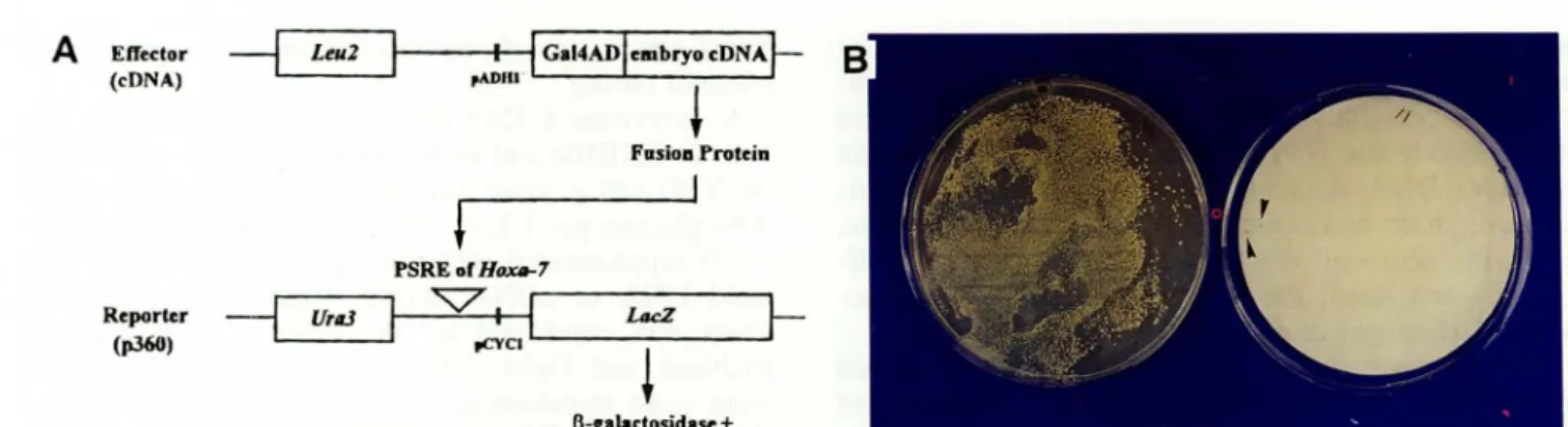
Screening of effectors interacting with PSRE of murine Hoxa-7
A
Effedor~
Leu2 (eDNA)Reporter
--j
Urll3(p.360)
t----fl--i! Gal4ADlelllbryo
<DNA~
,ADltI'
+
FasioD Protein PSRE of Ht»Ut-7"'V'
pCYCl L..-_ _ _ _ _I!
LIICZ -..JI-~
l3-cabdosidase +Figure 1. Strategy for screening effectors through their interaction with the PSRE of the Hoxa-7 gene. A) Schematic draw-ing of effector and reporter construct. The effector fusion proteins, yeast GAL4 activation domain (GaIAD) and embryonic cDNA fused in frame, which interact with the PSRE of Hoxa-7 were designed to be selected by activating lacZ expression in the reporter. Effector and reporter (P360) contain an LEU2 and URA3 marker for selection, respectively. The promoters, pADHl (alcohol dehydrogenase) and pCYCl (iso-I-cytochrome c) used in each effector and reporter construct are written. B) Screening LacZ positive clones from cDNA transformants. Colonies were lifted on filter paper and exposed to substrate X -gal after the filter was soaked into liquid nitrogen. Colonies showing a blue color were selected for further analysis.
region of murine Hoxa-7 specifies an anterior boun-dary of expression. The minimal region sufficient for directing the position specific expression of murine Hoxa-7 has been mapped to be a 470 bp long frag-ment (AX470) (Knittel et aI., 1995). Recently, we have cloned and determined the nucleotide sequence of a 3.9 kb human homologue from a human genom-ic library and revealed that the most 3' 1.1 kb frag-ment in this region had a similar function in transgen-ic mice (Min et aI., 1996). This portion has a se -quence homology of over 70% of that of the murine sequence, indicating that the regulatory mechanism for the position-specific expression of Hoxa-7 is con-served between two species, which strongly suggests the existence of regulatory factors having a conserved DNA binding motif. We further deleted the 5' region of the AX470 based on Min el al. (1996) and used a 357 bp DNA fragment (from nt - 1710 to nt - 1354 of Parikh et aI., 1995) as the minimal portion of the position-specific regulatory element (PSRE) required to set the anterior boundary of the Hoxa-7 expression and applied for screening the regulatory factors.
In order to isolate factors which interact with PSRE, we used a yeast one-hybrid system in which a reporter and an effector are present in a single cell. The reporter plasmid p360 contains the bacterial lacZ gene under the yeast CYC1 promoter as well as H oxa-7 PSRE (Fig. lA). Since no p-Gal activity was ob-served with the reporter plasmid alone, it was as-sumed that the endogenous yeast proteins do not bind to or activate PSRE. The effector embryonic cDNA li-brary was designed to be expressed as fusion proteins to the transcription activation domain of yeast GAL4 (Fig. lA), so that any fusion protein which can in-teract with Hoxa-7 PSRE could be isolated after X -gal stam1l1g. Since the transcriptional activation ability is supplied by the N-terminal GAL4 activation domain in the fusion protein, a fused gene encoding a protein which can interact with the PSRE element
subsequently activates the lacZ gene expression in a reporter, no matter whether it is a repressor or an ac -tivator. We screened 5 X
to
'
yeast transformants and selected 28 positive clones (Fig. IB).Sequence analysis of PSRE responsive inserts
To determine the nature of the proteins encoded in the positive effector plasm ids, reporter plasm ids were cured out as described in Materials and Methods. The effector plasm ids were then transferred into E. coli and partial nucleotide sequences were determined us-ing the dideoxynucleotide sequencing method. Most of the Hoxa-7 PSRE responsive cDNAs were iden-tified to be novel genes when compared with the EMBL data bases (Table ]); 19 out of 28 clones (68%) were previously unreported sequences and Gen-Bank accession numbers of these clones are shown in Table 1. Five clones, c134, c143, c161, c166, and c168, showed a strong homology to the reported eDNA sequences whose functions have been partially characterized, and four clones showed significant se-quence homologies to some of the ribosomal protein coding genes.
Effector responsiveness to PSRE
In order to quantitate the interaction between the ef-fector fusion protein and the PSRE of Hoxa-7, p-Gal activity was measured (see Materials and Methods). Six LacZ positive clones (c124, c131, c134, c143, c153, and c171), which turned blue a short time after X-gal treatment during screening, were chosen. As shown in Figure 2, most of the clones tested ex-hibited rather low p-Gal activities, about 1.5 to 3.5-fold higher than that of the control cells except for c153. When these clones were cotransformed with a plasmid lacking the PSRE, no actIvItIes were observed (data not shown). The clones c131, c124 having a sequence homology with ESTI07511 (H 34975; Lee el al., 1995) which is similar to the
ri-Vol. 7 (1997) Myungsun Cho et ai. 223
Table
1.
Sequence analysis of Hoxa-7 PSRE response cDNAsNo. Size (bp) Accession No. Putative identification No. Size (bp) Accession No. Putative identification
c101 157 Z82914 unknown c158 137 Z82924 unknown
c104 175 Z82915 98% homology to X76772" c159 137 Z82925 unknown
cl14 137 Z82916 unknown c160 227 Z82926 unkown
c1l7 73 Z82913 unknown c161 102 Z82927 80% homology to M26651e
c124 92 Y09519 90% homology to H34975b c162 179 Z82934 unknown
cl31 181 Z82917 unknown c163 133 Y09520 94% homology to U13369'
cl34 201 Z82918 80% homology to H36700c c165 138 Y09521 84% homology to N393118
cl37 209 Z82919 unknown c166 107 Y09522 83% homology to N20634"
c143 181 Z82920 70% homology to H72555" c167 163 Z28928 unknown
cl51 215 Z82921 unknown c168 164 Y09523 88% homology to
D1877i
cl53 221 Z82933 unknown c170 146 Z82929 unknown
cl54 160 Z82922 unknown c171 215 Z82930 unknown
cl56 147 Z82923 unknown c172 136 Z82931 unknown
clS7 218 Z82935 unknown c175 67 Z82932 unknown
"ribosomal protein S3; "ribosomal protein S9 (Lee el al., 1995); <ratlus sp. cDNA (Lee el ai., 1995); "human fetal cDNA; '5_
region of mouse epo gene (Bera el aI., 1989); fribosomal DNA repeating unit (Gonzalez el al., 1985); gribosomal protein S6; "human EST; fmouse EST
c171
o
clS3
Z
c143
a
c134
~ e131
c124
c
o
1
2
Activity(fold)
~,
J3
,
36
Figure 2. Transactivation of the lacZ gene by interaction between the PSRE of Hoxa-7 and putative effectors. Expression of the lacZ gene directed through effector interaction with PSRE was monitored by the ~-Gal assay using ONPG as a substrate. Yeast cells harboring a reporter and effector were lysed by immersing the cell pellet intoliquid nitrogen and subjecting them to the enzyme assay as described in Materials and Methods. Activities of each clone were given as ratios to the value of strain carrying only a reporter plasmid (C). Clone cl53 showed a 36-fold higher activity than that of the control.
bosomal protein S9, and c143 which has a sequence homology with an EST (H72555) isolated from hu-man fetal liver and spleen cDNA library (The WashU-Merck EST project, Unpublished) showed rather low
~-Gal activity compared to other tested clones.
It is generally believed that a clone having higher
~-Gal activity has a higher binding affinity to PSRE.
Therefore, clone c153 showing the highest ~-Gal activity (about 36 fold) was chosen for electro
-phoretic mobility shift assay. Unfortunately, how-ever, the signal was not strong enough to be seen (data not shown). It could be due to the low amount
of c153 proteins existing in crude yeast extract or
that the binding protein is provided by the host and
the fusion protein which associates with it by protein
-protein interaction is selected through this method. Although the latter case is very unlikely since we did not see any shifted band with the host cell extract, this problem could be solved by purification of the fusion protein.
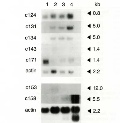
Stage-specific
expression
of PSRE responsive inserts
Since it is known that the expression of
Ho
xa
-7
is temporally regulated during embryogenesis like otherHox
genes, it was of particular interest to characterize the expression mode of positive clones during early mouse development. Therefore, 7 clones (6 tested ~Gal activity plus c158) were chosen and Northern hy
-bridization was conducted with the mouse embryonic
stage-specific RNA blot of Cion tech.
As shown in Figure 3, all the clones except c124 exhibited temporally restricted expression patterns. Clone c124, which has sequence homology with ri-bosomal protein S9, did not show any significant dif-ference (or slight difference, if any) at the level of ex -pression during development.
On the other hand, for example, clones c158 and c171 showed an extreme stage-specific expression
mode, such as a restricted expression, at day 17.0 and
7.0 p.c., respectively. A protein like c171, which is restrictively expressed at day 7.0 p.c., might be im
-portant for an onset of initial
Hox
gene expression. SinceHo
xa-7
gene starts expression at the beginning of mesoderm formation around day 7.5 p.c., the re-gulatory protein necessary for initial expression
somehow should be provided before gastrulation. Clones c131 and c134 are also interesting. They were not expressed at day 7.0 p.c., but abruptly syn-thesized at· day 11.0 p.c. and were maintained con-stantly throughout the development in the case of c134, or gradually increased in the case of c131.
1 2 3 4
kb
c124
<l1li 0.8
c131
<l1li 5.0
c134
<l1li5.0
c143
<l1li1.4
c171
<l1li1.4
actin
<l1li2.2
c153
<l1li 12.0
c158
<l1li 5.5
actin
<l1li 2.2
Figure 3. Stage-specific expression of putative factors in-teracting with PSRE. Northern blot analysis of poly(A)'
RNA isolated at different stages of mouse embryo was per-formed with several LacZ positive clones. The filter was
hybridized with EcoRI inserts isolated from each positive clone and labeled by a random priming method. Clone
numbers are shown on the left side and the size of the tra
n-scripts are given in kb on the right side. Human ~-actin gene was used as a control probe. Lane 1, poly(A)' RNA
from day 7 p.c.; lane 2, poly(A)' RNA from day 11 p.c.;
lane 3, poly(A)' RNA from day 15 p.c.; and lane 4, poly (A)' RNA from day 17 p.c.
Because gastrulation starts around day 7.5 p.c., it is reasonable to postulate that these are likely to be the mesoderm or neuroectoderm specific genes which are expressed after gastrulation. Especially c134, which
has some sequence homology with one of the rattus cDNA isolated from 9 day NGF treated PC12 cells (Lee et aI., 1995), might be a regulatory signal for
Hoxa-7 expression in the ectoderm-derived tissue,
since PC12 cells differentiate into sympathetic-like
neurons after NGF induction. Since Hox genes have a
different anterior boundary of expression between
ectoderm- and mesoderm-derived tissues, it has been thought that the regulatory mechanism for
position-specific expression could be different between two
tis-sue layers. It is also interesting to point that the PSRE used here has been postulated as being a
position-spe-cific regulatory element in the ectoderm derived
neur-al
tubeand s
pin
a
l
gang
li
a
(Min
etai.,
1996).Most transcripts of the clones c143 and c153 were detected at day 11.0 p.c., the mid-gestation stage, dur-ing which most Hox genes are strongly expressed.
The proteins expressing later in gastrulation might be
involved in the maintenance mechanism for Hox expression, for which an autoregulatory mechanism has been known in the case of Ultrabithorax and
Deformed genes of Drosophila (Beachy et aI., 1988;
Regulski et al., 1991). Among these, clone c143 has some sequence homology with an EST (H72555)
iso-lated from a liver and spleen cDNA "library made with a 20 week-post conception male fetus. Since
lineage-specific expression of Hox genes has been reported in human hematopoietic systems (Vieille-Grosjean et al., 1992), it is interesting to presume that clone c143 could be a Hox gene regulator involving murine erythropoiesis, which actively occurs in 20-week old fetal liver, in the case of humans.
It was surprising that most of the positive clones
analyzed here showed a temporally restricted expression
pattem during mouse development, suggesting that this method could provide an efficient method for isolating novel genes whose expressions are temporally regulated during embryogenesis. So far, various stragtegies have
been developed to isolate novel genes participating in the regulation of vertebrate development; cloning the
homOlogous genes from other species with specific sequence motifs (Kessel and Gruss, 1990), screening dif-ferentially expressing genes after treatment with certain
inducers such as growth factors or steroid hormones
(Lee et aI., 1995), gene trapping experiments (Friedrich and Soriano, 1991; Skames et al., 1992), and selection
by whole mount in situ hybridizations (Bettenhausen
and Gossler, 1995). Many genes isolated by these methods exhibited a spatially or temporally controlled expression pattern. Here, we used a yeast one-hybrid system to screen putative factors which interact with the PSRE of murine Hoxa-7. Interestingly, this method
turn-ed out to be a rapid and efficient way not only for
iso-lating novel factors associated with the specific motif but also for screening novel genes whose expressions are temporally regulated during development.
Compre-hensive characterization of these genes and factors in the future will help us to understand the molecular
mechanism underlying vertebrate development, includ-ing the regulatory cascade for Hox gene expression.
Acknowledgments
We thank Dr. P. Gruss for providing the plasmid pAX-L680, and Dr.
H.
S. Yoo for yeast strain L3262 and plasmid pHY100. Especially we would like to thank Dr. M. Won for critical advice and numerousassistances.
This work was supported by a 1995 and 1996 Genome Program Grant from the Ministry of Science and Technology of Korea. Dr. C. Shin was supported by a KOSEF postdoctoral fellowship.
Reference
s
Beachy, P. A., Krasnow, M. A., Gavis, E. R., and Hogness,
Vo!. 7 (1997) Myungsun Cho et al. 225
Bett
e
nh
a
u
se
n
,
B.
,
and
Gossler
,
A.
(1995)
Genomics 28,436-441.
Breeden, 1., and Nasmyth, K. (1985) in Cold Spring
Sym-posia 011 Quantitative Biology, Vo!. 50, pp. 643-650, Cold Spring Harbor Laboratory, Cold Spring Harbor, NY. Chomczynski, P., and Sacchi, N. (1987) Anal. Biochem.
162,156-159.
Friedrich, G., and Soriano, P. (1991) Genes Dev. 5, 1513
-1523.
Gstaiger, M., Knoepfel, L., Georglev, 0., Schaffner, W.,
and Hovens, C. M. (1995) Nature 373, 360-362. Hoffman, C. S., and Winston, F. (1987) Gene 57,267-272.
Kessel, M., and Gruss, P. (1990) Science 249, 374-379.
Kim, M. H., and Kessel, M. (1993) AgBiotech. News Info.
5, 189-194.
Knittel, T, Kessel, M., Kim, M. H., and Gruss, P. (1995) Development 121, 1077-1088.
Krumlauf, R. (1994) Cell 78, 191-201.
Lee, N. H., Weinstock, K. G., Kirkness, E. F., Ear!e-Hughes, J. A., Fuldner, R. A., Marmaros, S., Glodek, A.,
Gocayne, J. D., Adams, M. D., Ker!avage, A. R., Fraser, C. M., and Venter, J. C. (1995) Proc. Natl. A cad. Sci.
USA 92, 8303-8307.
Li, J. J., and Herskowitz, 1. (1993) Science 262, 1870-1874. Mahon, K. A., Westphal, H., and Gruss, P. (1988) De
-I'elopmell/ 104 (supp!.), 187-195.
Marshall, H., Studer, M., Popper!, H., Aparicio, S.,
Kuroiwa, A., Brenner, S., and Krumlauf, R. (1994)
Na-ture 370, 567-571.
Miller, J. H. (1972) E.>.perimellts in Molecular Genetics, Cold Spring Harbor Laboratory Press, Cold Spring Harbor,
NY.
Min, W., Park, 1. H., Chung, W. J., Park, H. Y., and Kim,
M. H. (1995) Korean]. Genetics 17,123-132.
Min, W., Cho, M., Jang, S. 1., Chang, H.-H., Lee, c.-S., Jun, M.-H., and Kiln, M. H. (1996) Gene 182, 1-6.
Parikh, H., Shah, S., Hilt, D., and Peterkofsky, A. (1995)
Gene 154, 237-242.
Puschel, A. W., Balling, R., and Gruss, P. (1990) De
-velopment 108, 435-442.
Puschel, A. W., Balling, R., and Gruss, P. (1991) De
-velopment 112, 279-287.
Regulski, M., Dessain, S., McGinnis, N., and McGinnis, W.
(1991) Genes Dev. 5, 278-286.
Sambrook, J., Fritsch, E. F., and Maniatis, T (1989) Molec
-ular Cloning: A Laboratory Manual, Cold Spring Harbor
Laboratory Press, Cold Spring Harbor, NY.
Schiestl, R. H., and Geitz, R. D. (1989) Curro Genet. 16,
339-346.
Sham, M. H., Vesque, c., Nonchev, S., Marshall, H., Frain, M., Gupta, R. D., Whiting, J., Wilkinson, D., Charnay,
P., and Krumlauf, R. (1993) Cell 72, 183-196.
Sikorski, R. S., and Boeke, 1. D. (1991) Methods Enzymol.
194, 302-318.
Skarnes, W. c., Auerbach, B. A., and Joyner, A. L. (1992)
Genes Dev. 6, 903-918.
Studer, M., Popper!, H., Marshall, H., Kuroiwa, A., and
Krumlauf, R. (1994) Science 265, 1728-1732.
Vieille-Grosjean, 1., Roullot, V., and Courtois, G. (1992)
Biochem. Biophys. Res. Commun. 183, 124-1130.
Wang, M. M., and Reed, R. R. (1993) Nature 364,
121-126.
Yoo, H. S., and Cooper, T G. (1989) Mol. Cell. BioI. 9,