JOURNAL OFCLINICALMICROBIOLOGY, Feb. 2009, p. 481–484 Vol. 47, No. 2 0095-1137/09/$08.00⫹0 doi:10.1128/JCM.01253-08
Copyright © 2009, American Society for Microbiology. All Rights Reserved.
Evaluation of an Immunochromatographic Assay Kit for Rapid
Identification of Mycobacterium tuberculosis Complex in
Clinical Isolates
䌤
Mi Young Park,
1* Young Jin Kim,
1Sang Hyun Hwang,
1Hyoung Hoi Kim,
1Eun Yup Lee,
1Seok Hoon Jeong,
2and Chulhun L. Chang
1,3Department of Laboratory Medicine, School of Medicine,1and Medical Research Institute,3Pusan National University, Busan, and Department of Laboratory Medicine, Yonsei University College of Medicine, Seoul,2Korea
Received 2 July 2008/Returned for modification 16 August 2008/Accepted 22 November 2008
We evaluated a new immunochromatographic assay (ICA) using mouse monoclonal anti-MPT64 antibody for rapid discrimination between Mycobacterium tuberculosis and nontuberculous mycobacteria in clinical isolates. A study with mycobacteria and other organisms showed excellent sensitivity (⬵99%) and specificity (100%) and an appropriate detection limit (105
CFU/ml) when tested with M. tuberculosis H37Rv. This ICA can simplify the identification of M. tuberculosis in clinical laboratories.
Tuberculosis is a global problem and the single most com-mon cause of death from any bacterial agent (15, 22).
Myco-bacterium tuberculosis and nontuberculous mycobacteria
(NTM) are different clinically, so prompt detection, isolation, and discrimination are essential for appropriate management (3, 7).
The MPT64 protein is highly specific for M. tuberculosis complex, including M. tuberculosis, Mycobacterium africanum,
Mycobacterium bovis, and some, although not all, substrains of M. bovis BCG (1, 6, 16, 18, 23), and can be detected in culture
isolates and biopsy samples (1, 7, 17, 18, 20). Recently, Stan-dard Diagnostics (SD, Yongin, Korea) developed a simple and rapid assay using a mouse monoclonal anti-MPT64 antibody to discriminate between M. tuberculosis complex and NTM by immunochromatography. Mouse monoclonal anti-MPT64 an-tibodies (SD Bioline TB Ag MPT64; SD) are immobilized on a nitrocellulose membrane as the capture material. Another antibody, which recognizes another epitope of MPT64 and has been conjugated with colloidal gold particles, is used for anti-gen capture and detection in a sandwich-type assay.
We evaluated the clinical usefulness of the kit using myco-bacteria and other organisms. To determine specificity, 137 bacterial isolates (68 species), 20 fungal isolates (10 species), 53 reference mycobacterial isolates (40 species), and 51 NTM isolates from clinical samples were tested (Tables 1 to 3). To determine sensitivity, 159 M. tuberculosis complex strains grown on 3% Ogawa medium (isolated at Pusan National University Hospital), 60 strains from Bactec MGIT 960 culture tubes (isolated at Kosin University Gospel Hospital), and one reference strain, M. tuberculosis H37Rv, were tested. All bac-terial, fungal, and mycobacterial isolates were stock cultures that had been kept in a ⫺4°C refrigerator or a ⫺72°C deep freezer for as long as 18 months. The cultured mycobacteria
were identified by acid-fast bacillus stain, nucleic acid amplifi-cation, and DNA microarray (10, 14). Finally, to determine the detection limit, a series of diluted suspensions of M.
tubercu-losis H37Rv were inoculated onto Middlebrook 7H10 agar and
the resulting colonies were counted (19). One hundred micro-liters of sample taken from liquid medium was applied directly to the sample well without preparation. Three or four colonies were scraped from the solid medium and suspended in 300l of extraction buffer (SD); then, 100l of the suspension was added to the sample well. If there was condensation fluid in egg-based medium, 100l of the fluid was applied directly to the sample well, instead of using extraction buffer. Tests were interpreted 15 min after sample application. The presence of a control band alone indicates a negative result, whereas the presence of two color bands (control and test bands), no mat-ter which band appears first, indicates a positive result. A color band of any intensity was read as a positive reaction (Fig. 1). If the control band was not visible after 15 min, the result was considered invalid, and the sample was retested.
All bacterial, fungal, and NTM isolates, including reference strains, were negative by the immunochromatographic assay (ICA) (specificity, 100%). One hundred fifty-eight of 159 M.
tuberculosis complex strains grown on solid medium and 59 of
60 strains from liquid medium were positive by the ICA (over-all sensitivity, 98.6%). The 1:128-diluted suspension (5.5⫻ 105
CFU/ml) revealed 10% reaction intensity, and the band inten-sity gradually weakened with serial dilutions until the 1:1,024 suspension (6.8⫻ 104CFU/ml) was negative. The detection
limit thus was determined to approximate 105CFU/ml.
Although most culture-positive mycobacteria are M.
tuber-culosis in regions where tubertuber-culosis is highly prevalent, NTM
isolates have been increasing gradually, such that now 20 to 30% of mycobacteria found in clinical specimens in Korea are NTM (13). These organisms trigger diseases and true infec-tions and thus can be important clinically (21). Because of the complexity of test methods, many small hospital laboratories do not discriminate between M. tuberculosis and NTM (4, 11, 12), meaning that NTM are inappropriately managed with first-line antituberculosis drugs (12, 24), worsening the
pa-* Corresponding author. Mailing address: Department of Labora-tory Medicine, School of Medicine, Pusan National University, Busan 602-739, Korea. Phone: 82-51-240-7417. Fax: 82-51-247-6560. E-mail: [email protected].
䌤Published ahead of print on 3 December 2008.
tient’s condition and raising the risk of drug resistance. Thus, exact and rapid identification of mycobacteria is important, and a simple, sensitive, and specific identification method is necessary. Direct staining of a colony is simple and fast but does not discriminate between M. tuberculosis and NTM, and traditional biochemical tests not only can produce equivocal results but also take a long time (2). Chemiluminescent DNA probes, nucleic acid amplification, high-performance liquid chromatography, and sequencing of 16S rRNA genes are more
TABLE 1. List of bacterial and fungal strains
Species (no. of strains) Bacteria
Acinetobacter baumannii (4) Aeromonas caviae (2) Aeromonas hydrophila (3) Aeromonas veronii (2)
Aeromonas veronii biovar sobria (1) Alcaligenes faecalis (2) Alcaligenes xylosoxidans (2) Bacillus cereus (2) Bordetella bronchiseptica (2) Branhamella catarrhalis (2) Brevundimonas vesicularis (2) Chryseobacterium indologenes (2) Chryseobacterium meningosepticum (2) Citrobacter freundii (4) Comamonas acidovorans (2) Escherichia coli (2) Enterobacter aerogenes (2) Enterobacter agglomerans (2) Enterobacter cloacae (2) Enterobacter intermedius (2) Enterococcus avium (2) Enterococcus casseliflavus (2) Enterococcus faecalis (2) Enterococcus faecium (2) Enterococcus gallinarum (2) Enterococcus raffinosis (2) Flavimonas oryzihabitans (2) Flavobacterium indologenes (2) Klebsiella ornithinolytica (2) Klebsiella oxytoca (2) Klebsiella ozaenae (2) Klebsiella pneumoniae (2) Kocuria rosea (1) Kocuria varians (2) Leclercia adecarboxylata (2) Morganella morganii (2) Myroides spp. (2) Neisseria gonorrhoeae (2) Ochrobactrum anthropi (2) Pichia anomala (2) Pichia ohmeri (2) Plesiomonas shigelloides (2) Proteus vulgaris (2) Providencia rettgeri (3) Providencia stuartii (2) Pseudomonas aeruginosa (2) Pseudomonas fluorescens (2) Ralstonia pickettii (2) Salmonella group D (1) Salmonella paratyphi A (2) Serratia marcescens (2) Shigella sonnei (2) Sphingobacterium spiritivorum (2) Sphingomonas paucimobilis (2) Staphylococcus aureus (4) Staphylococcus epidermidis (2) Staphylococcus haemolyticus (2) Staphylococcus saprophyticus (2) Staphylococcus, coagulase negative (2) Stenotrophomonas maltophilia (2) Streptococcus agalactiae (2) Streptococcus dysgalactiae (2) Streptococcus mitis (2) Streptococcus pneumoniae (2) Streptococcus pyogenes (2) Streptococcus viridans group (2) Subtotal (137) Fungi Candida albicans (3) Candida dubliniensis (2) Candida glabrata (2) Candida krusei (1) Candida pelliculosa (2) Candida tropicalis (2) Cryptococcus humicolus (2) Cryptococcus neoformans (2) Trichosporon asahii (2) Trichosporon beigelii (2) Subtotal (20) Total (157)
TABLE 2. List of reference mycobacterial strains
Mycobacterium species and strain M. abscessus ATCC 19977 ATCC 23003 M. acapulcensis KTCC9501 M. agri KTCC9502 M. asiaticum KTCC9503 M. austroafricanum KTCC9504 M. avium ATCC 25291 M. branderi ATCC 51788 M. celatum ATCC 51131 M. chelonae KTCC9505 M. diernhoferi KTCC9506 M. flavescens ATCC 14474 ATCC 23008 M. fortuitum KTCC1122 KTCC9510 M. gastri ATCC 15754 M. gilvum KTCC9512 M. gordonae KTCC3036 KTCC9513 M. interjectum ATCC 51457 M. intermedium ATCC 51846 M. intracellulare KIT41105 KTCC9514 M. kansasii KTCC9515 M. marinum ATCC 11564 ATCC 927 M. malmoense ATCC 29571 M. morikaense KTCC9516 M. mucogenicum KTCC19088 ATCC 49650 M. neoaurum ATCC 25795 M. nonchromogenicum ATCC 19530 M. peregrinum KTCC9615 ATCC 14467 M. phlei KTCC2192 M. porcinum KTCC9517 M. pulveris KTCC9518 M. scrofulaceum KTCC9519 ATCC 19981 M. senegalense ATCC 35796 M. shimoidei ATCC 27962 M. simiae ATCC 15080 ATCC 25275 M. smegmatis KTCC1057 M. sphagni ATCC 33027 M. szulgai KTCC9520 M. terrae KTCC9614 ATCC 15755 M. triviale ATCC 23290 ATCC 23292 M. tuberculosis H37Rv M. vaccae KTCC19087 ATCC 15483 M. xenopi ATCC 19250
sophisticated methods that require expensive equipment (2, 5, 10, 14). Although our study was conducted with culture spec-imens and needs further direct testing with clinical specspec-imens, the ICA was shown to be rapid and easy and to have high sensitivity and specificity.
In this study, one isolate of M. tuberculosis complex from solid medium was negative. This organism was subcultured twice on 3% Ogawa medium with failure of growth, suggesting that the culture was kept too long in a slant culture format in a refrigerator before testing and lost its viability. The negative isolate that was grown in liquid medium was subcultured on 3% Ogawa medium, and a repeat test also was negative. Ac-cording to a recent study, MPT64, once secreted into the me-dium, is stable, as the test remains positive even if performed 1 year after the detection of growth in either solid or liquid medium (7). This suggests another reason for the negative test results besides storage. Some M. bovis BCG substrains lack MPT64 production (1, 6, 16, 18), and this could have been a nonproducing strain. Another possible explanation for the
neg-ative test results is that the strain had mutations within the
mpt64 gene, leading to the production of an incomplete
pro-tein. By sequencing, Hirano et al. identified several such mu-tations, including deletion of nucleotides, point mumu-tations, and an IS6110 insertion mutation at nucleotide position 501 (8, 9). The ICA is rapid and easy, is applicable for specimens from both liquid and solid media, and does not require any special equipment. It showed excellent sensitivity (⬵99%) and speci-ficity (100%) and an appropriate detection limit (105CFU/ml).
It can simplify the identification of M. tuberculosis complex strains, avoiding the technical complexity of PCR and similar identification techniques in clinical laboratories.
C. L. Chang participated in the development of the MPT64 ICA kit as a scientific consultant.
REFERENCES
1. Abe, C., K. Hirano, and T. Tomiyama. 1999. Simple and rapid identification of the Mycobacterium tuberculosis complex by immunochromatographic as-say using anti-MPB64 monoclonal antibodies. J. Clin. Microbiol. 37:3693– 3697.
2. American Proficiency Institute. 2005. Educational commentary. Detection and identification of mycobacteria. American Proficiency Institute, Traverse City, MI.
3. American Thoracic Society. 1997. Diagnosis and treatment of disease caused by nontuberculous mycobacteria. Am. J. Respir. Crit. Care Med. 156(Suppl.):S1–S25.
4. Chang, C. L., T. H. Park, M.-N. Kim, N. Y. Lee, H.-J. Lee, and J.-T. Suh. 2001. Survey on changes in mycobacterial testing practices in Korean labo-ratories. Korean J. Clin. Microbiol. 4:108–114.
5. French, A. L., D. A. Benator, and F. M. Gordin. 1997. Nontuberculous mycobacterial infections. Med. Clin. N. Am. 81:361–379.
6. Harboe, M., S. Nagai, M. E. Patarroyo, M. L. Torres, C. Ramirez, and N. Cruz.1986. Properties of proteins MPB64, MPB70, and MPB80 of
Myco-bacterium bovis BCG. Infect. Immun. 52:293–302.
7. Hasegawa, N., T. Miura, K. Ishii, K. Yamaguchi, T. H. Lindner, S. Merritt, J. D. Matthews, and S. H. Siddiqi.2002. New simple and rapid test for culture confirmation of Mycobacterium tuberculosis complex: a multicenter study. J. Clin. Microbiol. 40:908–912.
8. Hillemann, D., S. Rusch-Gerdes, and E. Richter. 2005. Application of the Capilia TB assay for culture confirmation of Mycobacterium tuberculosis complex isolates. Int. J. Tuberc. Lung Dis. 9:1409–1411.
9. Hirano, K., A. Aono, M. Takahashi, and C. Abe. 2004. Mutations including IS6110 insertion in the gene encoding the MPB64 protein of Capilia TB-negative Mycobacterium tuberculosis isolates. J. Clin. Microbiol. 42:390–392. 10. Ichiyama, S., Y. Iinuma, S. Yamori, Y. Hasegawa, K. Shimokata, and N. Nakashima.1997. Mycobacterium growth indicator tube testing in conjunc-tion with the AccuProbe or the AMPLICOR-PCR assay for detecting and identifying mycobacteria from sputum samples. J. Clin. Microbiol. 35:2022– 2025.
11. Kim, M.-N., S. H. Lee, S. E. Yang, and C. H. Pai. 1999. Mycobacterial testing in hospital laboratories in Korea: results of a survey of 40 university or tertiary-care hospitals. Korean J. Clin. Pathol. 19:86–91.
12. Koh, W.-J., and O. J. Kwon. 2004. Treatment of tuberculosis patients in the private sector in Korea. Tuberc. Respir. Dis. 56:443–449.
13. Koh, W. J., O. J. Kwon, C. M. Yu, K. M. Jeon, G. Y. Suh, M. P. Chung, H. J. Kim, S. W. Han, S. Y. Park, and N. Y. Lee.2003. Recovery rate of nontu-berculous mycobacteria from acid-fast-bacilli smear-positive sputum speci-mens. Tuberc. Respir. Dis. 54:22–32.
14. Kusunoki, S., T. Ezaki, M. Tamesada, Y. Hatanaka, K. Asano, Y. Hashi-moto, and E. Yabuuchi.1991. Application of colorimetric microdilution plate hybridization for rapid genetic identification of 22 Mycobacterium spe-cies. J. Clin. Microbiol. 29:1596–1603.
15. Laughon, B. E. 2007. New tuberculosis drugs in development. Curr. Top. Med. Chem. 7:463–473.
16. Li, H., J. C. Ulstrup, T. O. Jonassen, K. Melby, S. Nagai, and M. Harboe. 1993. Evidence for absence of the MPB64 gene in some substrains of
My-cobacterium bovis BCG. Infect. Immun. 61:1730–1734.
17. Mustafa, T., H. G. Wiker, S. G. Mfinanga, O. Morkve, and L. Sviland. 2006. Immunohistochemistry using a Mycobacterium tuberculosis complex specific antibody for improved diagnosis of tuberculous lymphadenitis. Mod. Pathol. 19:1606–1614.
18. Nagai, S., H. G. Wiker, M. Harboe, and M. Kinomoto. 1991. Isolation and partial characterization of major protein antigens in the culture fluid of
Mycobacterium tuberculosis. Infect. Immun. 59:372–382.
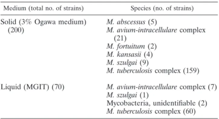
19. Oettinger, T., and A. B. Andersen. 1994. Cloning and B-cell-epitope mapping TABLE 3. List of mycobacteria isolated from clinical specimens
Medium (total no. of strains) Species (no. of strains) Solid (3% Ogawa medium)
(200) M. abscessus (5) M. avium-intracellulare complex (21) M. fortuitum (2) M. kansasii (4) M. szulgai (9) M. tuberculosis complex (159)
Liquid (MGIT) (70) M. avium-intracellulare complex (7) M. szulgai (1)
Mycobacteria, unidentifiable (2)
M. tuberculosis complex (60)
FIG. 1. Identification of the M. tuberculosis complex by the MPT64 ICA kit. Top, strong positive; middle, weak positive; bottom, negative.
of MPT64 from Mycobacterium tuberculosis H37Rv. Infect. Immun. 62:2058– 2064.
20. Purohit, M. R., T. Mustafa, H. G. Wiker, O. Morkve, and L. Sviland. 2007. Immunohistochemical diagnosis of abdominal and lymph node tuberculosis by detecting Mycobacterium tuberculosis complex specific antigen MPT64. Diagn. Pathol. 2:36.
21. Wagner, D., and L. S. Young. 2004. Nontuberculous mycobacterial infec-tions: a clinical review. Infection 32:257–270.
22. World Health Organization. 1996. Report of the tuberculosis epidemic. World Health Organization, Geneva, Switzerland.
23. Yamaguchi, R., K. Matsuo, A. Yamazaki, C. Abe, S. Nagai, K. Terasaka, and T. Yamada. 1989. Cloning and characterization of the gene for immunogenic protein MPB64 of Mycobacterium bovis BCG. Infect. Im-mun. 57:283–288.
24. Yim, J. J., and S. K. Han. 2005. Diagnosis and treatment of nontuberculous mycobacterial pulmonary diseases. J. Korean Med. Assoc. 48:563–570.