Disc tissue is adaptable to loading and this adaptation re-sults in consequent disc remodeling. It has been reported that articular disc and bony changes are related to various kinds of extracellular matrix and expression of certain enzymes1.
Matrix metalloproteinase (MMP) affects progress of TMJ disorders and also serves as a crucial indicator of early stage degenerative joint changes. It has been demonstrated that MMPs and their inhibitors are involved in both physiologi-cal and pathologiphysiologi-cal conditions in rabbit study2. In human
studies, MMP-1, 2, 3, 9, and 13 have been detected in the sy-novial fluid of osteoarthritis (OA) patients3-6, and expression
and concentration of MMP-2, 3, and 9 increased in synovial fluid of patients with internal derangement (ID) or OA4,7.
Therefore, it can be suggested that upregulation of MMP-1, 2, 3, 8, 9, and 13 in the synovial fluid from TMJ ID or in OA patients means pathological degradation and destruction of TMJ tissue8.
The MMP family contains the only mammalian proteinases
I. Introduction
Discrepancies between clinical symptoms, radiographic findings, and operative findings for temporomandibular joint (TMJ) disorders sometimes confuse clinicians. For this rea-son, many studies have tried to discover markers of TMJ dis-orders with resected tissue or molecules in synovial fluid and cartilage to make accurate diagnoses.
Jong-Ki Huh
Department of Oral and Maxillofacial Surgery, Gangnam Severance Hospital, Yonsei University College of Dentistry, 211 Eonju-ro, Gangnam-gu, Seoul 06273, Korea
TEL: +82-2-2019-4560 FAX: +82-2-3463-4052 E-mail: omshuh@yuhs.ac
ORCID: https://orcid.org/0000-0002-7381-3972
This is an open-access article distributed under the terms of the Creative Commons Attribution Non-Commercial License (http://creativecommons.org/ licenses/by-nc/4.0/), which permits unrestricted non-commercial use, distribution, and reproduction in any medium, provided the original work is properly cited.
CC
Expression of collagenases (matrix metalloproteinase-1, 8, 13)
and tissue inhibitor of metalloproteinase-1 of retrodiscal tissue in
temporomandibular joint disorder patients
Won Gyung Gho1, Yuri Choi2, Kwang-Ho Park2, Jong-Ki Huh2
1Department of Dentistry, Myongji Hospital, Goyang,
2Department of Oral and Maxillofacial Surgery, Yonsei University College of Dentistry, Seoul, Korea
Abstract(J Korean Assoc Oral Maxillofac Surg 2018;44:120-127)
Objectives: The aim of this study was to reveal how collagenases (matrix metalloproteinase [MMP]-1, 8, 13) and tissue inhibitor of metalloproteinase 1 (TIMP-1) are expressed in immunohistochemistry of retrodiscal tissue in temporomandibular joint disorder patients.
Materials and Methods: This study was conducted on 39 patients who underwent discoplasty or discectomy. Immunohistochemical staining was undertaken and expression levels of MMP-1, 8, 13, and TIMP-1 were evaluated. The status of internal derangement of disc, osteoarthritis, and joint ef-fusion were analyzed using magnetic resonance imaging (MRI). Disc status observed during operation was also categorized.
Results: The more severe disc derangement was observed on MRI, the more increased expression of MMPs and TIMP-1 appeared. Regarding MMP-13 expression, 86.7% of late-stage disc displacement patients showed grade II or III. Expression level of MMPs or TIMP was not statistically signifi-cant associated with joint effusion level. In perforation and/or adhesion groups, all patients showed grade II or III expression of MMP-13. Once perfo-ration occurred, MMP-13 showed increased expression with statistical significance.
Conclusion: MMP-1 and MMP-13 expression seem to be related to progression of osteoarthritis whereas MMP-8 does not seem to have a specific role with regard to temporomandibular joint disorders. TIMP-1 is considered to be partly related to internal derangement rather than osteoarthritis, but it is not significant.
Key words: Collagenases, Matrix metalloproteinase, Tissue inhibitor of metalloproteinase, Temporomandibular joint disorders, Immunohistochemistry
[paper submitted 2017. 12. 20 / revised 2018. 2. 9 / accepted 2018. 2. 12]
Copyright © 2018 The Korean Association of Oral and Maxillofacial Surgeons. All rights reserved.
cam, Cambridge, UK) and MMP-8 antibody (EP1252Y; Ab-cam) were rabbit monoclonal to origins with synthetic pep-tide corresponding to residues in human MMP-1 or MMP-8. 13 antibody (Abcam) was a rabbit polyclonal to MMP-13 human recombinant MMP-MMP-13 protein. TIMP-1 antibody (102D1; Abcam) was a mouse monoclonal origin from which BALB/C mice injected with recombinant human TIMP-1.
After washing in TBS, sections were incubated with a biotinylated secondary antibody for ten minutes at room temperature and rinsed. Then, they were rewashed after ten minutes of incubation with streptavidin-peroxidase at room temperature. In the above process, Histostain-plus kit (Zymed, Camarillo, CA, USA) was used. Finally they were colorized by diaminobenzidine tetrahydrochloride (DAB), counter-stained with Meyer’s hematoxylin, and sealed with glycerol.
2) Evaluation of immunohistochemical staining
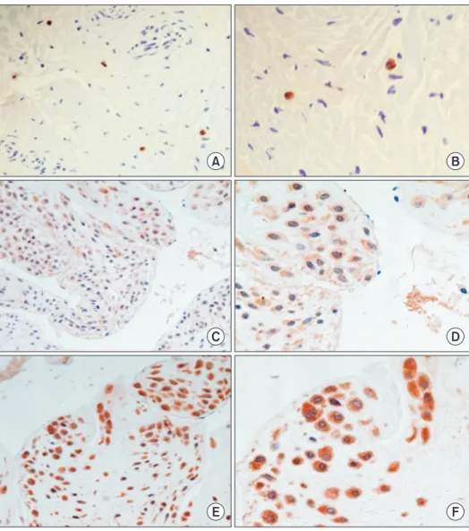
Staining patterns were classified into three grades according to expression levels of MMP-1, 8, 13 and TIMP-1. Those with diffuse expression less than 20 cells were classified as grade I; multiple group expression from 20 to 100 cells, grade II; and positive expression more than 100 cells, grade III.(Fig. 1)
3) Analysis of magnetic resonance imaging findings MRI findings for ID were classified into four groups—nor-mal position, disc displacement with reduction (DDcR), early stage of disc displacement without reduction (EDDsR), and late stage of disc displacement without reduction (LDDsR), based upon the position and status of joint disc on T-1 weighted MRI9.
(1) Normal: The disc is biconcave with the posterior band lying over the condyle, and the central thin zone is located between the condyle and the posterior part of the articular eminence.
(2) DDcR: When the jaw closes, the disc displaces ante-riorly, anterolaterally, or anteromedially. But when the jaw opens, the disc is reduced to normal position.
(3) EDDsR: The disc displaced relative to the condylar head, regardless of jaw position. Disc slightly thickens at its posterior edge or begins to be deformed anatomically. When the jaw closes, the disc displaces slightly forward. When the jaw opens, disc deformity is observed.
(4) LDDsR: The disc displacement is similar to EDDsR, and also significant anatomical disc deformity is seen such as spectacles shape or amorphous form. When the jaw opens, the disc follows anterior movement of the condyle without anatomical deformity.
known as ‘collagenases’. The ‘classical’ collagenases-1, 2, and 3 (MMP-1, 8, 13) are specific for certain collagens. Col-lagenases can affect matrix changes in the retrodiscal tissue which is composed of collagen type I and II. Meanwhile, activated MMPs are mainly regulated by tissue inhibitor of metalloproteinase (TIMP), classified as TIMP-1, 2, 3, and 4. Among TIMPs, TIMP-1 resists collagenase, stromelysin, and gelatinase.
The retrodiscal tissue from TMJ disorder patients who underwent TMJ surgery usually shows fibrous degeneration, congestion, adhesion, or perforation. But because of difficul-ties in subject selection, current studies are insufficient to find out extracellular matrix-related factors for these changes. Therefore in this study, MMP-1, 8, 13 and TIMP-1 expres-sion were investigated from retrodiscal tissue sections ob-tained during TMJ open surgery. To understand the relation-ship between expression of these enzymes and TMJ structural changes, immunohistochemical findings, magnetic resonance imaging (MRI), and operative findings were analyzed.
II. Materials and Methods
1. Materials
This study was conducted on 39 TMJs of 39 patients who had undergone discoplasty or discectomy. Patients were di-agnosed as ID or OA based upon clinical examination and MRI at the Department of Oral and Maxillofacial Surgery at Gangnam Severance Hospital (Seoul, Korea).
The wedge-shaped sample was obtained from the lateral side of retrodiscal tissue during discoplasty. During discec-tomy, tissue samples were obtained from both articular disc and retrodiscal tissue.
2. Methods
1) Immunohistochemistry
Each 5 µm-thick tissue section, which had been fixed in 10% neutral-buffered formalin and embedded in paraffin, was placed on a silanized slide. Sections were deparaffinized with xylene and soaked in ethanol. The sections were incubated in 3% hydrogen peroxide for 10 minutes to quench endogenous peroxidase activity and washed with Tris-buffered saline (TBS). After blocking the binding of nonspecific protein us-ing serum blockus-ing solution for ten minutes, sections were incubated with a primary antibody, diluted 1:50, for an hour at room temperature. Each MMP-1 antibody (EP1247Y;
Ab-teophytosis, resorption bone marrow signal change
• Temporal bone: generalized sclerosis and flattening, ir-regular bony surface
• Shortening in ramal length or no change in ramal length MRI findings for joint effusion were classified into level 0, 1, 2, or 3 based upon the amount of fluid collection or signal intensity on T-2 weighted MRI9.
(1) Level 0: No evidence of high signal intensity in the joint space
(2) Level 1: Fluid collection is seen in the boundary of tem-poral bone and disc or high signal intensity is visible within the folded disc
(3) Level 2: High signal intensity is visible in the anterior recess of upper or lower joint space. It extends outside the boundary of the disc
(4) Level 3: High signal intensity is seen in the whole re-gion of the upper or lower joint space
The diagnosis of degenerative joint depended on whether a patient has OA which includes erosion of mandibular condyle or condylar fossa, sclerosis, or absorption of cortical bone and signal change of bone marrow. The diagnostic criteria of OA on MRI are as follows:
(1) Normal bony structure
• No abnormal cortical bony changes such as flattening, sclerosis, spurring, osteophytosis and erosion in the condylar head and temporal part of joint.
• No abnormal bone marrow signal change of the condyle (2) Mild stage of OA
• Condylar head: localized cortical erosion or sclerosis, bony spurring without evidence of bone marrow signal change
• Temporal bone: localized cortical erosion or sclerosis • No change in ramal length
(3) Severe stage of OA
• Condylar head: generalized flattening and sclerosis,
os-Fig. 1. Immunohistochemistry. A, B.
Grade I expression, with extremely small portions of retrodiscal tissue re-moved after discoplasty was colorized (A: ×200, B: ×400). C, D. Grade II ex-pression, with higher numbers of posi-tive tissue cells and clearer expression than grade I (C: ×200, D: ×400). E, F. Grade III expression, with even expres-sion in cytoplasm and broad staining (E: ×200, F: ×400).
Won Gyung Gho et al: Expression of collagenases (matrix metalloproteinase-1, 8, 13) and tissue inhibitor of metalloproteinase-1 of retrodiscal tissue in temporomandibular joint disorder patients. J Korean Assoc Oral Maxillofac Surg 2018
A B
C D
4) Analysis of operative findings
The patients were categorized into four groups. The first group showed no signs of adhesion or perforation. The sec-ond group showed partial or entire adhesion of the articular disc or the retrodiscal tissue to the condylar eminence or fossa. The third group showed perforation with upper joint cavity connected to the lower joint cavity. The fourth group showed both adhesion and perforation.
5) Statistical analysis
Data analysis was performed by chi-square test and Fish-er’s exact test of cross tabulation analysis using IBM SPSS Statistics ver. 12.0 for Windows (SPSS Inc., Chicago, IL, USA).
This study was conducted with approval from the Institu-tional Review Board at Yonsei University Gangnam Sever-ance Hospital (IRB no. 3-2015-0076) and in compliSever-ance with the Declaration of Helsinki.
III. Results
Three males (7.7%) and 36 females (92.3%) were included in the present study with the average of age 33 years (range, 21-70 years). Twenty-eight patients underwent discoplasty while the other 11 underwent discectomy. The expression levels of MMPs and TIMP-1 are shown in Table 1. MMP-1 and MMP-8 were expressed in all cases, and 64% of cases showed grade I expression. MMP-13 was expressed in all cases; however, 44% and 41% of cases showed grade II and III expression.
1. Comparison with MRI findings
Normal positioned disc was absent in this study and 2, 7, and 30 joints were diagnosed as DDcR, EDDsR, and LDDsR respectively based on MRI. In MMP-13 expression, 86.7% of the LDDsR group showed grade II or III. With more severe disc derangement status, more frequent MMPs and TIMP-1 expression was observed.(Table 1)
With regard to OA, 5, 28, and 6 joints were categorized as normal, mild, and severe stage respectively. Interestingly, in mild and severe OA stages MMP-13 grade II and III showed higher percentages. But among mild stage patients, grade I were dominant in other MMPs and TIMP-1.(Table 1)
In joint effusion, 11 cases were level 0, 16 cases were level 1, and 10 cases were level 2. Level 3 was not found in this research. There was not statistical significance in comparison Table 1.
MMPs & TIMP-1 expr
ession levels of r
etr
odiscal tissue accor
ding to MRI and operative findings
Grade
MRI finding of disc status (n=39)
MRI finding of osteoarthritis (n=39)
MRI finding of joint effusion (n=39)
Operative finding of disc status (n=39)
No. of patients (%) DDcR EDDsR LDDsR Normal Mild Severe Level 0 Level 1 Level 2 Normal Perforation
Adhesion & perforation
MMP-1 MMP-8 MMP-13 TIMP-1 No. of patients
I II III I II III I II III I II III
1 (4.0) 0 1 (25.0) 1 (4.0) 1 (9.1) 0 0 1 (5.9) 1 (6.3) 0 2 (10.5) 0 2 7 (28.0) 0 0 6 (24.0) 1 (9.1) 0 2 (33.3) 5 (29.4) 0 4 (25.0) 3 (15.8) 0 7 17 (68.0) 10 (100) 3 (75.0)* 18 (72.0) 9 (81.8) 3 (100) 4 (66.7) 11 (64.7) 15 (93.7) 12 (75.0) 14 (73.7) 4 (100) 30 5 (20.0) 0 0 5 (20.0) 0 0 1 (16.7) 4 (23.5) 0 3 (18.8) 2 (10.5) 0 5 19 (76.0) 8 (80.0) 1 (25.0) 18 (72.0) 9 (81.8) 1 (33.3) 5 (83.3) 12 (70.6) 11 (68.8) 13 (81.3) 12 (63.2) 3 (75.0) 28 1 (4.0) 2 (20.0) 3 (75.0)* 2 (8.0) 2 (18.2) 2 (66.7) 0 1 (5.9) 5 (31.3) 0 5 (26.3) 1 (25.0) 6 5 (20.0) 4 (40.0) 2 (50.0) 5 (20.0) 5 (45.5) 1 (33.3) 0 5 (29.4) 6 (37.5) 4 (25.0) 6 (31.6) 1 (25.0) 11 13 (52.0) 3 (30.0) 0 12 (48.0) 3 (27.3) 1 (33.3) 5 (83.3) 6 (35.3) 5 (31.3) 8 (50.0) 7 (36.8) 1 (25.0) 16 7 (28.0) 3 (30.0) 2 (50.0) 8 (32.0) 3 (27.3) 1 (33.3) 1 (16.7) 6 (35.3) 5 (31.3) 4 (25.0) 6 (31.6) 2 (50.0) 12 11 (44.0) 0 2 (50.0) 7 (28.0) 5 (45.5) 1 (33.3) 4 (66.7) 7 (41.2) 2 (12.5) 6 (37.5) 4 (21.1) 3 (75.0) 13 8 (32.0) 7 (70.0) 1 (25.0) 11 (44.0) 3 (27.3) 2 (66.7) 2 (33.3) 5 (29.4) 9 (56.3) 5 (31.3) 10 (52.6) 1 (25.0) 16 6 (24.0) 3 (30.0) 1 (25.0) 7 (28.0) 3 (27.3) 0 0 5 (29.4) 5 (31.3) 5 (31.3) 5 (26.3) 0 10 25 (64.1) 10 (25.6) 4 (10.3) 25 (64.1) 11 (28.2) 3 (7.7) 6 (15.4) 17 (43.6) 16 (41.0) 16 (41.0) 19 (48.7) 4 (10.3) (MMP: matrix met alloproteinase,
TIMP: tissue inhibitor of metalloproteinase,
MRI: magnetic
resonance imaging, DDcR: disc displacement
with reduction, EDDsR: early stage of disc displacement
without reduction, LDDsR: late stage of disc displacement without reduction) *P<0.05, level I & II versus level III were compared. Values are presented as number (%) or number only. Won Gyung Gho et al: Expr
ession of collagenases (matrix metallopr
oteinase-1, 8, 13) and tissue inhibitor of metallopr
oteinase-1 of r
etr
odiscal tissue in tempor
omandibular joint disor
der patients. J Kor
ean
Assoc Oral Maxillofac Sur
and the state of TMJ such as ID, OA, adhesion or perforation of disc.
Collagena1 (MMP-1) is produced and immediately se-creted from some mesenchymal cells in response to specific inducers12,13. In contrast, collagenase-2 (MMP-8) is
primar-ily produced by neutrophils during their maturation in bone marrow, and it is then stored until the cells are stimulated to degranulate12,13. Collagenase-3 (MMP-13) is found in bone,
normal and pathologic cartilage, and various epithelial can-cers, and it is the predominant collagenase in mice and rats while collagenase-1 is undetectable in these species13-16.
Al-though each mammalian collagenase can degrade all fibrillar collagens, the preferred substrates for collagenases 1, 2 and 3 are collagens type III, I and II respectively17,18. Gepstein et
al.19 reported it has been established that although individual
enzymes have similar substrate specificities, their expression pattern is often distinct and characteristic in a certain tissue and cell type. In this study, all cases showed MMP-1, 8, 13 and TIMP-1 expression but the range or cell type of expres-sion were different. MMP-13, so-called ‘osteoblast collage-nase’, demonstrated high expression grade generally. With more expression of MMP-13, there was more degenerative ID or OA extent. This could verify the importance of collagen type II in retrodiscal tissue as well as articular cartilage of TMJ. As specificity of collagenases, MMP-1 called ‘fibroblast collagenase’ was expressed on fibroblast and MMP-8 called ‘neutrophil collagenase’ was expressed on neutrophils. The expression site and level of each collagenase showed diver-sity.
Some conflicting results could be explained partially by differences in the sensitivity of the assays used for detect-ing MMPs and TIMPs such as immunohistochemistry, in situ hybridization, zymography, western blots, and northern blots. One study reported that different results can be related to detected location of MMPs that isolated chondrocytes, sy-novial fluids, cultured explants, or intact cartilages20. Huh et
al.21 reported that they found MMP-1 mRNA from seven out
of eight joints with OA and suggested that MMP-1 from ret-rodiscal tissue can play a role in destruction of cartilage and bone structure in TMJ. Another study showed that MMP-1 is active in synovial fluid of patients with pain in most cases and that a high concentration of MMP-1 can increase joint inflammation22. As mentioned above, the role of MMP-1 is
not yet clear; however, it can be suggested that MMP-1 is in-volved in disc displacement or other related diseases because collagen is a major component of the retrodiscal tissue. In this study, 5/6 cases in the severe OA group expressed grade between expression of MMPs or TIMP and joint effusion
level.(Table 1)
2. Comparison with operative findings
In 13 cases, adhesion and perforation was not found. In 17 cases, only perforation was observed and in 10 cases both ad-hesion and perforation were observed. There were no cases of only adhesion in this research. In perforation and/or adhesion groups, all patients showed grade II or III MMP-13 expres-sion. Once perforation occurred, expression of MMP-13 was increased with statistical significance.(Table 1)
IV. Discussion
MMP is an endogenous proteinase whose activity depends on Zn2. It plays an important role in degeneration of all kinds
of collagen, proteoglycan, extracellular matrix, and particu-larly cartilage. It also contributes to breakdown of basement membrane and metabolism of normal matrix tissue10. MMPs
are classified by their function into five groups including in-terstitial collagenases (MMP-1, 8, 13), gelatinases (MMP-2, 9), stromelysins (MMP-3, 10), membrane-type MMPs, and other MMPs.
Activated MMPs can be inhibited by a common proteinase inhibitor such as α2-macroglobulin, but are mainly inhibited
by TIMPs as the chief endogenous MMP regulator, classified as TIMP-1, 2, 3, and 4. Among them, TIMP-1 is produced by most connective tissue cells and macrophages, and it resists against collagenase, stromelysin, and gelatinase.
The combination of extracellular matrix, including colla-gen, relieves pressure and tensile force put on joints. Howev-er, once these forces start to hurt articular cells, various kinds of degenerative enzymes are secreted and degrade connective tissue. Thus, to detecting TMJ disorders, some studies on synovial fluid have been conducted with the hypothesis that damaged extracellular matrix and degenerative enzymes will be released into the synovial fluid in TMJ disorder patients. These studies already have suggested involvement of MMPs in TMJ disease by increased MMP-13 levels in synovial fluid from patients with ID11. Since most human studies have
been carried out using synovial fluid, the production site of these enzymes has never been investigated in degenerated disc tissue. The purpose of this study was to identify MMPs and TIMP-1 immunoreactive patterns with retrodiscal tissue samples obtained during surgery. This study also evaluated the association between these enzymes from retrodiscal tissue
been reported that there is a correlation between the concen-tration of TIMP-1 and MMP-1, and the imbalance between them in the progression of OA results in unrecoverable deg-radation of cartilage matrix26. During OA, the production of
TIMP-1 is correlated to the level of MMPs. Their imbalance in the progression of OA may lead to the destruction of car-tilage27. Whereas overexpression of collagenage-1, coupled
with decreased production of TIMP-1, may impair healing, insufficient proteinase activity may lead to accumulation of wound-associated tissue and delays in wound closure. Thus, the properly regulated, site-specific expression of col-lagenase, as well as other MMPs, may be needed to promote efficient wound repair13. This study shows lower expression
of TIMP-1 than MMPs, because it might be that the experi-mental groups were patients with TMJ disorder. Four cases of grade III expression of TIMP-1 was applied to LDDsR and mild or severe stage of OA. TIMP-1 expression seemed to affect ID in comparison with OA results, but it was not significant. Nevertheless our study assumes that TIMP-1 can be active if MMP expression pattern fits into grade II or III without any change or destruction in joint structures.
The imbalance between MMP-1 and TIMP-1 also plays an important role in the degradation of extracellular matrix28.
This imbalance and reduced expression of TIMP-1, which suppresses the degradation of extracellular matrix, can ex-plain aggressive patterns against bone or cartilage. Follow-up surveys on OA-related symptoms among TMJ disorder, in-cluding interactions of MMP-1, 13 and TIMP-1, are required for clinical applications.
This study was aimed to observe how collagenases (MMP-1, 8, 13) and TIMP-1 are expressed in immunohistochemistry of retrodiscal tissue sections in order to understand how their expression pattern is associated with ID, OA, joint effusion, and adhesion or perforation among TMJ disorder patients.
V. Conclusion
In conclusion, MMP-13 plays a critical role in degrading of OA as it is the predominant collagenase in collagen type II and can worsen ID as the collage breakdown enzyme of retrodiscal tissue in TMJ. MMP-1 is related to degeneration of OA. MMP-8 does not characterize specificity with regard to TMJ disorder. TIMP-1 seems to affect ID in comparison with OA expression, but it is not significant. In this study, the causal relation of the imbalance between MMP and TIMP could not be explained, but TIMP activity, which shows a difference with degeneration of TMJ disorders, suggests that II and III 1 expression and it also suggested that
MMP-1 is associated with destruction of bone and cartilage of TMJ even with the small experimental group. But the character of MMP-8 expression was unclear in comparison with ID, OA, and surgical findings.
Among MMPs, MMP-13 (collagenase-3) appears to play a key role in joint connective tissue remodeling. MMP-13 can cleave type II collagen 10 times faster than MMP-1. MMP-13 expression has also been demonstrated in articular cartilage in OA patients and in synovium of rheumatoid arthritis patients. In this study, control disc cells were rarely immunopositive for MMP-13, although a few scattered fibroblast-like cells showed faint positive immunoreactions. MMP-13 was upreg-ulated in TMJ disc tissue from IDs patients. One study also reported that MMP-13 immunolabelling increased with TMJ disc degeneration. Thus MMP-13 has been thought to play an active role in the synthesis of ECM-degrading proteinases23.
In this study, with more MMP-13 in retrodiscal tissue, there was more degenerative ID or OA. The particularly severe OA group showed clear expression as grade II or III and this can explain that MMP-13 as well as MMP-1 is related with OA of TMJ. In comparison of MMP-13 expression with the posi-tion and status of articular disc, 86.7% of a group of LDDsR showed grade II or III, which means that breakdown enzymes involved in articular disc degeneration become more active as ID progresses. The case of grade III expression with DDsR in MRI was an OA patient who went through discectomy due to severe adhesion and perforation of articular disc.
Marchetti et al.’s study24 demonstrated that structural
modi-fications of articular disc could be specific responses to func-tional change of TMJ and variations in extrinsic stimuli may activate intrinsic factors, such as MMPs, that induce struc-tural modifications in discal tissue. The examination found that only the areas with myxoid change or hyalinization were stained or these areas resulted in much more staining than others, and that cell numbers increased in areas with myxoid change. Such differences appeared when the retrodiscal tissue deteriorated or responded to passed-on loading caused by ar-ticular disc displacement, which suggests an association with MMPs.
In normal physiological processes, MMP activity is con-trolled at several levels. Loss of control of MMP activity appears to have serious consequences, and aberrations in MMP expression have been associated with several diseases. Though there is no general awareness of the expression of TIMP-1 in osteoarthritic cartilage and synovium, it is thought that TIMP-1 correlates with the production of MMPs25. It has
TIMP should be considered in potential criteria for preven-tion of progressive disease via controlling collagenases.
ORCID
Won Gyung Gho, https://orcid.org/0000-0001-5980-5602 Yuri Choi, https://orcid.org/0000-0002-6018-496X Kwang-Ho Park, https://orcid.org/0000-0003-1942-2986 Jong-Ki Huh, https://orcid.org/0000-0002-7381-3972
Authors’ Contributions
W.G.G. participated in data collection and wrote the manu-script. Y.C. participated in the study design and performed the statistical analysis. K.H.P. and J.K.H. participated in the study design and coordination and helped to draft the manuscript. All authors read and approved the final manuscript.
Acknowledgements
This study was supported by a faculty research grant from the Yonsei University College of Dentistry (6-2007-0069).
Ethics Approval and Consent to Participate
This study was conducted with approval from the Institu-tional Review Board at Yonsei University Gangnam Sever-ance Hospital (IRB no. 3-2015-0076) and in compliSever-ance with the Declaration of Helsinki.
Conflict of Interest
No potential conflict of interest relevant to this article was reported.
References
1. Ishibashi H, Takenoshita Y, Ishibashi K, Oka M. Expression of extracellular matrix in human mandibular condyle. Oral Surg Oral Med Oral Pathol Oral Radiol Endod 1996;81:402-14.
2. Kapila S, Lee C, Richards DW. Characterization and identification of proteinases and proteinase inhibitors synthesized by temporo-mandibular joint disc cells. J Dent Res 1995;74:1328-36.
3. Väätäinen U, Lohmander LS, Thonar E, Hongisto T, Agren U, Rönkkö S, et al. Markers of cartilage and synovial metabolism in joint fluid and serum of patients with chondromalacia of the pa-tella. Osteoarthritis Cartilage 1998;6:115-24.
4. Kubota E, Kubota T, Matsumoto J, Shibata T, Murakami KI. Sy-novial fluid cytokines and proteinases as markers of temporoman-dibular joint disease. J Oral Maxillofac Surg 1998;56:192-8.
5. Imai K, Ohta S, Matsumoto T, Fujimoto N, Sato H, Seiki M, et al. Expression of membrane-type 1 matrix metalloproteinase and ac-tivation of progelatinase A in human osteoarthritic cartilage. Am J Pathol 1997;151:245-56.
6. Imai S, Konttinen YT, Jumppanen M, Lindy O, Ceponis A, Kemp-pinen P, et al. High levels of expression of collagenase-3 (MMP-13) in pathological conditions associated with a foreign-body reaction. J Bone Joint Surg Br 1998;80:701-10.
7. Kubota E, Imamura H, Kubota T, Shibata T, Murakami K. Inter-leukin 1 beta and stromelysin (MMP3) activity of synovial fluid as possible markers of osteoarthritis in the temporomandibular joint. J Oral Maxillofac Surg 1997;55:20-7; discussion 27-8.
8. Tiilikainen P, Pirttiniemi P, Kainulainen T, Pernu H, Raustia A. MMP-3 and -8 expression is found in the condylar surface of tem-poromandibular joints with internal derangement. J Oral Pathol Med 2005;34:39-45.
9. Huh JK, Kim HG, Ko JY. Magnetic resonance imaging of temporo-mandibular joint synovial fluid collection and disk morphology. Oral Surg Oral Med Oral Pathol Oral Radiol Endod 2003;95:665-71.
10. McDonnell S, Morgan M, Lynch C. Role of matrix metallopro-teinases in normal and disease processes. Biochem Soc Trans 1999;27:734-40.
11. Mizui T, Ishimaru J, Miyamoto K, Kurita K. Matrix metallopro-teinase-2 in synovial lavage fluid of patients with disorders of the temporomandibular joint. Br J Oral Maxillofac Surg 2001;39:310-4.
12. Dioszegi M, Cannon P, Van Wart HE. Vertebrate collagenases. Methods Enzymol 1995;248:413-31.
13. Jeffrey JJ. Interstitial collagenases. In: Parks WC, Mecham RP, eds. Matrix metalloproteinases. San Diego: Academic Press; 1998. 14. Freije JM, Díez-Itza I, Balbín M, Sánchez LM, Blasco R, Tolivia J,
et al. Molecular cloning and expression of collagenase-3, a novel human matrix metalloproteinase produced by breast carcinomas. J Biol Chem 1994;269:16766-73.
15. Reboul P, Pelletier JP, Tardif G, Cloutier JM, Martel-Pelletier J. The new collagenase, collagenase-3, is expressed and synthesized by human chondrocytes but not by synoviocytes. A role in osteoar-thritis. J Clin Invest 1996;97:2011-9.
16. Mitchell PG, Magna HA, Reeves LM, Lopresti-Morrow LL, Yocum SA, Rosner PJ, et al. Cloning, expression, and type II col-lagenolytic activity of matrix metalloproteinase-13 from human osteoarthritic cartilage. J Clin Invest 1996;97:761-8.
17. Hasty KA, Jeffrey JJ, Hibbs MS, Welgus HG. The collagen sub-strate specificity of human neutrophil collagenase. J Biol Chem 1987;262:10048-52.
18. Welgus HG, Jeffrey JJ, Eisen AZ. The collagen substrate specificity of human skin fibroblast collagenase. J Biol Chem 1981;256:9511-5.
19. Gepstein A, Shapiro S, Arbel G, Lahat N, Livne E. Expression of matrix metalloproteinases in articular cartilage of temporomandib-ular and knee joints of mice during growth, maturation, and aging. Arthritis Rheum 2002;46:3240-50.
20. Gepstein A, Arbel G, Blumenfeld I, Peled M, Livne E. Association of metalloproteinases, tissue inhibitors of matrix metalloprotein-ases, and proteoglycans with development, aging, and osteoarthritis processes in mouse temporomandibular joint. Histochem Cell Biol 2003;120:23-32.
21. Huh JK, Park KK, Choi MA, Kim HG. Expression of matrix metalloproteinase-1 and -2 mRNA in retrodiscal tissue of the temporomandibular joint. J Korean Assoc Oral Maxillofac Surg 2003;29:212-8.
22. Ishimaru JI, Oguma Y, Goss AN. Matrix metalloproteinase and tissue inhibitor of metalloproteinase in serum and lavage synovial fluid of patients with temporomandibular joint disorders. Br J Oral Maxillofac Surg 2000;38:354-9.
Musumeci G, et al. MMP-13 (collagenase 3) localization in human temporomandibular joint discs with internal derangement. Acta Histochem 2008;110:314-8.
24. Marchetti C, Cornaglia I, Casasco A, Bernasconi G, Baciliero U, Stetler-Stevenson WG. Immunolocalization of gelatinase-A (matrix metalloproteinase-2) in damaged human temporomandibular joint discs. Arch Oral Biol 1999;44:297-304.
25. Gomis-Rüth FX, Maskos K, Betz M, Bergner A, Huber R, Suzuki K, et al. Mechanism of inhibition of the human matrix metallopro-teinase stromelysin-1 by TIMP-1. Nature 1997;389:77-81. 26. Naito K, Takahashi M, Kushida K, Suzuki M, Ohishi T, Miura
M, et al. Measurement of matrix metalloproteinases (MMPs) and tissue inhibitor of metalloproteinases-1 (TIMP-1) in patients with knee osteoarthritis: comparison with generalized osteoarthritis. Rheumatology (Oxford) 1999;38:510-5.
27. Wang YL, Li XJ, Qin RF, Lei DL, Liu YP, Wu GY, et al. Matrix metalloproteinase and its inhibitor in temporomandibular joint os-teoarthrosis after indirect trauma in young goats. Br J Oral Maxil-lofac Surg 2008;46:192-7.
28. Sawatsubashi M, Mizokami H, Tokunaga O, Shin T. Expression of MMP-1, TIMP-1, and type I collagen in laryngeal carcinoma. Mod Pathol 1998;11:878-85.