Regulation of invasion and migration
by PKCK2 in Helicobacter pylori
infected gastric cancer cells
Yeo Song Lee
Department of Medical Science
Regulation of invasion and migration
by PKCK2 in Helicobacter pylori
infected gastric cancer cells
Directed by Professor Yong Chan Lee
The Doctoral Dissertation
submitted to the Department of Medical Science,
the Graduate School of Yonsei University
in Partial Fulfillment of the Requirements for the
Degree of Doctor of Philosophy
Yeo Song Lee
감사의 글
학위를 마치며 감사의 마음을 전하고자 합니다. 먼저 학위 과정 동안 제가 할 수 있는 이 상의 것을 도출하게끔 지원해주시고, 기대에 부응치 못 했을 때도 조용히 기다려 주신 이용 찬 지도교수님께 무한한 감사의 마음 전합니다. 바쁘신 와중에도 논문을 꼼꼼히 확인 해 주 시고 지도와 격려를 아낌없이 해주신 김건홍 교수님, 라선영 교수님, 형우진 교수님과 늘 가까이서 냉철한 조언을 주신 이상길 교수님께도 깊은 감사 올립니다. 또한 미국연수 동안 물심양면으로 도와주시고, 과학자로서 한 층 더 성장하게 해 주신 남기택 교수님께도 진심 으로 감사 드립니다. 8년동안 항상 친언니같이 저를 위해 바쁜 시간을 비워 상담해 주고 위로해 주던 사랑하는 양은정 선생님과 늘 실험적 필요를 채워 주었던 서진원 선배님에게도 감사와 기쁨을 나누고 싶습니다. 실험실에서 기쁨과 고충을 함께 나눈 이도연 박사님과 실험실의 분위기 메이커 김신 학생, 유다연 학생, 짧은 시간이었지만 여러모로 내게 힘이 되어 준 김가희 학생과 곁 에서 웃음과 간식을 함께 나눴던 이나금 선생님, 이정화 선생님께도 감사의 마음을 전하며, 앞으로 훌륭한 결과를 내길 바랍니다. 늘 내 편이 되어주었던 둘도 없는 친구 신순주, 남민 선과 끝까지 옆에서 응원해 준 경미 언니, 자주 못 봐 보고 싶은 우미, 갈수록 예뻐지는 영 미에게도 한없는 사랑과 고마운 마음을 전하며 늘 건강과 행운이 가득하길 기도합니다. 끝이 보이지 않는 학위의 기나긴 터널을 기도로 응원해 주신 사랑하고 존경하는 부모님과 며느리의 부족함을 사랑으로 감싸고 기다려 주신 사랑하는 시부모님께 글로는 표현할 수 없 는 무한한 감사의 마음 올립니다. 또한 누나의 부족함을 200백배 채워주는 믿음직한 동생 이석재와 늘 상냥한 웃음으로 마음을 즐겁고 편안하게 해주신 최인영 형님에게 감사의 마음 전합니다. 마지막으로 늘 옆에서 아낌없는 사랑과 위로로 수많은 어려움을 이길 수 있도록 지지해준 하나뿐인 평생의 동반자, 남편 최새롬에게 감사와 사랑을 전합니다. 학위과정의 처음과 마지막이 되신 하나님께 모든 영광 돌리며, 지면을 통해 일일이 언급하 지 못한 저를 아껴주고 사랑해 주신 모든 분들께 다시 한 번 마음을 다해 감사 드립니다.TABLE OF CONTENTS
ABSTRACT ... 1
I. INTRODUCTION ... 3
II. MATERIALS AND METHODS ... 5
1. Bacteria, cell culture, and H. pylori infection ... 5
2. Western blot analysis ... 5
3. siRNA and plasmid DNA transfection ... 6
4. Immunoprecipitation ... 6
5. In vitro PKCK2 kinase assay ... 7
6. Quantitative reverse transcription-polymerase chain reaction (qRT-PCR) ... 7
7. CK2 stable knockdown using lentiviral short hairpin RNA against PKCK2α
catalytic subunit ... 7
8. Cell migration assay ... 8
9. Matrigel invasion assay ... 8
10. Immunofluorescence microscopy ... 8
11. Enzyme-linked immunosorbent assay (ELISA) ... 9
12. Immunohistochemical staining ... 9
13. Statistical analysis ... 10
III. RESULTS ... 11
1.
H. pylori induces the invasion and migration in gastric cancer cells through
PKCK2 activation ... 11
2. H. pylori up-regulates PKCK2 activity without affecting PKCK2α protein
expression levels in gastric cancer cells... 14
3. Phosphorylation of α-catenin by PKCK2 results in dissociation of the α/β-catenin
complex in H. pylori infected gastric cancer cells ... 17
4. Expression of PKCK2 subunits and junctional proteins in an in vivo gastric
cancer tissue model ... 21
5. H. pylori induced α/β-catenin dissociation resulted in β-catenin nuclear
translocation and increased MMP7 expressions ... 24
6. H. pylori induces PKCK2β degradation through PI3K/Akt pathway ... 27
IV. DISCUSSION ... 29
V. CONCLUSION ... 32
REFERENCES ... 33
LIST OF FIGURES
Figure 1. H. pylori induces the invasion and migration in gastric cancer
cells through PKCK2 activation ... 12
Figure 2. H. pylori upregulates PKCK2 activity without affecting PKCK2α
protein expression levels in gastric cancer cells ... 15
Figure 3. PKCK2-mediated phosphorylation of α-catenin results in
dissociation of the α/β-catenin complex in H. pylori-infected gastric cancer
cells ... 19
Figure 4. Immunohistochemical detection of PKCK2α, PKCK2β, α-catenin,
β-catenin and E-cadherin expression in H. pylori non-infected and infected
gastric cancer tissues ... 22
Figure 5. H. pylori promotes β-catenin/TCF promoter activity and
downstream MMP7 expression by increasing PKCK2 activity ... 25
Figure 6. H. pylori induced PKCK2β degradation through PI3K/Akt
1
ABSTRACT
Regulation of invasion and migration
by PKCK2 in Helicobacter pylori infected gastric cancer cells
Yeo Song Lee
Department of Medical Science
The Graduate School, Yonsei University
(Directed by Professor Yong Chan Lee)
Chronic infection with Helicobacter pylori (H. pylori) is causally linked with gastric inflammation and carcinogenesis. Virulent H. pylori strains harbor cag pathogenicity island (PAI) for delivery of the bacterial CagA into gastric epithelial cells. Induction of high motility and an elongated phenotype is considered to be CagA-dependent process. Epithelial-mesenchymal transition (EMT) is a complex cellular program involved in both development and cancer, and the induction of cell migration and invasion is the hallmark of the EMT. Protein kinase casein kinase 2 (PKCK2) plays a critical role in carcinogenesis through signaling pathways related to the epithelial mesenchymal transition (EMT). This study was aimed to investigate the effect of H. pylori infection on the PKCK2 mediated migration and invasion in gastric cancer cells. In in-vivo results, PKCK2α immunostaining revealed strong expression in
2
pylori-infected gastric cancer tissues. In in vitro data, H. pylori infection increases host cell
PKCK2 activity and decreases PKCK2β expression. Inhibition of PKCK2 with the chemical inhibitor 4,5,6,7-tetrabromo-2-azabenzimidazole (TBB), siRNA or shRNA significantly decreased both invasion / migration and dissociation of the membranous α/β-catenin complex in
H. pylori infected gastric cancer cells. These results suggest that H. pylori induces
PKCK2-mediated cell migration and invasion through α/β-catenin dissociation in gastric cancer cells.
3
Regulation of invasion and migration
by PKCK2 in Helicobacter pylori infected gastric cancer cells
Yeo Song Lee
Department of Medical Science
The Graduate School, Yonsei University
(Directed by Professor Yong Chan Lee)
I. INTRODUCTION
Helicobacter pylori (H. pylori) is a gram-negative, microaerophilic bacterium that selectively
colonizes the human stomach. Virulent H. pylori strains harbor a cag pathogenicity island (PAI) for the translocation of the bacterial oncoprotein, CagA into gastric epithelial cells 1-3. Induction of high motility and an elongated phenotype has been considered hallmark of cag PAI-dependent process 4. However, the pathogenic molecular mechanism which underlies the carcinogenesis and metastasis in gastric cancer by H. pylori remains largely unknown. The epithelial-mesenchymal transition (EMT) is a complex cellular program associated with induction of cell migration and invasion which plays critical roles in the development of H.
pylori induced gastric carcinogenesis. Protein kinase casein kinase 2 (PKCK2) is a
serine/threonine protein kinase 5, 6 that plays a key role in cell cycle control, cellular differentiation, transformation and tumorigenesis 7. PKCK2 can function as a monomer or a tetramer, composed of two catalytic subunits, CK2α and/or CK2α’, and two CK2β regulatory subunits 8. Although PKCK2 is traditionally considered a constitutively active kinase, studies
4
have shown that PKCK2 is activated in response to a diverse array of growth-factor stimuli, including EGF treatment 9, 10. PKCK2 phosphorylates various intracellular molecules involved in tumor invasion, progression, or metastasis in multiple cancers, including cancers of the stomach, breast, lung, prostate, liver, and colon11-13. Additionally, a selective inhibitor of PKCK2, 4,5,6,7-tetrabromo-2-azabenzimidazole (TBB), significantly inhibits invasion of human tongue cancer cells and lung adenocarcinoma cells 14, 15. Although aberrant expression of PKCK2 is known to be involved in many cancers, the mechanism by which PKCK2 promotes tumorigenesis remains obscure.
Adherence junctions (AJs) are a network of membrane proteins and associated molecules, including E-cadherin and catenins. AJs are essential for establishing the polarity of epithelial cells and maintaining the integrity of the epithelial layer 16, 17. The cytoplasmic domain of E-cadherin binds to the β-catenin, which in turn binds α-catenin 18-20
. Deleterious mutations in adhesion molecules correlate with tumor metastasis and invasion. Although it is obvious from several reports that cell elongation and migration are strongly enhanced by CagA 21-23, the precise role of CagA in the disruption of cellular AJs remains controversial and multiple mechanisms maybe involved in the disruption of cell adhesion during H. pylori infection. Phosphorylation of α-catenin at serine 641 by PKCK2 in response to EGF-ERK activation facilitates disruption of binding between α-catenin and β-catenin and intercellular adhesion, as well as promoting tumor cell migration 24. In this study, we report that CagA+ H. pylori strongly activates PKCK2 without affecting the endogenous level of CK2α protein. Furthermore, we show that CagA+ H. pylori induces PKCK2-mediated migration of gastric cancer cells by disrupting the membrane-bound α/β-catenin complex, resulting in loss of the E-cadherin complex and loss in cell to cell adhesive properties. Taken together, these data identify a new mechanistic link between CagA+ H. pylori and gastric cancer metastasis and invasion through activation of PKCK2 and subsequent induction of the EMT.
5
II. MATERIALS AND METHODS1. Bacteria, cell culture, and H. pylori infection
H. pylori strains Hp60190 (CagA+) and Hp8822 (CagA-) were used. H. pylori strains were cultured on agar plates containing 10% horse serum at 37°C in a microaerobic atmosphere using the Campy Container System (BBL, Sparks, MD, USA). Human gastric cancer cells (AGS and MKN28) were cultured in RPMI-1640 medium (Gibco, Grand Island, NY, USA) containing 10% fetal bovine serum (Gibco). The cells were incubated at 37°C in a humidified atmosphere with 5% CO2. Confluent cells were incubated overnight in serum-free and antibiotic-free media before experiments. Human gastric cancer cells with/without pretreatment with PKCK2 inhibitor (4,5,6,7-tetrabromo-2-azabenzimidazole [TBB]), PI3K inhibitor (LY294002), Akt inhibitor (Akt1/2 kinase inhibitor) were infected with H. pylori at multiplicity of infection (MOI) of 100:1 for various times.
2. Western blot analysis
Whole cell lysates were prepared with lysis buffer containing 50 mM Tris (pH 7.5), 5 mM EDTA, 100μM NaCl, 1% Triton X-100, 1 mM Na3VO4, and protease inhibitors (Roche Molecular Biochemical, Indianapolis IN, USA). Lysates were separated by 10% SDS-PAGE and transferred to polyvinylidene fluoride (PVDF) membranes. Immunodetection was performed using an enhanced chemiluminescence (ECL) reagent (Intron, Seoul, Korea), according to the manufacturer’s instructions. In experiments using the kinase inhibitor, cells were incubated with TBB for 1 hour before co-incubation with H. pylori. Anti-α-catenin, anti-β-catenin, and anti-CK2α antibodies were from Santa Cruz Biotechnology (Santa Cruz, CA, USA), anti-p-α-catenin antibody was from Signalway Antibody (Pearland, TX, USA) and anti-MMP-7 antibody was from Abcam (Cambridge, UK).
6
3. siRNA and plasmid DNA transfectionCK2α-specific siRNAs and control siRNA were purchased from Bioneer (Daejeon, Korea). In brief, cells were seeded in a 6-well plate at a density of 106 cells/well one day before transfection, with a target of 40-50% confluency at the time of transfection. Cells were transfected with 50 nM of siRNA using Lipofectamine 2000 (Invitrogen, Carlsbad, CA, USA) according to the manufacturer’s protocol. Adequate inhibition of the siRNA-mediated knockdown was confirmed by western blot analysis. The CK2α expression vector pRC/CMV-CK2α-HA or control pRC/CMV-HA plasmid vectors were transfected into AGS cells using Lipofectamine 2000 (Invitrogen, Carlsbad, CA, USA), according to the manufacturers protocol. Cells were harvested for western blot analysis or used in invasion and migration assays at the indicated time intervals.
4. Immunoprecipitation
Cells grown in a 100-mm dish were infected with H. pylori at an MOI of 100. Cells were washed twice with PBS and incubated in lysis buffer (50 mM Tris-Hcl pH 7.5, 5 mM EDTA, 100μM NaCl, 1% Triton X-100, 1 mM Na3VO4, and protease inhibitors (Complete, Roche Molecular Biochemical) for 30 minutes at 4°C. Lysates were incubated with the appropriate antibodies for 4 hours at 4°C and immune complexes were trapped on protein A/G-agarose beads (Santa Cruz Biotechnology) overnight. Beads were washed 3-5 times with cold lysis buffer (50 mM Tris-HCl pH 7.5, 150 mM NaCl, protease inhibitors). The total cell lysates and immunoprecipitated materials were subjected to SDS-PAGE and the proteins were transferred to PVDF membranes, which were incubated with solutions containing the appropriate antibodies and then visualized using ECL reagent (Intron).
7
5. In vitro PKCK2 kinase assayPKCK2 activity in cell lysates was determined using a PKCK2 assay kit (MBL International, Woburn, MA, USA) according to the manufacturer’s instructions. Each treatment was repeated in triplicate and statistical significance was determined as p<0.05.
6. Quantitative reverse transcription-polymerase chain reaction (qRT-PCR)
Total RNA was extracted with TRIzol (Invitrogen) according to the manufacturer’s instructions. First-strand complementary DNA was synthesized from 1μg total cellular RNA with random primers using a RNA PCR kit (Intron). Quantitative RT-PCR was performed on a 7500 fast real-time PCR system (Applied Biosystems, Foster City, CA, USA) using Power SYBR Green PCR Master Mix (Applied Biosystems). The oligonucleotide primers used for qRT-PCR were as follows: CK2α, 5'-CTTCTCAGGGGAGGCAGGA-3' and 5'- CACACTTCCACAAGAGCCACT-3'; MMP7, 5'-GGAGATGCTCACTTCGATGA -3' and 5'- ATACCCAAAGAATGGCCAAG -3'; β-actin, TTGCCGACAGGATGCAGAAGA-3' and 5'-AGGTGGACAGCGAGGCCAGGAT-3'.
7. CK2α stable knockdown using lentiviral short hairpin RNA
Four premade lentiviral CK2α short hairpin RNA (shRNA) constructs and a negative control construct in the same vector system (pLKO.1) were purchased from Open Biosystems (Huntsville, AL, USA). Lentiviral helper plasmids (pCMV-dR8.2 dvpr and pCMV-VSV-G) were obtained from Addgene (Cambridge, MA, USA). Transient lentivirus stocks were prepared in 293T cells according to the manufacturer’s protocol. AGS cells that stably expressed shRNA constructs were selected with 0.5-2μg/ml puromycin 48 hours after lentivirus infection. After 2 weeks of selection, monolayers of stably infected clones were harvested for use and cryopreservation.
8
8. Cell migration assayCell migration assays were performed using μ-Dish 35-mm Culture Inserts (Ibidi, Martinsried, Germany) according to the manufacturer’s protocols. In brief, cells were seeded into each well of culture inserts and incubated at 37°C in a humidified atmosphere with 5% CO2. On the day before experiment, confluent cells were incubated overnight with serum-free RPMI 1640. After cell attachment, the culture inserts were gently removed using sterile tweezers and cells were infected with H. pylori for 24 hours at an MOI of 100.
9. Matrigel invasion assay
Invasion of cells was measured using Matrigel (BD Biosciences, San Jose, CA, USA)-coated Transwell inserts (6.5 mm, Costar, Cambridge, MA, USA) containing polycarbonate filters with 8-μm pores. After exposure to H. pylori for 24 hours, cells (2 × 10⁵ cells in 200 μl of serum-free medium) were plated in the upper chamber, and 600 μl of medium containing 10% FBS was added to the lower well. After incubation for 24 hours at 37°C, non-invaded cells on top of the transwell were scraped off with a cotton swab. The filters (with attached invaded cells on the lower side) were washed with PBS, fixed with Diff Quik fixative (Sysmex corporation, Kobe, Japan), and stained with Diff Quik solutions I, II (Sysmex corporation). The invaded cells were counted under a light microscope (Olympus BX40) in 10 randomly selected fields at ×200 magnification. Each experiment was performed in triplicate. Invasion of cells under different conditions was normalized to the control and expressed as fold change.
10. Immunofluorescence microscopy
After co-incubation with H. pylori, cells were washed twice with cold PBS, fixed in methanol for 10 minutes at 4°C, washed with PBS, and permeabilized with 0.1% Triton X-100 in PBS for 10 minutes. Non-specific binding was blocked with 3% BSA in 0.1% Triton X-100/PBS for 30
9
minutes, followed by incubation with primary antibody against α-catenin, E-cadherin or MMP-7 in 1% BSA/0.1% Triton X-100/PBS at 4°C overnight. After washing with PBS, immunolabeled proteins were visualized by treatment with fluorescence-conjugated secondary antibodies for 60 minutes at room temperature. Cells were washed with PBS, mounted with DAPI mounting medium, sealed with cover slips, and examined using a confocal laser scanning microscope (LSM 510; Carl Zeiss, Thornwood NY, USA).
11. Enzyme-linked immunosorbent assay (ELISA)
On the day before experimentation, confluent cells were incubated overnight with serum-free RPMI 1640 and then infected with Hp60190 or Hp8822 for 8 hours with or without pretreatment with TBB for 1 hour. MMP-7 concentrations in culture supernatant were determined using commercially available ELISA kit (Abcam, Cambridge, UK). Method was as described in the manufacturer's protocol. The optical density at 450 nm was read using an automated microplate photometer, and concentrations of MMP-7 were determined by comparison with the MMP-7 standard curve.
12. Immunohistochemical staining
Immunohistochemistry (IHC) for α-catenin, β-catenin, and E-cadherin was performed on 4-mm sections of frozen clinically obtained gastric cancer tissues from our hospital tissue bank. The study protocol and expression of inferred consents were approved by the Institutional Review Board of the Severance Hospital (4-2010-0265). Tissue sections were deparaffinized with xylene, hydrated in serial dilutions of alcohol, and immersed in 3% H2O2. Following antigen retrieval in citrate buffer (pH 6.0), the tissue sections were incubated with protein blocking agent (Immunotech, Marseille, France) to block non-specific antibody binding for 30 minutes at room temperature and then incubated overnight at 4°C with primary antibody against CK2α,
10
CK2β, α-catenin, β-catenin, or E-cadherin (Santa Cruz Biotechnology; 1:200), in a humidified chamber. After washing with PBS three times, the sections were incubated with a biotinylated secondary antibody and streptavidin conjugated to horseradish peroxidase (Immunotech) for 60 minutes at room temperature, followed by a PBS wash. The chromogen was developed for five minutes with liquid 3, 3’-diaminobenzidine (Immunotech) followed by counterstaining with Meyer’s hematoxylin. Slides were examined under a light microscope.
13. Statistical analysis
The resulting data from invasion, migration, In vitro PKCK2 kinase assay, reporter assay, quantitative RT-PCR and ELISA were analyzed using GraphPad Prism 5.0 software (GraphPad Software, Inc., La Jolla, CA, USA), applying one-way ANOVA with post hoc analysis using Bonferroni post hoc test. To evaluate the immunohistochemistry results, statistical analyses were carried out using SPSS 20.0 software (SPSS, Chicago, IL, USA), applying fisher’s exact test.
11
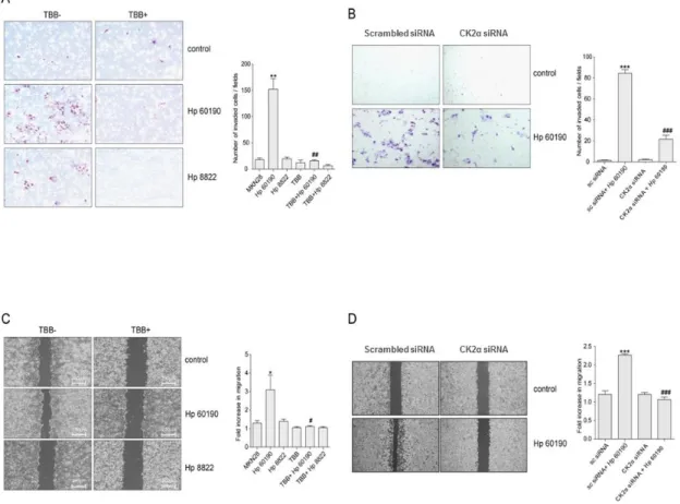
III. RESULTS1. H. pylori induces the invasion and migration in gastric cancer cells through PKCK2 activation
H. pylori induces an elongated ‘hummingbird’ morphologic phenotype in epithelial cells that is
associated with the loss of cell polarity and enhanced cell migration 22, 25. To determine whether
H. pylori activates PKCK2 to induce cancer cell invasion and migration, we treated MKN28
(Fig. 1A, C) and AGS (Fig. 1B, D) cells with the PKCK2 inhibitor TBB or CK2α-specific siRNA before infection with CagA+ (Hp60190) and CagA- (Hp8822) H. pylori. The invasiveness (Fig. 1A, B) and migration (Fig. 1C, D) of cells were more pronounced after infection with CagA+ than CagA- H. pylori. Inhibition of PKCK2 by TBB in H. pylori infected MKN28 cells profoundly suppressed cell invasiveness (Fig. 1A) and motility (Fig. 1C). Similarly, down regulation of CK2α using siRNA resulted in a significant decrease in the invasiveness (Fig. 1B) and migratory activities in AGS cells (Fig. 1D). MKN28 and AGS cells with down-regulated PKCK2 showed significantly impaired invasion and migration compared with control cells (P<0.05).
These results indicate that increased cellular invasion and
migratory activities induced by H. pylori are mediated through PKCK2.
12
Figure 1. H. pylori induces the invasion and migration in gastric cancer cells through PKCK2 activation. (A) MKN28 cells were pretreated with TBB (100 μM) for 1 hour followed by infection with CagA+ (Hp60190) and CagA- (Hp8822) H. pylori for 24 hours. (B) The repeated experiments in AGS cells infected only with Hp60190 were performed following transfection with scrambled siRNA and CK2α siRNA. After invasion, the membranes were fixed and stained, and the invaded cells were counted. (C) MKN28 cells were pretreated with TBB (100 μM) for 1 hour followed by infection with Hp60190 and Hp8822 for 24 hours. (D) The repeated experiments except for infection with only Hp60190 were performed following transfection of
13
AGS cells with scrambled siRNA and CK2α siRNA. Quantitative analysis of the invasive and migratory cells was performed (right panels). Data was assessed by one-way ANOVA followed by Bonferroni method (* p < 0.05, ** p < 0.01, *** p < 0.001 vs control, # p < 0.05, ## p < 0.01,
###
14
2. H. pylori activates PKCK2 without affecting CK2α protein expression levels in gastric cancer cells
PKCK2 is a ubiquitous, highly conserved serine, threonine kinase that is up-regulated in rapidly dividing cells including most human tumors. However, the mechanism by which PKCK2 is activated in gastric cancer remains unclear. CK2α was expressed in all five gastric cancer cell lines tested with variation in the expression level among these cell lines (Fig. 2A). Most importantly, CK2α protein and mRNA expression levels were not altered in H. pylori infected MKN28 cell lines (Fig. 2B). To examine whether CagA translocation by H. pylori infection increases PKCK2 activity, we infected AGS cells (Fig. 2C) and MKN28 cells (Fig. 2D) with CagA+ (Hp60190), CagA- (Hp8822) H. pylori. Although there were no significant differences in CK2α protein and mRNA expression levels in H. pylori infected AGS and MKN28 cells,
infection with CagA+ (Hp60190) H. pylori significantly increased PKCK2 activities while CagA- (Hp8822) H. pylori did not. As expected, analysis of PKCK2 activity confirmed that inhibition of PKCK2 by TBB in H. pylori infected gastric cancer cells profoundly suppressed the activity of PKCK2. These data indicate that CagA affects host cell PKCK2 activity without affecting the transcriptional or translational level of CK2α.
16
Figure 2. H. pylori activates PKCK2 without affecting CK2α expression levels in gastric cancer cells. (A) Western blot and RT-PCR analysis of CK2α expression in five gastric cancer cell lines. (B) MKN28 cells were infected with H. pylori for the indicated times. Actin was used as a loading control. AGS (C) and MKN28 (D) cells were pretreated with 100 μM TBB for 1 hour followed by infection with H. pylori for 6 hours. The PKCK2 activity was determined using a Cyclex PKCK2 activity kit. Data was assessed by one-way ANOVA followed by Bonferroni method (** p < 0.01 vs control, ##
p < 0.01 vs Hp60190-infected cells without TBB).
17
3. Phosphorylation of α-catenin by PKCK2 results in dissociation of the α/β-catenin complex in H. pylori infected gastric cancer cells
It was previously reported that, EGF associated ERK activates PKCK2 to phosphorylates α-catenin at serine 641. Phosphorylation of α-α-catenin promotes disruption of α/β-α-catenin complex, resulting in EMT 24. Like EGF, H. pylori is known to induce ERK activation, which may be dependent or independent of CagA in gastric epithelial cells. Therefore, we examined whether H.
pylori disrupts the α/β-catenin complex by selective measurement of membranous α-catenin
expression. As expected, infection of MKN28 cells with CagA+ (Hp60190), but not CagA -(Hp8822), significantly depleted the membranous α-catenin levels in a time-dependent manner (Fig. 3A). The E-cadherin complex is composed of E-cadherin, β-catenin, α-catenin and other compounds. Dissociation of α/β-catenin enhances disruption of the E-cadherin complex and induces cellular invasion and migratory activities of epithelial cells 26. To investigate whether H.
pylori influences α/β-catenin disruption through activation of PKCK2, MKN28 cells were
pretreated with TBB (100 μM) for 1 hour before infection with H. pylori for 24 hours. Cell lysates were immunoprecipitated with antibody against α-catenin, β-catenin or CK2α and then immunoblotted with antibody against β-catenin or α-catenin as indicated. Through immunoprecipitation analyses, we found that Hp60190 increased the interaction between CK2α and α-catenin and induced disruption of the α/β-catenin interaction (Fig. 3B). Next, to determine whether H. pylori increases α-catenin phosphorylation at serine 641 through activation of PKCK2, we performed Western blot analysis in gastric cancer cells that were pretreated with TBB prior to H. pylori infection. Infection with Hp60190 resulted in an increased level of p-α-catenin at serine 641 compared with Hp8822 infection. TBB pretreatment decreased the up-regulation of p-α-catenin in cells infected with Hp60190 (Fig. 3C). Considering that H. pylori infected gastric cancer cells expressed the phosphorylated form of α-catenin through the activity of PKCK2, we next determined whether inhibition of CK2α expression directly affects
α-18
catenin phosphorylation. We used CK2α-specific shRNA lentivirus and puromycin to select for stable pools of AGS cells with CK2α down-regulation and control lines transfected with pLKO.1. Analysis of CK2α protein expression by immunoblotting confirmed that CK2α protein expression was significantly suppressed in the AGS cells expressing CK2α shRNA (sh1 and sh4), but not in the cells transfected with control pLKO.1. Although CK2α knockdown had no effect on total α-catenin expression in AGS cell lines, the level of p-α-catenin was prominently decreased in CK2α knockdown cell lines (Fig. 3C, lower left panel). Moreover, overexpression of CK2α protein resulted in significant up-regulation of α-catenin phosphorylation (Fig. 3C, lower right panel). Immunofluorescence studies showed that H. pylori infection for 24 hours resulted in disruption of membranous α-catenin and E-cadherin. In addition, more pronounced effects were observed for infection with Hp60190 compared with Hp8822 (Fig. 3D). These results show that PKCK2 mediated phosphorylation of α-catenin leads to membranous α-catenin depletion through dissociation of the α/β-catenin complex in H. pylori infected gastric cancer cells in CagAdependent manner.
20
Figure 3. PKCK2-mediated phosphorylation of α-catenin results in dissociation of the α/β-catenin complex in H. pylori-infected gastric cancer cells. (A) MKN28 cells were infected with Hp60190 or Hp8822 for the indicated times. The change in membranous α-catenin level was analyzed by immunoblotting. (B) MKN28 cells were pretreated with TBB (100 μM) for 1 hour followed by infection with Hp60190 or Hp8822 for 24 hours. Endogenous CK2α, α-catenin or β-catenin was immunoprecipitated, and binding of CK2α to α-catenin (first panel), β-catenin to α-catenin (second panel) and α-catenin to β-catenin (third panel) was analyzed by immunoblotting. (C) MKN28 cells were pretreated with TBB for 1 hour followed by infection with Hp60190 or Hp8822 for 3 hours (upper panel). Western blot analysis was performed to determine the α-catenin and phospho-α-catenin levels in shRNA-mediated CK2α knockdown AGS cells (lower left panel) and the expression of α-catenin and phospho-α-catenin in AGS cells transfected with pRC-CK2α-HA (lower right panel). (D) MKN28 cells were pretreated with TBB (100 μM) for 1 hour followed by infection with Hp60190 and Hp8822 for 24 hours at a MOI of 1:100. The samples were subjected to immunofluorescent staining with antibody against α-catenin or E-cadherin and mounted in DAPI-containing mounting solution; 400× magnification.
21
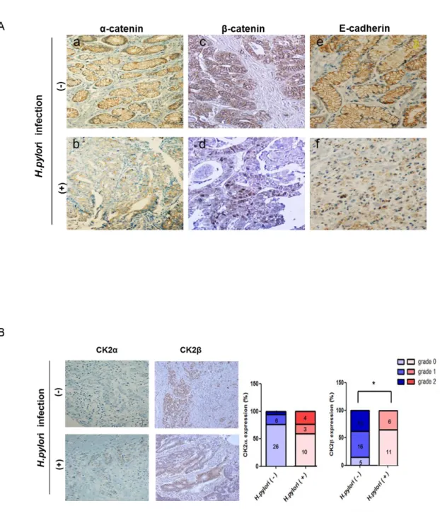
4. Expression of CK2 subunits and junctional proteins in an in vivo gastric cancer tissue model
To examine the relationship between H. pylori infection and expression of junctional proteins and cellular CK2α, CK2β, gastric cancer tissues of patients with H. pylori infection (n=17) or without H. pylori infection (n=34) were subjected to immunohistochemical analysis using antibodies to α-catenin, β-catenin, E-cadherin (Fig. 4A) and CK2α, CK2β (Fig. 4B). Membranous α-catenin immunostaining revealed strong expression in non-infected gastric cancer tissues (Fig. 4A,a) and low expression in H. pylori-infected gastric cancer tissues (Fig. 4A,b).Although membranous β-catenin expression was dominant (Fig. 4A,c), nuclear β-catenin immunostaining was frequently seen in H. pylori-infected gastric cancer tissues (Fig. 4A,d). Membranous expression of E-cadherin (Fig. 4A,e) was also decreased in H. pylori-infected gastric cancer tissue (Fig. 4A,f). CK2α immunostaining revealed more strong expression in H.
pylori-infected gastric cancer tissues than non-infected gastric cancer tissues and nuclear
expression of CK2β in H. pylori -infected gastric cancer tissues (Fig. 4d) were lower than non-infected gastric cancer tissues (Fig. 4B). We performed statistical analysis by fisher’s exact test to examine the relation between H. pylori-infection and CK2α expression according to the staining grade (0<10%, 1=10-49%, 2=50-100% in CK2α scoring and 0= no, 1=weak, 2=modest in CK2β scoring). Although CK2α expression had no statistically significant correlation with H.
pylori infection (p=0.2127), CK2α expression tended to be higher in H. pylori infected gastric
cancer tissues. In contrast, there was an significant inverse relationship between CK2β expression and H. pylori infection (p=0.0002).
22
Figure 4. Immunohistochemical detection of α-catenin, β-catenin, E-cadherin, CK2α and CK2β expression in H. pylori non-infected and infected gastric cancer tissues.
23
Gastric cancer tissues of patients with or without H. pylori infection were subjected to immunohistochemical analysis using antibodies to (A) α-catenin, β-catenin and E-cadherin and (B) CK2α, CK2β. Magnification, ×200. Bar graphs represented percent expression of CK2α (left panel) and CK2β (right panel) according to the staining grade. Numbers of patients are defined in the graphs (p<0.05).
24
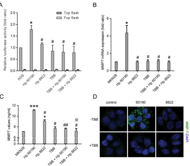
5. H. pylori induced α/β-catenin dissociation resulted in β-catenin nuclear translocation and increased MMP7 expressions
To examine the effect of H. pylori-induced dissociation of the α/β-catenin complex on β-catenin transactivation, we next examined T-cell factor-1 (TCF1) and lymphoid enhancing factor-1 (LEF1) transcriptional activity profiles. TCF/LEF-1 luciferase reporter analysis showed that Hp60190 induced greater β-catenin transactivation than Hp8822 in gastric cancer cells. Furthermore, Hp60190-enhanced TCF/LEF-1 transcriptional activity was blocked by treatment with the PKCK2 inhibitor TBB, showing that activation of PKCK2 by H. pylori plays an important role in β-catenin transactivation (Fig. 5A). Recent studies have shown that the nuclear translocation of β-catenin increases expression of the MMP7 gene expressions 27 in lung and gastric epithelial cell lines and that MMP7 is strongly involved in EMT 28, 29. Therefore, we examined expression of the β-catenin/Tcf downstream target gene MMP7 and production of MMP7 by real-time qRT-PCR and ELISA, respectively in MKN28 cells that were pretreated with TBB (100 μM) before H. pylori infection. As expected, the level of MMP7 mRNA expression and MMP7 production were increased only in CagA+ H. pylori infected MKN28 cells. Moreover, H. pylori-induced MMP7 expression and production of MMP7 were blocked by TBB pretreatment (Fig. 5B, C). Next, to determine whether H. pylori increases MMP7 through PKCK2 activation, we performed immunofluorescence studies in MKN28 cells that were pretreated with TBB prior to H. pylori infection. Infection with Hp60190 resulted in an increased level of MMP7 compared with Hp8822 infection. TBB pretreatment decreased the up-regulation of MMP7 in cells infected with Hp60190 (Fig. 5D). Taken together, these findings indicate that up-regulation of PKCK2 by CagA+ H. pylori plays an important role in β-catenin nuclear translocation and EMT-related MMP7 expression in gastric cancer cells.
25
Figure 5. H. pylori promotes β-catenin/TCF promoter activity and downstream MMP7 expression by increasing PKCK2 activity. (A) AGS cells were co-transfected with TopFlash (multiple copies of an optimal TCF-binding site) and FopFlash (mutant Tcf binding sites) vectors together with TK-Renilla (control) vector and then infected with Hp60190 or Hp8822 for 24 hours with or without pretreatment with 100 μM TBB for 1 hour. (B) MKN28 cells were infected with Hp60190 or Hp8822 for 6 hours at a MOI of 100. MMP7 expression was measured by qRT-PCR. (C) MKN28 cells were pretreated with TBB (100 μM) for 1 hour
26
followed by infection with Hp60190 and Hp8822 for 8 hours at a MOI of 1:100. MMP-7 secretions were measured by ELISA kit. (D) The repeated experiments in MKN28 cells were subjected to immunofluorescent staining with antibody against MMP-7 (green) and mounted in DAPI-containing mounting solution. Data was assessed by one-way ANOVA followed by Bonferroni method (* p < 0.05, *** p < 0.001 vs control, # p < 0.05, ## p < 0.01 vs
27
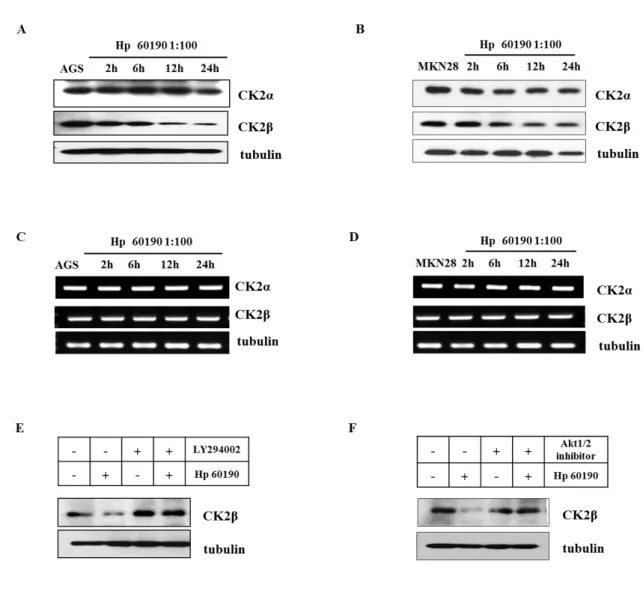
6. H. pylori induces CK2β degradation through PI3K/Akt pathway
Recent studies have shown that low CK2β expression correlated with characteristic EMT markers, including Snail1, Zeb2 or Twist130 and leads to morphological transformations associated with activation of EMT pathways. Therefore, we examined whether H. pylori increases CK2β degradation by measurement of CK2β protein expression. As expected, infection of AGS and MKN28 cells with CagA+ (Hp60190), significantly depleted the CK2β levels in a time-dependent manner while CK2α level did not change (Fig. 6A, B). However, both CK2α and CK2β mRNA expression levels were not altered in H. pylori infected AGS and MKN28 cell lines (Fig. 6C, D). To investigate which signaling pathway involved in CK2β degradation, we used PI3K-specific inhibitor, LY294002 and Akt specific inhibitor, Akt1/2 kinase inhibitor. As expected, H. pylori-induced CK2β degradation were blocked by LY294002 (Fig. 6E) and Akt1/2 kinase inhibitor (Fig. 6F) in AGS cells. Taken together, these results indicate that H. pylori infection increases EMT-related CK2β degradation through PI3K-Akt pathway.
28
Figure 6. H. pylori induced CK2β degradation through PI3K/Akt pathway. AGS cells (A, C) were infected with CagA+ (Hp60190) at multiplicity of infection (MOI) of 100:1 for various times. (B, D) The repeated experiments in MKN28 cells. (E) AGS cells were pretreated with LY294002 (10μM) for 1 hour followed by infection with CagA+
(Hp60190) H pylori for 4 hours. (F) The repeated experiments except for pretreatment with Akt1/2 kinase inhibitor were performed.
29
IV. DISCUSSIONThe Helicobacter pylori (H. pylori) cag pathogenicity island (cag PAI) is a 35-40-kb genetic element that encodes a type IV secretion system and is strongly associated with gastric malignant progression 31-33. Virulent H. pylori strains harbor a PAI for the translocation of the bacterial oncoprotein, CagA into gastric epithelial cells 1-3. Differences have been observed in the abilities of CagA+ and CagA- H. pylori strains to activate various signaling cascades in infected gastric epithelial cells 34, 35. Using CagA-inducible gastric epithelial model, Murata-Kamiya et al. demonstrated that intracellular CagA interacts with E-cadherin and disrupts the formation of E-cadherin–catenin complexes, as well as inducing nuclear accumulation of β-catenin 36, suggesting that inhibition of cell-cell adhesion may be an intracellular target of CagA. In our study, H. pylori infection induced β-catenin transactivation and down-regulation of E-cadherin expressions which induced cell invasiveness and motility in gastric epithelial cells in CagA-dependent manner. These findings are consistent with previous reports that investigated the mechanisms regulating invasiveness and motility in H. pylori-infected gastric epithelial cells. Protein kinase casein kinase 2 (PKCK2) is a serine/threonine protein kinase 5, 6 that plays a key role in cell cycle control, cellular differentiation andtransformation7. Many studies have shown that dysregulation of PKCK2 is associated with tumorigenesis 12, 37, 38. Moreover, Zou et al. reported that CK2α modulates the process of epithelial mesenchymal transition (EMT), thereby affecting regulation of cell migration and invasion in colorectal cancer cells and PKCK2-specific inhibitors significantly inhibit membrane invasion, adhesion, and migration of ovarian carcinoma cells 39. In agreement with these results, our study demonstrated that activity of PKCK2 in gastric epithelial cells increased in a CagA-dependent manner during H. pylori infection.
In the current study we show for the first time that CagA+ (Hp60190) H. pylori up-regulates cellular invasiveness and migration activity through PKCK2 activation whereas CagA
-30
(Hp8822) H. pylori does not induce cell invasiveness or migration. Furthermore, H. pylori affects PKCK2 activity in gastric cancer cells without affecting the transcriptional or translational expression of CK2α. Although EMT is typically observed in the invasive progression of cancer cells, in vivo evidence suggests that loss of epithelial adhesion is sufficient to initiate invasive phenotype in the pancreas and stomach. In adherens junctions, α-catenin links the cadherin-beta-α-catenin complex to the actin-based cytoskeleton and stabilizes cell polarity20. Our results showed that CagA-mediated PKCK2 activation disrupts these adherens junction complexes through α/β–catenin dissociation in gastric epithelial cells. Because it has been suggested that CK2α disrupts α-catenin binding to β-catenin through CK2α/α-catenin interaction 24, it is tempting to speculate that CagA is a mediator of this interaction. However, many questions remain unanswered, mainly concerning the association between CagA and CK2α. In any case, the results presented here have revealed new perspectives on the role of H. pylori CagA and epithelial PKCK2 activation in the regulation of cellular migration and invasion in gastric carcinogenesis.
Immunohistochemical analysis revealed that membranous α-catenin, β-catenin and E-cadherin expression was lowered in H. pylori-infected gastric cancer tissues. Although CK2α expression in gastric cancer tissues had no statistically significant correlation with H. pylori infection, CK2α expression tended to be higher in H. pylori infected gastric cancer tissues. The lack of significance in CK2α expression and H. pylori infection may be due to the small number of patients in the H. pylori infected group. However, the expression of CK2β significantly lowered in H. pylori infected gastric cancer tissues and these results matched with our in-vitro results. The MMP-7 gene is one of target gene of β-catenin transactivation signals, which is critical for remodeling of the extracellular matrix for a variety of physiological and pathological processes27. It has been suggested that H. pylori-induced MMP expression plays a role in inflammation, carcinogenesis and cancer metastasis28, 29, and our findings clearly showed that H.
31
pylori being able to induce MMP-7 protein expression inducing epithelial cell invasiveness and
motility in vitro. In this study, we observed that H. pylori-induced α/β-catenin dissociation resulted in β-catenin nuclear translocation and increased MMP7 expression. Furthermore, we found that up-regulation of PKCK2 by CagA+ H. pylori played an important role in β-catenin nuclear translocation and EMT-related MMP7 expression in gastric cancer cells.
Evidence of the mechanistic interplay between CagA and PKCK2-induced α/β-catenin dissociation along with β-catenin transactivation provides important insight into cell invasion and migration in gastric cancer. Taken together, these data suggest that targeting oncogenic PKCK2 activity may be a potential therapeutic strategy for treating human gastric cancer associated with H. pylori infection.
Recent studies have shown that unbalanced expression of PKCK2 subunits is sufficient to drive epithelial-to-mesenchymal transition by Snail1 induction40. We show for the first time that CagA+ (Hp60190) H. pylori infection increases CK2β degradation through PI3K-Akt pathway. Reduced CK2β may represent a novel molecular alteration during malignant tumor progression. Interactions between H. pylori and cancer cells play an important role in the development of epithelial mesenchymal transition. Our finding will not only improve our understanding of H.
pylori-induced epithelial mesenchymal transition but also provide mechanistic insight into other
32
V. CONCLUSIONIn this study, we showed for the first time that CagA+ (Hp60190) H. pylori up-regulates cancer cell invasiveness and migration activity through PKCK2 activation, whereas CagA- (Hp8822) H.
pylori does not induce cell invasiveness or migration. Evidence of the mechanistic interplay
between CagA and PKCK2-induced α/β-catenin dissociation along with β-catenin transactivation provides important insight into cell invasion and migration in gastric cancer. Taken together, these data suggest that targeting oncogenic PKCK2 activity may be a potential therapeutic strategy for treating human gastric cancer associated with H. pylori infection.
33
REFERENCES1. Amieva MR, Vogelmann R, Covacci A, Tompkins LS, Nelson WJ, Falkow S. D isruption of the epithelial apical-junctional complex by Helicobacter pylori CagA. Scienc e 2003;300(5624):1430-4.
2. Backert S, Moese S, Selbach M, Brinkmann V, Meyer TF. Phosphorylation of t yrosine 972 of the Helicobacter pylori CagA protein is essential for induction of a scatt ering phenotype in gastric cancer cells. Mol Microbiol 2001;42(3):631-44.
3. Churin Y, Al-Ghoul L, Kepp O, Meyer TF, Birchmeier W, Naumann M. Helico bacter pylori CagA protein targets the c-Met receptor and enhances the motogenic respo nse. J Cell Biol 2003;161(2):249-55.
4. Moese S, Selbach M, Kwok T, Brinkmann V, Konig W, Meyer TF, et al. Helic obacter pylori induces AGS cell motility and elongation via independent signaling pathw ays. Infect Immun 2004;72(6):3646-9.
5. Padmanabha R, Chen-Wu JL, Hanna DE, Glover CV. Isolation, sequencing, and disruption of the yeast CKA2 gene: casein kinase II is essential for viability in Sacchar omyces cerevisiae. Molecular and cellular biology 1990;10(8):4089-99.
6. Kikkawa U, Mann SK, Firtel RA, Hunter T. Molecular cloning of casein kinase II alpha subunit from Dictyostelium discoideum and its expression in the life cycle. Mol ecular and cellular biology 1992;12(12):5711-23.
7. McElhinny JA, Trushin SA, Bren GD, Chester N, Paya CV. Casein kinase II p hosphorylates I kappa B alpha at S-283, S-289, S-293, and T-291 and is required for its degradation. Molecular and cellular biology 1996;16(3):899-906.
8. Filhol O, Martiel JL, Cochet C. Protein kinase CK2: a new view of an old mo lecular complex. EMBO Rep 2004;5(4):351-5.
9. Litchfield DW, Dobrowolska G, Krebs EG. Regulation of casein kinase II by gr owth factors: a reevaluation. Cell Mol Biol Res 1994;40(5-6):373-81.
10. Litchfield DW. Protein kinase CK2: structure, regulation and role in cellular dec isions of life and death. Biochem J 2003;369(Pt 1):1-15.
11. Christensen B, Nielsen MS, Haselmann KF, Petersen TE, Sorensen ES. Post-tran slationally modified residues of native human osteopontin are located in clusters: identifi cation of 36 phosphorylation and five O-glycosylation sites and their biological implicati ons. Biochem J 2005;390(Pt 1):285-92.
12. Trembley JH, Wang G, Unger G, Slaton J, Ahmed K. Protein kinase CK2 in he alth and disease: CK2: a key player in cancer biology. Cell Mol Life Sci 2009;66(11-1 2):1858-67.
13. Giusiano S, Cochet C, Filhol O, Duchemin-Pelletier E, Secq V, Bonnier P, et al. Protein kinase CK2alpha subunit over-expression correlates with metastatic risk in breast
34
carcinomas: quantitative immunohistochemistry in tissue microarrays. Eur J Cancer 2011; 47(5):792-801.
14. Chen YY, Chiang SY, Lin JG, Ma YS, Liao CL, Weng SW, et al. Emodin, alo e-emodin and rhein inhibit migration and invasion in human tongue cancer SCC-4 cells through the inhibition of gene expression of matrix metalloproteinase-9. Int J Oncol 201 0;36(5):1113-20.
15. Kim J, Hwan Kim S. CK2 inhibitor CX-4945 blocks TGF-beta1-induced epitheli al-to-mesenchymal transition in A549 human lung adenocarcinoma cells. PloS one 2013; 8(9):e74342.
16. Gumbiner BM. Cell adhesion: the molecular basis of tissue architecture and mor phogenesis. Cell 1996;84(3):345-57.
17. Yap AS, Brieher WM, Gumbiner BM. Molecular and functional analysis of cad herin-based adherens junctions. Annu Rev Cell Dev Biol 1997;13:119-46.
18. Drubin DG, Nelson WJ. Origins of cell polarity. Cell 1996;84(3):335-44.
19. Gumbiner B. Cadherins: a family of Ca2+-dependent adhesion molecules. Trends in biochemical sciences 1988;13(3):75-6.
20. Pokutta S, Weis WI. Structure of the dimerization and beta-catenin-binding regio n of alpha-catenin. Mol Cell 2000;5(3):533-43.
21. Kang DW, Hwang WC, Park MH, Ko GH, Ha WS, Kim KS, et al. Rebamipid e abolishes Helicobacter pylori CagA-induced phospholipase D1 expression via inhibition of NFkappaB and suppresses invasion of gastric cancer cells. Oncogene 2013;32(30):353 1-42.
22. Kikuchi K, Murata-Kamiya N, Kondo S, Hatakeyama M. Helicobacter pylori sti mulates cancer cell migration via CagA-mediated perturbation of host cell signaling. Mic robes Infect 2012;14(5):470-6.
23. Krueger S, Hundertmark T, Kuester D, Kalinski T, Peitz U, Roessner A. Helico bacter pylori alters the distribution of ZO-1 and p120ctn in primary human gastric cance r cells. Pathol Res Pract 2007;203(6):433-44.
24. Ji H, Wang J, Nika H, Hawke D, Keezer S, Ge Q, et al. EGF-induced ERK a ctivation promotes CK2-mediated disassociation of alpha-Catenin from beta-Catenin and t ransactivation of beta-Catenin. Mol Cell 2009;36(4):547-59.
25. Saadat I, Higashi H, Obuse C, Umeda M, Murata-Kamiya N, Saito Y, et al. He licobacter pylori CagA targets PAR1/MARK kinase to disrupt cancer cell polarity. Nature 2007;447(7142):330-3.
26. Ozawa M, Kemler R. Altered cell adhesion activity by pervanadate due to the dissociation of alpha-catenin from the E-cadherin.catenin complex. The Journal of biologi cal chemistry 1998;273(11):6166-70.
35
27. Barnes EA, Kenerson HL, Mak BC, Yeung RS. The loss of tuberin promotes c ell invasion through the ss-catenin pathway. Am J Respir Cell Mol Biol 2010;43(5):617-27.
28. Yin Y, Grabowska AM, Clarke PA, Whelband E, Robinson K, Argent RH, et al. Helicobacter pylori potentiates epithelial:mesenchymal transition in gastric cancer: links to soluble HB-EGF, gastrin and matrix metalloproteinase-7. Gut 2010;59(8):1037-45.
29. Fanelli MF, Chinen LT, Begnami MD, Costa WL, Fregnami JH, Soares FA, et al. The influence of transforming growth factor-alpha, cyclooxygenase-2, matrix metallopr oteinase (MMP)-7, MMP-9 and CXCR4 proteins involved in epithelial-mesenchymal tran sition on overall survival of patients with gastric cancer. Histopathology 2012;61(2):153-6 1.
30. MacPherson MR, Molina P, Souchelnytskyi S, Wernstedt C, Martin-Perez J, Port illo F, et al. Phosphorylation of serine 11 and serine 92 as new positive regulators of h uman Snail1 function: potential involvement of casein kinase-2 and the cAMP-activated kinase protein kinase A. Molecular biology of the cell 2010;21(2):244-53.
31. Censini S, Lange C, Xiang Z, Crabtree JE, Ghiara P, Borodovsky M, et al. Ca g, a pathogenicity island of Helicobacter pylori, encodes type I-specific and disease-assoc iated virulence factors. Proc Natl Acad Sci U S A 1996;93(25):14648-53.
32. Huang JQ, Zheng GF, Sumanac K, Irvine EJ, Hunt RH. Meta-analysis of the re lationship between cagA seropositivity and gastric cancer. Gastroenterology 2003;125(6):1 636-44.
33. Peek RM, Jr. Helicobacter pylori strain-specific modulation of gastric mucosal c ellular turnover: implications for carcinogenesis. J Gastroenterol 2002;37 Suppl 13:10-6. 34. Keates S, Keates AC, Warny M, Peek RM, Jr., Murray PG, Kelly CP. Differenti al activation of mitogen-activated protein kinases in AGS gastric cancer cells by cag+ a nd cag- Helicobacter pylori. J Immunol 1999;163(10):5552-9.
35. Glocker E, Lange C, Covacci A, Bereswill S, Kist M, Pahl HL. Proteins encod ed by the cag pathogenicity island of Helicobacter pylori are required for NF-kappaB ac tivation. Infect Immun 1998;66(5):2346-8.
36. Murata-Kamiya N, Kurashima Y, Teishikata Y, Yamahashi Y, Saito Y, Higashi H, et al. Helicobacter pylori CagA interacts with E-cadherin and deregulates the beta-catenin signal that promotes intestinal transdifferentiation in gastric cancer cells. Oncogene 2007; 26(32):4617-26.
37. Gapany M, Faust RA, Tawfic S, Davis A, Adams GL, Ahmed K. Association o f elevated protein kinase CK2 activity with aggressive behavior of squamous cell carcino ma of the head and neck. Mol Med 1995;1(6):659-66.
38. Faust M, Montenarh M. Subcellular localization of protein kinase CK2. A key t o its function? Cell Tissue Res 2000;301(3):329-40.
36
39. Ruzzene M, Di Maira G, Tosoni K, Pinna LA. Assessment of CK2 constitutive activity in cancer cells. Methods Enzymol 2010;484:495-514.
40. Deshiere A, Duchemin-Pelletier E, Spreux E, Ciais D, Combes F, Vandenbrouck Y, et al. Unbalanced expression of CK2 kinase subunits is sufficient to drive epithelial-to-mesenchymal transition by Snail1 induction. Oncogene 2013;32(11):1373-83.
37
ABSTRACT (IN KOREAN)