저작자표시-비영리-변경금지 2.0 대한민국 이용자는 아래의 조건을 따르는 경우에 한하여 자유롭게 l 이 저작물을 복제, 배포, 전송, 전시, 공연 및 방송할 수 있습니다. 다음과 같은 조건을 따라야 합니다: l 귀하는, 이 저작물의 재이용이나 배포의 경우, 이 저작물에 적용된 이용허락조건 을 명확하게 나타내어야 합니다. l 저작권자로부터 별도의 허가를 받으면 이러한 조건들은 적용되지 않습니다. 저작권법에 따른 이용자의 권리는 위의 내용에 의하여 영향을 받지 않습니다. 이것은 이용허락규약(Legal Code)을 이해하기 쉽게 요약한 것입니다. Disclaimer 저작자표시. 귀하는 원저작자를 표시하여야 합니다. 비영리. 귀하는 이 저작물을 영리 목적으로 이용할 수 없습니다. 변경금지. 귀하는 이 저작물을 개작, 변형 또는 가공할 수 없습니다.
A Profile of Patients Presenting with
Non-odontogenic Pain to a
Orofacial Pain Clinic in Korea
Jeong Il Seo
Department of Dentistry
A Profile of Patients Presenting with
Non-odontogenic Pain to a
Orofacial Pain Clinic in Korea
Directed by Professor Seong Taek Kim, D.D.S., M.S.D., Ph.D.
The Doctoral Dissertation
submitted to the Department of Dentistry,
the Graduate School of Yonsei University
in partial fulfillment of the requirements for the degree of
Doctor of Philosophy in Dental Science
Jeong Il Seo
This certifies that the doctoral dissertation
of Jeong IL Seo is approved.
Thesis Supervisor : Seong Taek Kim
Thesis Committee Member : Hyung-Joon Ahn
Thesis Committee Member : Jeong-Seung Kwon
Thesis Committee Member : Chang-Sung Kim
Thesis Committee Member : Il-Young Jung
The Graduate School
Yonsei University
감사의 글
오랜 기간의 연구가 드디어 그 결실을 맺게 되었습니다. 논문이 완성되기까지 부족한 저를 이끌어주신 많은 분들께 감사의 인사를 전합니다. 먼저 논문의 시작부터 끝맺음 까지 오랜 기간 성심 성의껏 지도해 주시고 신경 써 주신 김성택 지도교수님께 진심으로 감사 드립니다. 또한 대학원 과정 동안 많은 가르침을 주시고 인자한 미소로 대해주신 최종훈 교수님, 항상 좋은 강의로 많은 것을 배우게 해 주시고 이번 논문의 심사에도 신경 써 주신 안형준 교수님, 논문의 방향을 잡는 데에 많은 도움을 주시고 고민해 주신 권정승 교수님께 깊은 감사를 드립니다. 부족한 저의 논문의 심사와 지도를 맡아주신 정일영 교수님, 김창성 교수님께도 깊은 감사를 드리고 싶습니다. 바쁜 임상 과정 중에 논문을 준비하고 완성하는 동안 격려와 배려를 아끼지 않은 고운미소 강호성, 김성현, 김정석, 박재한 원장님과 우은영 실장님을 비롯한 스텝들, 바쁘신 와중에도 여러 도움을 주신 모든 의국원 분들에게도 진심으로 감사 드립니다. 마지막으로 제가 이 자리에 올 때까지 항상 같은 마음으로 희생해주신 감사하는 아버님, 어머님, 늘 사랑으로 보살펴주시는 장인, 장모님, 저의 인생 동반자로서 제가 하는 일을 항상 믿고 끝없는 내조로 힘을 주는 나의 사랑하는 아내 자형이와 예쁜 딸 한슬이, 듬직한 아들 한승이에게 감사의 마음을 전하며 학위 취득의 기쁨을 같이 하고자 합니다. 2016 년 6 월TABLE OF CONTENTS
TABLE OF CONTENTS ··· i
LIST OF FIGURES···ii
LIST OF TABLES ···iii
ABSTRACT··· iv
I. INTRODUCTION···1
II. MATERIALS AND METHODS···3
Patient Chart Review···3
III. RESULTS···5
1. Analysis of Subjective Symptoms and Pain History ···5
2. Analysis of Diagnoses and Treatments ··· 12
IV. DISCUSSION ··· 18
V. CONCLUSION··· 27
REFERENCES ··· 29
LIST OF FIGURES
Figure 1. Distribution of intraoral pain locations
···
8
Figure 2. Number of endodontically treated tooth for same
patient (n=93)
···
11
Figure 3. Diagnosis in the Department of Orofacial Pain and
Oral Medicine (n=210)
···
14
Figure 4. Efficacy of drugs administered for non-odontogenic
pain
···
16
LIST OF TABLES
Table 1. Distribution of Gender and age in non-odontogenic
pain subjects
···
5
Table 2. Baseline Characteristics of Patients
···
6
Table 3. Pain and treatment history characteristics of
non-odontogenic pain
···
9
Table 4. Pain and treatment history characteristics of
non-odontogenic pain
···
11
Table 5. Diagnosis methods in the Department of Orofacial Pain
and Oral Medicine
···
13
Table 6. Treatment in the Department of Orofacial Pain and
Abstract
A Profile of Patients Presenting with Non-odontogenic Pain to a
Orofacial Pain Clinic in Korea
Jeong Il Seo
Department of Orofacial Pain and Oral Medicine / Dentistry
The Graduate School, Yonsei University
(Directed by Professor Seong Taek Kim, D.D.S., M.S.D., Ph.D.)
Etiology as well as diagnosis and treatment methods for non-odontogenic (NO) pain have not yet been clearly established. Patients with NO pain generally have complex histories of dental treatment without improvement of symptoms. Patients often suffer from persistent pain because of failure to establish the NO etiology, resulting in incorrect diagnosis and inappropriate dental treatment unnecessary to resolving NO pain. Despite this issue, NO pain has not been systemically evaluated in Korea. This study therefore investigated the various clinical profiles of patients with NO pain as well as diagnosis and treatment methods of NO pain.
May 2003 and April 2013. Chart reviews were conducted on all 210 patients. The profiles were evaluated using patient charts (gender, age, smoking, alcohol consumption, systemic disease, pain duration, pain location, and areas affected by pain) and surveys (pain intensity, spontaneity of pain, referred pain, and dental history associated with the NO pain).
Based on the obtained results, we report the following findings:
1. NO pain was diagnosed mainly as neuropathic pain (53.8%) and myofascial pain (MFP) (23.3%).
2. There were 65.7% without medical history, 20.5% cardiovascular diseases, 6.7% with GI troubles and 4.3% with psychogenic diseases. And only few of them have smoking (male 6.7%, female 1.0%) and drinking habits (male 8.6%, female 5.7%). 3. The common site of NO pain is left maxilla (30.0%), and right maxilla (27.6%).
Distribution of affected area were generally in accordance with previous reports but was not predominant in maxillary posterior region. Single area was affected in 90 patients (42.9%) with similar occurrence with multiple lesions (41.0%).
4. 17 patients (8.1%) had referred pain. 63.8% of patients have spontaneous pain. And the number of patients who have severe pain and who have moderate pain were 24.3%, 51.9% each. The average period of treatment was 6.7 months.
5. NO pain occurs more often to the female (74.8%) than to the male (25.2%). The average age is 49.0 years and the average pain duration is 31 months.
6. Considering the departments referring to the Department of Orofacial Pain and Oral Medicine, treatments related to NO pain, previous dental treatment history, and visiting dental departments, NO pain is very closely related to the RCT. 48.6% of patients have treated more than 3 dentists.
And the effectiveness of anticonvulsants (79.2%) is better than that of antidepressants (75.3%).
8. Mainly the treatment drugs of neuropathic pain used amitriptyline (18.9%) and gabapentin (14.1%). And topical capsaicin has a limited effect.
9. Mainly the treatment drugs of MFP used eperisone HCl (24.3%) and cyclobenzaprine HCl (13.6%).
A Profile of Patients Presenting with Non-odontogenic Pain to a
Orofacial Pain Clinic in Korea
Jeong Il Seo
Department of Dentistry
The Graduate School, Yonsei University
(Directed by Professor Seong Taek Kim, D.D.S., M.S.D., Ph.D.)
I. INTRODUCTION
One of the most common types of orofacial pain is the toothache, or pain deriving from the tooth. The prevalence of non-odontogenic pain (NO pain) has been reported at 2.1% and the average duration of pain lasting 33 months (Ram S et al., 2009). However, there are types of pain that mimic the toothache, but derive from non-odontogenic sources. As patients with NO pain often present to the dentist mistakenly for toothache, NO pain poses a real diagnostic challenge (Mattscheck D et al., 2011). Identifying all the causes, effects, and symptoms that overlap with those of odonotogenic pain remains difficult and for this reason, NO pain is often considered one of the most frustrating facial pain conditions confronting medical and dental clinicians today (Melis M et al., 2003). In a previous study, of patients who visited the dental
office, 88% had come for toothache-like pain; 3% for NO pain, and 9% for a mixed condition of odontogenic and NO pain (Yatani H et al., 2014). In addition, NO pain can at times be iatrogenic, caused by general dental treatments like root canal treatments (RCT) or extractions. To differentiate between the many types of NO pain, including pain-related phenomena of the head, face and neck, it is essential to use a systematic approach.
Conceptually, NO pain in the dentoalveolar region can arise from four potential processes: referred musculoskeletal pain, neuropathic pain, neurovascular pain, or pathological process outside of but referring pain to the immediate dentoalveolar region. Examples of such pathological processes include sinus disease, brain tumor, angina, throat cancer, as well as salivary gland, and craniofacial vascular disorders (Mattscheck D et al., 2011).
Patients with NO pain generally have complex histories of dental treatment without improvement of symptoms. Patients often suffer from persistent pain because of failure to establish the NO etiology, resulting in incorrect diagnosis and inappropriate dental treatment unnecessary to resolving NO pain. Despite this issue, NO pain has not been systemically evaluated in Korea. To investigate the management of NO pain in Korea, this study profiles various clinical characteristics of patients with NO pain as well as diagnosis and treatment methods of NO pain.
II. MATERIALS AND METHODS
Subjects included 210 patients with orofacial pain complaints who were admitted to the Department of Orofacial Pain and Oral Medicine of Yonsei University Dental Hospital between May 2003 and April 2013. Chart reviews were conducted on all 210 patients.
Patient Chart Review
Gender, age, smoking, alcohol consumption, systemic disease, pain duration, pain location, and areas affected by pain were reviewed using patient charts.
Surveys evaluating pain intensity, spontaneity of pain, and referred pain were distributed to all subjects. Dental history associated with the NO pain prior to admission were also tracked, including the number of dental practitioners involved, previous departments visited for treatment, and sources by which patients were referred to the Department of Orofacial pain and Oral Medicine. Whether the NO pain had been initiated by a dental treatment was also evaluated. If symptoms were found to be iatrogenic, or caused by previous dental treatment, the number of teeth involved in the root canal treatment (RCT) and the number of visits required for treatment were recorded.
Data related to diagnoses and treatments were also investigated through chart review. Diagnostic methods, diagnosis, treatment methods, treatment duration, frequency of medication intake, and treatment effects conducted by the Department of Orofacial and Oral Medicine were examined. Diagnostic methods included diagnostic anesthesia, brain computerized tomography (CT), magnetic resonance imaging (MRI), and diagnostic oral
appliance. Diagnostic anesthesia was conducted with topical anesthesia and infiltration and the visual analogue scale (VAS) to measure pain was used before and after anesthesia. Aspects of diagnosis examined include diagnosis, frequency of neuropathic pain, myofascial pain (MFP), sleep bruxism and clenching, psychogenic pain, maxillary sinusitis and rhinitis, migraine and pain from other origins. Treatment methods included trigger point injection (TPI) and oral appliance. Frequency and effects of each treatment method were also evaluated. TPI was delivered using 0.5% procaine and 1% lidocaine while anticonvulsant, anxiolytics, antidepressant, nonsteroidal anti-inflammatory drug (NSAIDs), muscle relaxant, topical capsaicin, and other medications were used for treatment.
III. RESULTS
1. Analysis of Subjective Symptoms and Pain History
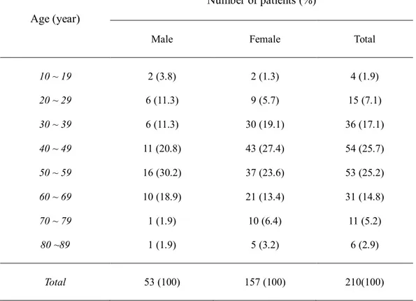
157 patients were females (74.8%), 53 patients were males (25.2%) and NO pain was more common in females (Table 1). The average age of patients was 49.0 years. Average ages for male patients and female patients were 48.6 years (17 ~ 86) and 49.1 years (15 ~ 85), respectively (Table 1).
Table 1. Distribution of Gender and age in non-odontogenic pain subjects
Age (year)
Number of patients (%)
Male Female Total
10 ~ 19 2 (3.8) 2 (1.3) 4 (1.9) 20 ~ 29 6 (11.3) 9 (5.7) 15 (7.1) 30 ~ 39 6 (11.3) 30 (19.1) 36 (17.1) 40 ~ 49 11 (20.8) 43 (27.4) 54 (25.7) 50 ~ 59 16 (30.2) 37 (23.6) 53 (25.2) 60 ~ 69 10 (18.9) 21 (13.4) 31 (14.8) 70 ~ 79 1 (1.9) 10 (6.4) 11 (5.2) 80 ~89 1 (1.9) 5 (3.2) 6 (2.9) Total 53 (100) 157 (100) 210(100)
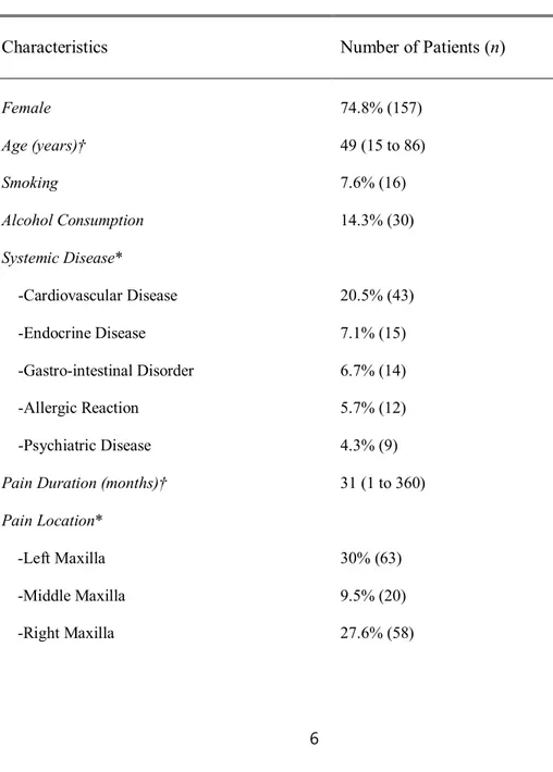
Regarding medical history, the most commonly reported medical disorders reported were cardiovascular diseases (20.5%), followed by endocrine diseases (7.1%), gastro-intestinal disorders (6.7%), psychiatric diseases (4.3%), and allergic reactions (5.7%) (Table 2).
Table 2. Baseline Characteristics of Patients
Characteristics Number of Patients (n)
Female 74.8% (157) Age (years)† 49 (15 to 86) Smoking 7.6% (16) Alcohol Consumption 14.3% (30) Systemic Disease* -Cardiovascular Disease -Endocrine Disease -Gastro-intestinal Disorder -Allergic Reaction -Psychiatric Disease 20.5% (43) 7.1% (15) 6.7% (14) 5.7% (12) 4.3% (9) Pain Duration (months)† 31 (1 to 360) Pain Location* -Left Maxilla -Middle Maxilla -Right Maxilla 30% (63) 9.5% (20) 27.6% (58)
-Left Mandible -Middle Mandible -Right Mandible -Temporomandibular Joint -Buccal Cheek -Lip 25.7% (54) 13.8% (29) 25.2% (53) 7.1% (15) 10.0% (21) 5.2% (11) Number of Affected Areas
-One -Two -Three or more 42.9% (90) 41.0% (86) 16.2% (34) †Mean (Range) *Permission of repetition
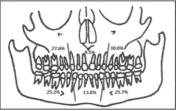
Pain duration varied from 1 month to 30 years (mean = 31 months). Commonly affected areas were right maxilla (27.6%) and left maxilla (30.0%) (Figure 1). 42.9% (n=90) of patients complained of symptoms in a single tooth or area, 41.0% (n=86) patients in two teeth or areas, and 16.2% (n=34) in three or more teeth or areas (Table 2).
Figure 1. Distribution of intraoral pain locations. Of the patients, 50.9% reported pain in the maxilla and 49.1% reported pain in the mandible. 35 patients(16.7%) had pain in both the maxilla and the mandible. Some patients had pain in more than one area of the jaw.
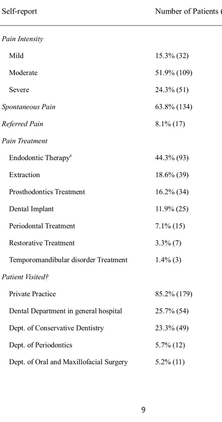
Spontaneous and referred pain was reported in 63.8% (n=134) and 8.1% (n=17) of patients, respectively. Treatments received prior to admission for patient’s chief complaint were RCT (44.3%), extraction (18.6%), prosthodontic treatment (16.2%), implant (11.9%), periodontal treatment (7.1%), restorative treatment (3.3%), and TMD treatment (1.4%) (Table 3).
Table 3. Pain and treatment history characteristics of non-odontogenic pain
Self-report Number of Patients (n)
Pain Intensity Mild Moderate Severe 15.3% (32) 51.9% (109) 24.3% (51) Spontaneous Pain 63.8% (134) Referred Pain 8.1% (17) Pain Treatment Endodontic Therapy# Extraction Prosthodontics Treatment Dental Implant Periodontal Treatment Restorative Treatment
Temporomandibular disorder Treatment
44.3% (93) 18.6% (39) 16.2% (34) 11.9% (25) 7.1% (15) 3.3% (7) 1.4% (3) Patient Visited† Private Practice
Dental Department in general hospital Dept. of Conservative Dentistry Dept. of Periodontics
Dept. of Oral and Maxillofacial Surgery
85.2% (179) 25.7% (54) 23.3% (49) 5.7% (12) 5.2% (11)
Dept. of Prosthodontics Dept. of Orthodontics Other‡ 2.9% (6) 1.0% (2) 32.1% (68) Dental Practitioners involved in pain treatment* 2.7 (2 to 7) Referral Source
Private Dental Practice Operative Dentistry
Oral & Maxillofacial Surgery Periodontal Dentistry Direct Visit (no referral)
36.7% (77) 28.6% (60) 4.3% (9) 3.8% (8) 26.7% (56)
#Included apicoectomy (n=9, 9.7%) and replantation (n=7, 7.5%).
†Permission of repetition.
‡Dept. of Neurology (n=25, 11.9%), Dept. of Otolaryngology (n=18, 8.6%), oriental medicine clinic (n=11, 5.2%), Dept. of Anesthesiology or pain Clinic (n=8, 3.8%), Dept. of Internal Medicine (n=4, 1.9%), and Dept. of Orthopedics (n=2, 1.0%).
*Mean (Range)
Notably, 60.5% (n=127) of patients reported developing symptoms following dental procedures. RCT was the common procedure (55.1%), followed by prosthodontic treatment (16.5%), extraction (13.4%), implant (13.4%), and periodontal surgery (1.6%) (Table 4).
Table 4. Pain and treatment history characteristics of non-odontogenic pain
Self-report Number of Patients (n)
Pain started with dental practice Root canal treatment
Prosthodontics treatment Extraction Dental implant Periodontal surgery 60.5% (127) 55.1% (70) 16.5% (21) 13.4% (17) 13.4% (17) 1.6% (2)
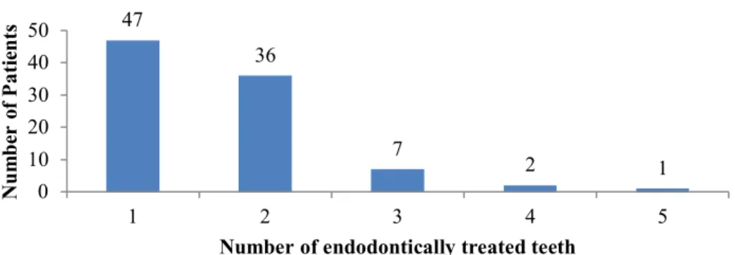
Among patients who received RCT, 50.5% (n=47) of patients received treatment for one tooth, 38.7% (n=36) for two teeth, 7.5% (n=7) for three teeth, 2.2% (n=2) for four teeth, and 1.1% (n=1) for five teeth. The mean number of teeth treated by RCT was 1.65 teeth (Figure 2).
47 36 7 2 1 0 10 20 30 40 50 1 2 3 4 5 N um be r of P at ie nt s
Number of endodontically treated teeth
Regarding the number of RCTs received per tooth, 59.1% (n=55) of cases received RCT once, 32.3% (n=30) received re-RCT after RCT, and 8.6% (n=8) received the same RCT three times. Of patients who received endodontic therapy, 17.2% received either apicoectomy or replantation.
Among patients who received periodontal treatment, 47.4% (n=9) received gingival
curettage, 36.8% (n=7) patients received scaling, and 15.8% (n=3) received periodontal surgery. Of the 36 teeth extraction cases studied, 66.7% (n=24) involved extraction of one tooth, 25.0% (n=9) involved two teeth, and 8.3% (n=3) involved three teeth.
When examining the total number of dental practitioners seen by patients for treatment of NO pain, 51.4% (n=108) of patients received treatment from two dental practitioners, 35.7% (n=75) from three dental practitioners, and 12.9% (n=27) from 4 or more dental practitioners (Table 3). The largest number of patients (36.7%) were referred to the Department of Orofacial Pain and Oral Medicine from private practice (36.7%), followed by the Department of Conservative Dentistry (28.6%), Department of Oral and Maxillofacial Surgery (4.3%), and Department of Periodontics (3.8%), while 26.7% of patients arrived directly to the Department of Oral Medicine without referral.
2. Analysis of Diagnoses and Treatments
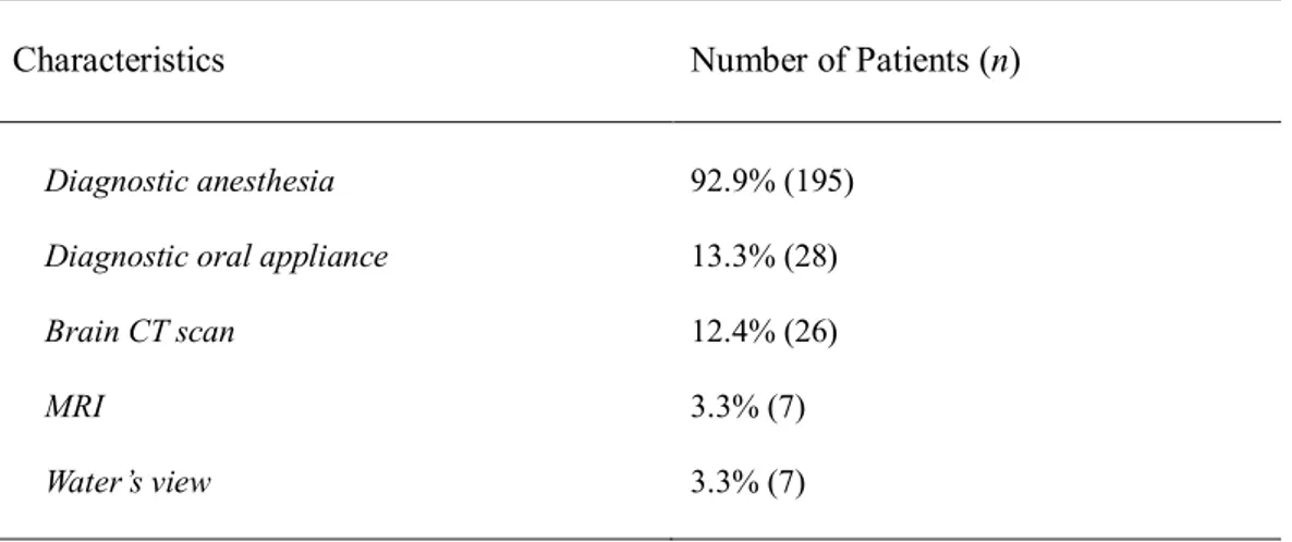
Diagnostic methods conducted by the Department of Orofacial Pain and Oral Medicine included diagnostic anesthesia (92.9%, n=195), brain CT scan (12.4%, n=26), MRI (3.3%, n=7), Water’s view (3.3%, n=7) and diagnostic oral appliance (13.3%, n=28) (Table 5).
Table 5. Diagnosis methods in the Department of Orofacial Pain and Oral Medicine*
Characteristics Number of Patients (n)
Diagnostic anesthesia Diagnostic oral appliance Brain CT scan MRI Water’s view 92.9% (195) 13.3% (28) 12.4% (26) 3.3% (7) 3.3% (7) *Permission of repetition.
CT, computerized tomography; MRI, magnetic resonance imaging.
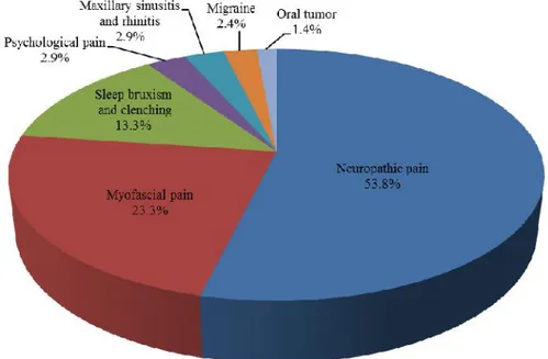
The two common diagnoses made were neuropathic pain (53.8%, n=113) and MFP (23.3%, n=49), followed by sleep bruxism and clenching (13.3%, n=28), psychological pain (2.9%, n=6), maxillary sinusitis and rhinitis (2.9%, n=6), migraine (2.4%, n=5), and oral tumor (1.4%, n=3) (Figure 3).
Figure 3. Diagnosis in the Department of Orofacial Pain and Oral Medicine (n=210)
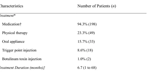
For treatment, 94.3% (n=198) of patients received medication; physical therapy (23.3%, n=49), oral appliance (15.7%, n=33), TPI (8.6%, n=18), and botulinum toxin injection (1.0%, n=2). Patients who received oral appliance and showed slight improvement (36.4%, n=12) were later diagnosed with MFP. Treatment duration ranged from 1 month to 5.7 years, with the mean treatment duration of 6.7 months (Table 6).
Table 6. Treatment in the Department of Orofacial Pain and Oral Medicine
Characteristics Number of Patients (n)
Treatment* Medication† Physical therapy Oral appliance Trigger point injection Botulinum toxin injection
94.3% (198) 23.3% (49) 15.7% (33) 8.6% (18) 1.0% (2) Treatment Duration (months)‡ 6.7 (1 to 68)
*Permission of repetition.
†Anticonvulsant, anxiolytics, antidepressant, NSAIDs, muscle relaxant, etc. ‡Mean (Range)
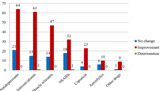
Medications used by the Department of Orofacial Pain and Oral Medicine included antidepressants (42.9%, n=85), anticonvulsants (38.9%, n=77), muscle relaxants (30.8%, n=61), NSAIDs (25.8%, n=51), topical capsaicin (13.6%, n=27) and anxiolytics (8.0%, n=16) (Figure. 4). The anticonvulsants were slightly more effective than antidepressants (79.2% vs. 75.3%) (Figure 4).
Figure 4. Efficacy of drugs administered for non-odontogenic pain
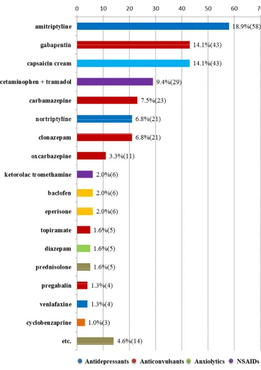
Drugs used for treatment of neuropathic pain included amitriptyline (18.9%) and gabapentin (14.1%) (Figure 5). For treatment of MFP, eperisone HCl (24.3%) and cyclobenzaprine HCl (13.6%) were used. Medications for treatment of sleep bruxism and clenching included nortriptyline (12.8%), cyclobenzaprine HCl (12.8%), and eperisone HCl (12.8%).
IV. DISCUSSION
Etiology as well as diagnosis and treatment methods for NO pain have not yet been clearly established. Thus, NO patients often suffer from chronic pain for extended periods and receive unnecessary treatment repeatedly when their symptoms fail to improve. Our study examined the population of patients with NO pain to provide a profile of the Korean patient population, comparing against the existing literature on NO pain.
We examined the demographics of the NO patient population in Korea versus those studied in the existing literature so as to investigate characteristics particular to the population in Korea. With regard to gender, our study found that the incidence of NO pain was greater in females (74.8%) than in males. It showed a small percentage compared to the previous studies (Pigg M et al., 2013, List T et al., 2007). Pigg et al. reported an incidence of NO pain in females of 83.8% (Pigg M et al., 2013) while List et al. described an incidence of 84.8% (List T et al., 2007).
Notably, concerning the relationship between NO pain and medical history, 20.5% (n=43) of patients had cardiovascular disease. The high prevalence of cardiovascular disease in the current study population may be due to the fact that hypertension is common in the general Korean population. 6.7% (n=14) of patients had gastrointestinal (GI) disorders. The prevalence of GI disease observed may also be explained by the relationship between NO pain and stress, with various studies describing associations between GI disorders and stress. Chronic pain may carry a close relationship to psychosocial problems, which have been shown to contain elements involved in pain (Dougall AL et al., 2012, Turk DC et al., 2010). In the current study,
psychological disorders were found in 4.3% (n=9) of patients and were relatively well managed, but did not seem to have any association with the presence of NO pain.
NO pain can also be iatrogenic, caused by general dental treatments. Polycarpou et al. reported that the prevalence of persistent pain after successful root canal treatment was 12% (Polycarpou N et al., 2005). 3.4% of patients experienced persistent pain of NO origin after root canal therapy, a number that likely represents about half of all persistent ‘‘tooth’’ pain (Nixdorf DR et al., 2010). In the current study, 60.5% of patients had NO symptoms that were initiated by dental treatment; among this sub-population, RCT occupied the largest proportion (55.1%), followed by prosthodontic treatment (16.5%), extraction (13.4%), periodontal surgery (1.6%). During RCT, the inferior alveolar nerve can be damaged from over instrumentation of the mandibular molar teeth, pressure within the inferior alveolar nerve canal exerted by the endodontic point or sealant, and neurotoxic effects of the used medications (Pogrel MA, 2007). Increased risks for damage to the inferior alveolar nerve during third molar operations have also been reported, with explanations including the roots’ close proximity to the third molar and mandibular canal, impaction depth, and neurotoxic materials used in surgery (Blondeau F and Daniel NG, 2007). NO pain may have been more frequently initiated following RCT, prosthodontic treatment, and extraction due to the induction of direct trauma to the peripheral nerve involved in this procedure.
This study also investigated the characteristics of pain experienced by NO patients, specifically, locations and severity of NO pain reported. With regard to locations of pain, the current study found that 30.0% of the study population were affected at the left maxilla and 27.6% at the right maxilla. This was somewhat different to the study by Ram et al., which found almost half the patients had NO pain localized to the maxillary posterior region, of which 50% were affected on the left side, 29.7% on the right side (Ram S et al., 2009). When
we compared the anterior teeth versus the posterior teeth, the posterior teeth was more frequently affected than the anterior teeth (82.3% vs. 17.7%). And the maxillary areas were also found to be slightly more affected (50.9% vs. 49.1%). The reason is considered to be due to the complex and large structure surrounding the posterior teeth. Maxillary sinusitis and rhinitis can be worsened by the adjacency of the sinus and roots of maxillary molars, inducing a pain similar to the toothache in the same area. Due to such cases of referred pain, TMJ inflammation and masticatory pain in the masseter and temporal muscles are often misperceived by patients as toothache. This seems to be the reason why NO pain is found to be present more at the maxilla than at the mandible, when in fact, this is not the case.
Regarding severity of NO pain, Pigg et al. reported on 20 patients diagnosed with NO pain, a mean pain intensity of 5.6 ± 1.8 on a scale of 0-10 and a mean pain duration of 4.3 ± 5.2 years (Pigg M et al., 2014). This present study found similar results in regard to pain intensity, with a mean pain intensity of 5.7 and 51.9% of patients reporting moderate pain, 24.3% severe pain, and 11.0% mild pain. However, the mean pain duration of 4.3 years was, notably, shorter by 2.6 years when compared to previous studies. This difference may be due to the relatively inexpensive cost for medical treatment as well as the wider accessibility to medical care due to the large number of hospitals in Korea.
Due to the difficulty in diagnosing NO pain, many NO patients often receive unnecessary treatments and multiple dental practitioners may become involved in the care of an NO patient. Thus, we investigated the frequency at which such patients receive unnecessary RCT and extraction procedures. In the current study, we found that the 59.1% of cases received RCT once, 32.3% of cases received re-RCT after RCT, and 8.6% of cases received the same RCT three times. It is considered that the result is performed without considering the different causes of dental pain. Among the 36 extraction cases studied, one tooth was extracted in 66.7%,
two teeth were extracted in 25.0%, and three teeth were extracted in 8.3% of patients. As seen by the high rate at which RCTs and extractions were performed, it seems that the main problem still remains misdiagnosis of NO pain, which may furthermore potentially lead to unnecessary treatments.
Notably, a large number of referrals made to the Department of Orofacial Pain and Oral Medicine were from the Department of Conservative Dentistry (DCD) (28.6%). The high referral rate is probably because patients suffering from persistent pain even following RCT are typically referred to the DCD, but a portion of such patients may have been referred back to the Department of Orofacial Pain and Oral Medicine after their pain is found to stem from NO origins. Thus, NO pain should be considered if persistent pain is not resolved following endodontic treatment.
Furthermore, NO patients with complex dental histories are often treated by multiple dental practitioners prior to the arrival of suspicion that symptoms may be a result of NO pain. Israel et al. published a report on 120 patients with chronic facial pain who sought treatment at the Center for Oral, Facial and Head Pain at New York Presbyterian Hospital for more than two years (Israel HA et al., 2003). They reported that on average, a patient with facial pain consulted with six specialists for their pain before reaching an orofacial pain clinic (Israel HA et al., 2003). In the current study, the total number of dentists involved in a single patient’s treatment ranged from 2 to 7, averaging out to 2.7 dental practitioners. A majority of patients (46.7%) were treated by 2 dental practitioners. The involvement of multiple dental practitioners and specialists required in the arrival to diagnosis and treatment of NO pain seen in our study reasserts the fact that diagnosis and treatment of NO pain still remains a difficult and complicated process. As seen by the involvement of multiple dental practitioners per NO
patient, the current study reasserts the fact that arrival to diagnosis and treatment of NO pain remains a difficult and complicated process.
Among the various diagnostic methods used for the differential diagnosis of NO pain, local anesthesia is one method thought to be a very valuable and effective tool in identifying the source of pain. List et al. evaluated the effect of local anesthesia (lidocaine) in a randomized controlled trial on 35 consecutive NO patients and found that administration of local anesthetics resulted in significant pain relief (List T et al., 2006). VAS was reduced by an average of 21.4% after topical anesthesia and an average of 70.6% after infiltration (List T et al., 2006). In the current study, the most commonly used diagnostic method was anesthesia (92.9%). Diagnostic anesthesia was also conducted using both topical and infiltration anesthesia, and VAS was measured before and after anesthesia. Consistent with the study by List et al., infiltration anesthesia reduced pain more than did topical anesthesia, though the degree of response varied by patient (List T et al., 2006). If the pain derives from the pulp, namely, from the tooth itself, the nerves of the affected teeth must be anesthetized as such pain will not decrease with just the use of topical anesthesia. When pain decreases following administration of local anesthetics, the cause of such continuous pain of the teeth or gums was often found to be neuropathic pain. These results substantiate the notion that diagnostic anesthesia is an effective and relatively simple diagnostic method, one that can be performed at the chair-side and can be satisfactorily used during differential diagnosis of NO pain.
NO pain is a type of heterotopic pain that includes projected nerve pain felt throughout the peripheral distribution of the affected nerve (e.g. TN, cluster headache, post herpetic neuralgia etc.) and referred pain perceived as a toothache as a result of convergence and central sensitisation (e.g. myofascial, sinus, or cardiac pain, etc.) (De Leeuw R, 2013). In accordance with these diagnoses of NO pain, Tomoyasu et al. retrospectively investigated the
characteristics of 221 dental patients who had suffered from persistent orofacial pain (Tomoyasu Y et al., 2014). The main diagnoses were neuropathic pain (30.3%), MFP (23.5%), psychogenic pain (20.4%), odontogenic toothache (17.2%), and others (7.7%) such as temporomandibular disorders (TMD) and glossitis (Tomoyasu Y et al., 2014). Wirz et al. reported that of 476 patients suffering from orofacial pain, 26.4% of patients suffered from one of the following secondary pain syndromes: TMD (MFP: 39.6%, involvement of temporomandibular joint: 33.5%); migraine, 15.2%; neuropathic pain, 5.9% (Wirz S et al., 2010). In the current study, neuropathic pain (53.8%) and MFP (23.3%) were the two common diagnoses. Compared to previous studies (Tomoyasu Y et al., 2014, Wirz S et al., 2010), the rates of MFP were smaller. This may be due to the fact that differential diagnosis of MFP was relatively easier compared to that of neuropathic pain. Thus, at the moment of diagnosis for patients complaining of NO pain, neuropathic pain should be considered with high-priority.
Once patients are diagnosed with NO pain, they may undergo treatment using medications, for which guidelines exist regarding types of medications to be used in various cases. Recently, a systematic review on the effectiveness of anticonvulsants for the management of orofacial pain was conducted using randomized controlled trials (Martin WJ et al., 2012). In this review, gabapentin, topical clonazepam, lamotrigine, and carbamazepine were found to significantly reduce pain intensity in patients with chronic masticatory myalgia, stomatodynia, and neuropathic pain. In another review on the clinical management of NO pain, most of experts recommended the use of tricyclic antidepressants for NO pain, though the use of anticonvulsants, such as phenytoin, carbamazepine, pregabalin and gabapentin, was also proposed (Baad-Hansen L, 2008). For trigeminal neuropathic pain, the consensus is that tricyclic antidepressants, especially nortriptyline, and calcium channel blockers like gabapentin and pregabalin, should be first-line therapies (Vedula SS et al., 2009). Selective
serotonin/norepinephrine reuptake inhibitors and specific selective norepinephrine reuptake inhibitors are considered as second-line therapies (O’Connor AB and Dworkin RH, 2009). Due to the overlap of both neuropathic and nociceptive mechanisms in trigeminal neuropathic pain, opioids and tramadol are considered the third-line therapies, though they should only be used on a limited basis and with close monitoring by the prescribing provider (Napeñas JJ and Zakrzewska JM, 2011).
Topical medicaments have been advocated as first-line treatments for peripheral neuropathic pain due to the minimal systemic side effects. They may be delivered to the injured or affected sites with the aid of shields or custom made intraoral stents made of a soft splint material (Napeñas JJ and Zakrzewska JM, 2011). In open labeled studies, topical amitriptyline was effective in peripheral neuropathy, and capsaicin 0.025% cream was effective in oral neuropathic pain and traumatic trigeminal dysesthesias (Lynch ME et al., 2005). Another study showed that local anesthesia provided some, but not complete NO pain relief (Ram S et al., 2009).
However, medication therapy for NO patients is complicated and diverse, requiring the use of a combination of several medications. In the current study, anticonvulsants such as gabapentin, carbamazepine, and clonazepam were widely use, however, antidepressants such as amitriptyline and nortriptyline were used slightly more often. Among anticonvulsants, gabapentin was the most commonly used medication. The efficacy of anticonvulsants was slightly stronger than that of antidepressants. Capsaicin topical agent was found to be more effective when combined with other medications. The mean treatment duration was 6.7 months. Because treatment of NO pain requires various specialized drug treatments and long-term treatment periods, it is recommended that when NO pain is suspected, patients are referred to either a pain specialist or the Department of Orofacial Pain and Oral Medicine.
Non-pharmacological approaches like behavioral modifications (soft food diet, resting of the jaw, self-massage, etc.) and physical therapy (stretching exercises, massage, thermotherapy, and posture correction) are effective against pain deriving from myofascial problems (De Leeuw R, 2013). TPI was performed with 2% lidocaine hydrochloride to the tender point in the masseter muscle. Handa et al. diagnosed MFP based on evidence of NO tooth pain caused by referred pain from the masseter muscle (Handa T et al., 2013). The VAS pain score dropped from 7~9 to 3~6, and the effects of TPI lasted for approximately 7 hours, after which severe throbbing pain returned (Handa T et al., 2013). In the current study, TPI was effective in reducing pain in only 5 patients of the 18 patients (6.7%) who received TPI. Muscle relaxants can be used for pain relief, but their use should be limited to a few weeks (De Leeuw R, 2013). Pharmacologic therapies that demonstrated efficacy for myofascial pain causing NO toothache involved ibuprofen and low-dose amitriptyline (De Leeuw R, 2013). Existing studies support the efficacy of occlusal splint therapy in treating MFP (Al-Ani Z et al., 2005, Fricton J, 2006). In the current study as well, 13 (39.4%) of the 33 patients treated with oral appliance reported slight improvement of symptoms and all were diagnosed with MFP, suggesting the limited effects of non-pharmacologic approaches in the mitigation of pain. This also puts forward the notion that the causes of NO pain may root not from the musculature, but rather, from the nervous system.
Etiology as well as diagnosis and treatment methods for NO pain have not yet been clearly established. Despite the fact that patients are often treated multiple times at various dental offices, such patients often fail to show improvement of symptoms. This study aimed to investigate clinical patterns and related characteristics of NO pain patients. Although a clear relationship was not found in all characteristics due to limited number of subjects and time constraints.
This study’s profiling of NO pain will potentially be of use in the diagnosis and treatment of patients who developed NO pain due to general dental treatments. Furthermore, our results will hopefully be beneficial in decrease the number unnecessary care performed to reduce NO pain. Further studies with larger number of subjects and extended length of study are required for more systemized diagnostic methods and development of future treatment guidelines.
V. CONCLUSION
The purpose of this study is to profile patients with NO pain, to investigate the relationship between NO pain and previous dental treatments, and to gain helpful information about the diagnosis and treatment of NO pain.
Based on the obtained results, we report the following findings:
1. NO pain was diagnosed mainly as neuropathic pain (53.8%) and MFP (23.3%).
2. There were 65.7% without medical history, 20.5% cardiovascular diseases, 6.7% with GI troubles and 4.3% with psychogenic diseases. And only few of them have smoking (male 6.7%, female 1.0%) and drinking habits (male 8.6%, female 5.7%).
3. The common site of NO pain is left maxilla (30.0%), and right maxilla (27.6%). Distribution of affected area were generally in accordance with previous reports but was not predominant in maxillary posterior region. Single area was affected in 90 patients (42.9%) with similar occurrence with multiple lesions (41.0%).
4. 17 patients (8.1%) had referred pain. 63.8% of patients have spontaneous pain. And the number of patients who have severe pain and who have moderate pain were 24.3%, 51.9% each. The average period of treatment was 6.7 months.
5. NO pain occurs more often to the female (74.8%) than to the male (25.2%). The average age is 49.0 years and the average pain duration is 31 months.
6. Considering the departments referring to the Department of Orofacial Pain and Oral Medicine, treatments related to NO pain, previous dental treatment history, and visiting dental departments, NO pain is very closely related to the RCT. 48.6% of patients have treated more than 3 dentists.
7. The used drugs were mainly antidepressants (42.9%) and anticonvulsants (38.9%). And the effectiveness of anticonvulsants (79.2%) is better than that of antidepressants (75.3%).
8. Mainly the treatment drugs of neuropathic pain used amitriptyline (18.9%) and gabapentin (14.1%). And topical capsaicin has a limited effect.
9. Mainly the treatment drugs of MFP used eperisone HCl (24.3%) and cyclobenzaprine HCl (13.6%).
In conclusion, patients in Korea suffering from NO pain often visit multiple dental practitioners and receive unnecessary treatments before arrival to the correct diagnosis. In this study, such NO patients were largely diagnosed to have neuropathic pain and MFP. Rather than performing irreversible treatments, such as RCT or extraction, this study provides grounds for encouraging reversible treatments such as medications or physical therapy that will better benefit NO patients. With the continual publishing of future studies, we believe that a guideline will eventually be made for the treatment of NO patients in Korea.
REFERENCES
Ram S, Teruel A, Kumar SK, Clark G: Clinical characteristics and diagnosis of atypical odontalgia: implications for dentists. J Am Dent Assoc. 2009 Feb;140(2):223-8.
Mattscheck D, Law AS, Nixdorf DR: Diagnosis of non-odonogentic toothache. In: Hargreaves KM, Cohen S, eds. Cohen’s Pathways of the Pulp, 10th ed. St Louis: Mosby; 2011:49–70.
Melis M, Lobo SL, Ceneviz C, Zawawi K, Al-Badawi E, Maloney G, Mehta N: Atypical odontalgia: a review of the literature. Headache. 2003;43:1060-74.
Yatani H, Komiyama O, Matsuka Y, Wajima K, Muraoka W, Ikawa M, Sakamoto E, De Laat A, Heir GM: Systematic review and recommendations for NO toothache. J Oral Rehabil. 2014 Nov;41(11):843-52.
Pigg M, Svensson P, Drangsholt M, List T: Seven-year follow-up of patients diagnosed with atypical odontalgia: a prospective study. J Orofac Pain. 2013 Spring;27(2):151-64.
List T, Leijon G, Helkimo M, Oster A, Dworkin SF, Svensson P: Clinical findings and psychosocial factors in patients with atypical odontalgia: a case-control study. J Orofac Pain. 2007 Spring;21(2):89-98.
Dougall AL, Jimenez CA, Haggard RA, Stowell AW, Riggs RR, Gatchel RJ: Biopsychosocial factors associated with the subcategories of acute temporomandibular joint disorders. J Orofac Pain. 2012;26:7–16.
Turk DC, Audette J, Levy RM, Mackey SC, Stanos S: Assessment and treatment of psychosocial comorbidities in patients with neuropathic pain. Mayo Clin Proc. 2010;85:S42– S50.
Polycarpou N, Ng YL, Canavan D, Moles DR, Gulabivala K: Prevalence of persistent pain after endodontic treatment and factors affecting its occurrence in cases with complete radiographic healing. Int Endod J. 2005 Mar;38(3):169-78.
Nixdorf DR, Moana-Filho EJ, Law AS, McGuire LA, Hodges JS, John MT: Frequency of nonodontogenic pain after endodontic therapy: a systematic review and meta-analysis. J Endod. 2010 Sep;36(9):1494-8.
Pogrel MA: Damage to the inferior alveolar nerve as the result of root canal therapy. J Am Dent Assoc. 2007;138(1):65-9.
Blondeau F, Daniel NG: Extraction of impacted mandibular third molars: postoperative complications and their risk factors. J Can Dent Assoc. 2007;73(4):325.
Pigg M, List T, Abul-Kasim K, Maly P, Petersson A: A comparative analysis of magnetic resonance imaging and radiographic examinations of patients with atypical odontalgia. J Oral Facial Pain Headache. 2014 Summer;28(3):233-42.
Israel HA, Ward JD, Horrell B, Scrivani SJ: Oral and maxillofacial surgery in patients with chronic orofacial pain. J Oral Maxillofac Surg. 2003;61(6):662-667.
List T, Leijon G, Helkimo M, Oster A, Svensson P: Effect of local anesthesia on atypical odontalgia--a randomized controlled trial. Pain. 2006 Jun;122(3):306-14.
de Leeuw R (ed). The American Academy of Orofacial Pain. Orofacial pain – guidelines for assessment, diagnosis, and management. Chicago (IL): Quintessence, 2013.
Tomoyasu Y, Higuchi H, Mori M, Takaya K, Honda Y, Yamane A, Yabuki A, Hayashi T, Ishii-Maruyama M, Jinzenji A, Maeda S, Kohjitani A, Shimada M, Miyawaki T: Chronic orofacial pain in dental patients: retrospective investigation over 12 years. Acta Med Okayama. 2014;68(5):269-75.
Wirz S, Ellerkmann RK, Buecheler M, Putensen C, Nadstawek J, Wartenberg HC: Management of chronic orofacial pain: a survey of general dentists in german university hospitals. Pain Med. 2010 Mar;11(3):416-24.
Martin WJ, Perez RS, Tuinzing DB, Forouzanfar T: Efficacy of antidepressants on orofacial pain: a systematic review. Int J Oral Maxillofac Surg. 2012 Dec;41(12):1532-9.
Baad-Hansen L: Atypical odontalgia -- pathophysiology and clinical management. J Oral Rehabil. 2008;35:1-11
Vedula SS, Bero L, Scherer RW, Dickersin K: Outcome reporting in industry-sponsored trials of gabapenitn for off-label use. N Engl J Med. 2009 Nov 12;361(20):1963-71.
O’Connor AB, Dworkin RH: Treatment of neuropathic pain: an overview of recent guidelines. Am J Med. 2009 Oct;122(10 Suppl):S22-32.
Napeñas JJ, Zakrzewska JM: Diagnosis and management of trigeminal neuropathic pains. Pain Manag. 2011 Jul;1(4):353-65.
Lynch ME, Clark AJ, Sawynok J, Sullivan MJ: Topical amitriptyline and ketamine in neuropathic pain syndromes: an open-label study. J Pain. 2005 Oct;6(10):644-9.
Handa T, Fukuda K, Ichinohe T: Effect of combination of trigger point injection and stellate ganglion block on non-odontogenic mandibular molar pain referred from masseter muscle: a case report. Bull Tokyo Dent Coll. 2013;54(3):171-5.
Al-Ani Z, Gray RJ, Davies SJ, Sloan P, Glenny AM: Stabilization splint therapy for the treatment of temporomandibular myofascial pain: a systematic review. J Dent Educ. 2005 Nov;69(11):1242-50.
Fricton J: Current evidence providing clarity in management of temporomandibular disorders: summary of a systematic review of randomized clinical trials for intra-oral appliances and occlusal therapies. J Evid Based Dent Pract. 2006 Mar;6(1):48-52.
ABSTRACT (in Korean)
비치성 통증으로 안면통증 클리닉에 내원한 환자의 특성
<지도교수 김 성 택>
연세대학교 대학원 치의학과
서 정 일
비치성 통증에 대한 원인 및 진단, 치료는 아직 명확하게 밝혀진 바는 없다. 일반적으로 비치성 통증을 호소하는 환자는 비교적 복잡한 치과치료 병력을 가지고 있으나 증의 변화가 없다. 결국 환자는 불필요한 치과치료를 받으면서도 주소부위에 대한 해결은 이루어지지 않고 만성적인 동통에 시달려 고통을 받게 된다. 그러나 우리나라에서는 아직 이러한 비치성 통증 환자에 대한 체계적인 연구조사가 부족한 상태이다. 이에 본 연구에서는 비치성 통증 환자의 임상적 특징과 병력조사를 통해 다양한 연관성을 조사하여 비치성 통증 환자가 불필요한 치과치료를 받지 않도록 진단과 치료에 새로운 지침을 마련하고자 다음과 같은 연구를 시행하였다2003 년 5 월부터 2013 년 4 월까지 연세대학교 치과대학병원 구강내과에 내원한 비치성 통증으로 여겨지는 환자 남자 53 명, 여자 157 명, 총 210 명을 대상으로 비치성 통증 환자의 진료기록부를 통하여 성별, 연령, 동통기간, 전신질환, 증상발현부위, 증상발현의 다발성 여부 등에 대해 조사하였다. 또한 구강내과에서 사용된 진단방법, 진단명, 치료 방법, 사용된 약물의 빈도, 그리고 치료 효과에 대해 분석을 시행하였다. 이 연구를 통해 다음과 같은 결과를 알 수 있었다. 1. 비치성 통증의 흔한 임상적 진단은 신경병증성 통증(53.8%)과 근막동통 (23.3%)이었다. 2. 전신질환 병력이 없는 환자가 65.7%로 가장 많았고, 다음으로 심혈관질환 20.5%, 위장장애 6.7%의 비유로 나타났다. 정신과 질환 병력은 4.3%로 연관성은 낮은 것으로 보이고 흡연은 7.6%, 음주는 14.3%이었다. 3. 증상 발현부위는 좌측상악구치부(30.3%)와 우측상악구치부(27.6%)에서 비슷하게 나타났고 대부분이 구치부에서 나타났다. 증상발현의 다발성 여부는 단독 부위에만 나타난 경우는 42.9%로 단독으로 나타나는 경우와 다발성으로 나타나는 경우가 비슷하다. 4. 연관통은 8.1%로 적게 나타났으며, 자발통은 63.8%로 다소 높게 나타났다. 통증양상은 중간의 동통이 51.9%로 많았으며, 평균 치료기간은 6.7 개월이었다.
5. 대상 환자 중 여자 157 명, 남자 53 명으로 여자에게 더 많이 발생하였으며, 평균연령은 49.0 세였고 동통기간은 평균 31 개월이었다. 6. 구강내과로 협의진료를 의뢰한 과, 증상발현과 연관된 치료, 이전 치과 병력 및 그 동안 환자가 내원한 과를 종합하여 보면 근관치료와의 연관성이 가장 높다고 볼 수 있다. 7. 약물의 사용은 다양하지만 항우울제(42.9%)와 항경련제(38.9%)가 주로 사용되고, 효과는 항경련제(79.2%)가 항우울제(75.3%)보다 다소 높게 나타났다. 8. 신경병증성 통증에는 항우울제인 amitriptyline(18.9%)과 항경련제인 gabapentin(14.1%)의 사용빈도가 높았고 capsaicin 도포제도 제한적이지만 효과를 보였다. 9. 근막동통에는 근이완제인 eperisone HCl(24.3%)과 cyclobenzaprine HCl (13.6%)의 사용빈도가 높았다.