저작자표시-비영리-변경금지 2.0 대한민국 이용자는 아래의 조건을 따르는 경우에 한하여 자유롭게 l 이 저작물을 복제, 배포, 전송, 전시, 공연 및 방송할 수 있습니다. 다음과 같은 조건을 따라야 합니다: l 귀하는, 이 저작물의 재이용이나 배포의 경우, 이 저작물에 적용된 이용허락조건 을 명확하게 나타내어야 합니다. l 저작권자로부터 별도의 허가를 받으면 이러한 조건들은 적용되지 않습니다. 저작권법에 따른 이용자의 권리는 위의 내용에 의하여 영향을 받지 않습니다. 이것은 이용허락규약(Legal Code)을 이해하기 쉽게 요약한 것입니다. Disclaimer 저작자표시. 귀하는 원저작자를 표시하여야 합니다. 비영리. 귀하는 이 저작물을 영리 목적으로 이용할 수 없습니다. 변경금지. 귀하는 이 저작물을 개작, 변형 또는 가공할 수 없습니다.
The molecular mechanisms of
hearing loss in tubby mice
Woongsu Han
Department of Medical Science
The Graduate School, Yonsei University
[UCI]I804:11046-000000514284
[UCI]I804:11046-000000514284
[UCI]I804:11046-000000514284
The molecular mechanisms of
hearing loss in tubby mice
Woongsu Han
Department of Medical Science
The Graduate School, Yonsei University
The molecular mechanisms of
hearing loss in tubby mice
Directed by Professor Chul Hoon Kim
The Doctoral Dissertation submitted to the
Department of Medical Science,
The Graduate School of Yonsei University
in partial fulfillment of the requirements for the degree of
Doctor of Philosophy
Woongsu Han
December 2017
This certifies that the Doctoral Dissertation
of Woongsu Han is approved.
Thesis Supervisor: Chul Hoon Kim
Thesis Committee Member#1: Dong Goo Kim
Thesis Committee Member#2: Jinwoong Bok
Thesis Committee Member#3: Jae Young Choi
Thesis Committee Member#4: Seok Jun Moon
The Graduate School
Yonsei University
ACKNOWLEDGEMENTS
학위 과정에 진학하기 위해 이 곳 의과대학 약리학교실
의 문을 두드린 지 어느덧 약 8년 이라는 시간이 흘렀습
니다. 오랜 시간이 지났지만 돌이켜 보면 처음 이 곳에
와서 제가 사용할 실험 테이블을 닦고 정리하던 기억이
생생하게 떠오릅니다. 다니던 회사를 정리하고 이곳에서
다시 공부를 하겠다고 마음을 다지고 시작했지만, 일이
잘 안 풀릴 때마다 마음가짐이 흔들릴 때도 있었습니다.
하지만 많은 분들의 도움으로 여기까지 올 수 있었습니다.
여러 우여곡절이 있었지만 부족한 저를 물심양면으로 지
원하고 지도해 주신 김철훈 교수님께 진심으로 감사의 말
씀을 드립니다. 그리고 개인적인 고민과 상담을 들어 주
시고 힘이 되는 조언과 함께 아낌없는 지원을 베풀어 주
신 김동구 교수님께도 진심으로 감사의 말씀을 드립니다.
연구 방향에 대한 좋은 의견과 격려의 말씀을 아낌없이
해 주신 약리학교실 교수님들과 선생님들께도 감사의 말
씀을 전합니다. 또한 현재 연구실에는 안계시지만, 연구
실을 거쳐 가신 여러 선생님들께도 감사하다는 말씀 전하
고 싶습니다.
본 프로젝트를 진행해 오면서 수많은 난관에 부딪혔지
만, 많은 분들의 도움으로 좋은 성과를 이룰 수 있었습니
다. 프로젝트를 위해 아낌없이 지원해 주신 복진웅 교수
님, 문석준 교수님, 최재영 교수님, 신정오 박사님, 마지
현 박사님께 정말 감사하다는 말씀 드리고 싶습니다.
끝으로 지금까지 저를 묵묵하게 믿고 지원해 준 아내와
부모님, 장인어른, 장모님, 그리고 아들 유준이에게 진심
으로 고맙고 사랑한다는 말을 전합니다.
2017년 12월 한웅수
TABLE OF CONTENTS
ABSTRACT∙∙∙∙∙∙∙∙∙∙∙∙∙∙∙∙∙∙∙∙∙∙∙∙∙∙∙∙∙∙∙∙∙∙∙∙∙∙∙∙∙∙∙∙∙∙∙∙∙∙∙∙∙∙∙∙∙∙∙∙∙∙∙∙∙∙∙∙∙∙∙∙∙∙∙∙∙∙∙∙∙∙∙∙∙∙∙∙∙∙∙∙∙∙∙∙∙∙∙∙1
I. INTRODUCTION∙∙∙∙∙∙∙∙∙∙∙∙∙∙∙∙∙∙∙∙∙∙∙∙∙∙∙∙∙∙∙∙∙∙∙∙∙∙∙∙∙∙∙∙∙∙∙∙∙∙∙∙∙∙∙∙∙∙∙∙∙∙∙∙∙∙∙∙∙∙∙∙∙∙∙∙∙∙∙∙∙∙∙∙∙∙4
1. Tubby and tubby-like proteins∙∙∙∙∙∙∙∙∙∙∙∙∙∙∙∙∙∙∙∙∙∙∙∙∙∙∙∙∙∙∙∙∙∙∙∙∙∙∙∙∙∙∙∙∙∙∙∙∙∙∙∙∙∙∙∙∙∙∙∙∙∙∙∙∙5
2. Phenotypes of tubby mice∙∙∙∙∙∙∙∙∙∙∙∙∙∙∙∙∙∙∙∙∙∙∙∙∙∙∙∙∙∙∙∙∙∙∙∙∙∙∙∙∙∙∙∙∙∙∙∙∙∙∙∙∙∙∙∙∙∙∙∙∙∙∙∙∙∙∙∙∙∙∙∙7
3. Aims of study∙∙∙∙∙∙∙∙∙∙∙∙∙∙∙∙∙∙∙∙∙∙∙∙∙∙∙∙∙∙∙∙∙∙∙∙∙∙∙∙∙∙∙∙∙∙∙∙∙∙∙∙∙∙∙∙∙∙∙∙∙∙∙∙∙∙∙∙∙∙∙∙∙∙∙∙∙∙∙∙∙∙∙∙∙∙∙∙∙∙∙9
II. MATERIALS AND METHODS∙∙∙∙∙∙∙∙∙∙∙∙∙∙∙∙∙∙∙∙∙∙∙∙∙∙∙∙∙∙∙∙∙∙∙∙∙∙∙∙∙∙∙∙∙∙∙∙∙∙∙∙∙∙∙∙∙∙∙∙10
1. Animals∙∙∙∙∙∙∙∙∙∙∙∙∙∙∙∙∙∙∙∙∙∙∙∙∙∙∙∙∙∙∙∙∙∙∙∙∙∙∙∙∙∙∙∙∙∙∙∙∙∙∙∙∙∙∙∙∙∙∙∙∙∙∙∙∙∙∙∙∙∙∙∙∙∙∙∙∙∙∙∙∙∙∙∙∙∙∙∙∙∙∙∙∙∙∙∙∙∙10
2. Hearing ability measurements∙∙∙∙∙∙∙∙∙∙∙∙∙∙∙∙∙∙∙∙∙∙∙∙∙∙∙∙∙∙∙∙∙∙∙∙∙∙∙∙∙∙∙∙∙∙∙∙∙∙∙∙∙∙∙∙∙∙∙∙∙∙∙10
A. ABR measurement∙∙∙∙∙∙∙∙∙∙∙∙∙∙∙∙∙∙∙∙∙∙∙∙∙∙∙∙∙∙∙∙∙∙∙∙∙∙∙∙∙∙∙∙∙∙∙∙∙∙∙∙∙∙∙∙∙∙∙∙∙∙∙∙∙∙∙∙∙∙∙∙∙∙∙∙11
B. DPOAE measurement∙∙∙∙∙∙∙∙∙∙∙∙∙∙∙∙∙∙∙∙∙∙∙∙∙∙∙∙∙∙∙∙∙∙∙∙∙∙∙∙∙∙∙∙∙∙∙∙∙∙∙∙∙∙∙∙∙∙∙∙∙∙∙∙∙∙∙∙∙∙∙12
3. Scanning electron microscopy∙∙∙∙∙∙∙∙∙∙∙∙∙∙∙∙∙∙∙∙∙∙∙∙∙∙∙∙∙∙∙∙∙∙∙∙∙∙∙∙∙∙∙∙∙∙∙∙∙∙∙∙∙∙∙∙∙∙∙∙∙∙∙13
4. Transmission electron microscopy∙∙∙∙∙∙∙∙∙∙∙∙∙∙∙∙∙∙∙∙∙∙∙∙∙∙∙∙∙∙∙∙∙∙∙∙∙∙∙∙∙∙∙∙∙∙∙∙∙∙∙∙∙∙∙∙∙13
5. FM1-43 dye uptake assay∙∙∙∙∙∙∙∙∙∙∙∙∙∙∙∙∙∙∙∙∙∙∙∙∙∙∙∙∙∙∙∙∙∙∙∙∙∙∙∙∙∙∙∙∙∙∙∙∙∙∙∙∙∙∙∙∙∙∙∙∙∙∙∙∙∙∙∙∙∙14
6. Generation of tubby and stereocilin antibodies∙∙∙∙∙∙∙∙∙∙∙∙∙∙∙∙∙∙∙∙∙∙∙∙∙∙∙∙∙∙∙∙∙∙∙∙∙15
7. Immunolabeling studies∙∙∙∙∙∙∙∙∙∙∙∙∙∙∙∙∙∙∙∙∙∙∙∙∙∙∙∙∙∙∙∙∙∙∙∙∙∙∙∙∙∙∙∙∙∙∙∙∙∙∙∙∙∙∙∙∙∙∙∙∙∙∙∙∙∙∙∙∙∙∙∙∙15
8. Semi-quantitative real-time polymerase chain reaction (PCR) ∙∙∙∙∙∙∙∙∙∙∙∙17
9. Statistical analysis∙∙∙∙∙∙∙∙∙∙∙∙∙∙∙∙∙∙∙∙∙∙∙∙∙∙∙∙∙∙∙∙∙∙∙∙∙∙∙∙∙∙∙∙∙∙∙∙∙∙∙∙∙∙∙∙∙∙∙∙∙∙∙∙∙∙∙∙∙∙∙∙∙∙∙∙∙∙∙∙∙∙17
III. RESULTS∙∙∙∙∙∙∙∙∙∙∙∙∙∙∙∙∙∙∙∙∙∙∙∙∙∙∙∙∙∙∙∙∙∙∙∙∙∙∙∙∙∙∙∙∙∙∙∙∙∙∙∙∙∙∙∙∙∙∙∙∙∙∙∙∙∙∙∙∙∙∙∙∙∙∙∙∙∙∙∙∙∙∙∙∙∙∙∙∙∙∙∙∙∙∙18
1. Tubby mice show elevated ABR threshold and no DPOAE responses
∙∙∙∙∙∙∙∙∙∙∙∙∙∙∙∙∙∙∙∙∙∙∙∙∙∙∙∙∙∙∙∙∙∙∙∙∙∙∙∙∙∙∙∙∙∙∙∙∙∙∙∙∙∙∙∙∙∙∙∙∙∙∙∙∙∙∙∙∙∙∙∙∙∙∙∙∙∙∙∙∙∙∙∙∙∙∙∙∙∙∙∙∙∙∙∙∙∙∙∙∙∙∙∙∙∙∙∙∙18
2. Horizontal top connectors and lateral links are disappeared in OHC hair
bundles of tubby mice∙∙∙∙∙∙∙∙∙∙∙∙∙∙∙∙∙∙∙∙∙∙∙∙∙∙∙∙∙∙∙∙∙∙∙∙∙∙∙∙∙∙∙∙∙∙∙∙∙∙∙∙∙∙∙∙∙∙∙∙∙∙∙∙∙∙∙∙∙∙∙∙∙∙∙∙21
3. MET channels of OHCs are functionally normal in tubby mice∙∙∙∙∙∙∙∙∙∙∙26
4. Tub proteins are localized to the stereocilia tip of OHC hair
bundles∙∙∙∙∙∙∙∙∙∙∙∙∙∙∙∙∙∙∙∙∙∙∙∙∙∙∙∙∙∙∙∙∙∙∙∙∙∙∙∙∙∙∙∙∙∙∙∙∙∙∙∙∙∙∙∙∙∙∙∙∙∙∙∙∙∙∙∙∙∙∙∙∙∙∙∙∙∙∙∙∙∙∙∙∙∙∙∙∙∙∙∙∙∙∙∙∙∙∙28
5. The OHC hair bundle localization of stereocilin is disrupted in tubby
mice∙∙∙∙∙∙∙∙∙∙∙∙∙∙∙∙∙∙∙∙∙∙∙∙∙∙∙∙∙∙∙∙∙∙∙∙∙∙∙∙∙∙∙∙∙∙∙∙∙∙∙∙∙∙∙∙∙∙∙∙∙∙∙∙∙∙∙∙∙∙∙∙∙∙∙∙∙∙∙∙∙∙∙∙∙∙∙∙∙∙∙∙∙∙∙∙∙∙∙∙∙∙∙31
6. Expression of stereocilin mRNA in cochlea of tubby mice is normal∙∙∙34
IV. DISCUSSION∙∙∙∙∙∙∙∙∙∙∙∙∙∙∙∙∙∙∙∙∙∙∙∙∙∙∙∙∙∙∙∙∙∙∙∙∙∙∙∙∙∙∙∙∙∙∙∙∙∙∙∙∙∙∙∙∙∙∙∙∙∙∙∙∙∙∙∙∙∙∙∙∙∙∙∙∙∙∙∙∙∙∙∙∙∙∙∙∙36
V. CONCLUSION∙∙∙∙∙∙∙∙∙∙∙∙∙∙∙∙∙∙∙∙∙∙∙∙∙∙∙∙∙∙∙∙∙∙∙∙∙∙∙∙∙∙∙∙∙∙∙∙∙∙∙∙∙∙∙∙∙∙∙∙∙∙∙∙∙∙∙∙∙∙∙∙∙∙∙∙∙∙∙∙∙∙∙∙∙∙∙∙41
REFERENCES∙∙∙∙∙∙∙∙∙∙∙∙∙∙∙∙∙∙∙∙∙∙∙∙∙∙∙∙∙∙∙∙∙∙∙∙∙∙∙∙∙∙∙∙∙∙∙∙∙∙∙∙∙∙∙∙∙∙∙∙∙∙∙∙∙∙∙∙∙∙∙∙∙∙∙∙∙∙∙∙∙∙∙∙∙∙∙∙∙∙∙∙∙42
ABSTRACT (IN KOREAN) ∙∙∙∙∙∙∙∙∙∙∙∙∙∙∙∙∙∙∙∙∙∙∙∙∙∙∙∙∙∙∙∙∙∙∙∙∙∙∙∙∙∙∙∙∙∙∙∙∙∙∙∙∙∙∙∙∙∙∙∙∙∙∙∙∙∙∙∙∙∙46
LIST OF FIGURES
Figure 1. Auditory phenotypes of wild-type and tubby mice∙∙∙∙∙∙20
Figure 2. OHCs hair bundle morphology of wild-type and tubby
mice∙∙∙∙∙∙∙∙∙∙∙∙∙∙∙∙∙∙∙∙∙∙∙∙∙∙∙∙∙∙∙∙∙∙∙∙∙∙∙∙∙∙∙∙∙∙∙∙∙∙∙∙∙∙∙∙∙∙∙∙∙∙∙∙∙∙∙∙∙∙∙∙∙∙∙∙∙23
Figure 3. OHCs hair bundle morphology of wild-type and tubby
mice during OHCs hair bundle maturation∙∙∙∙∙∙∙∙∙∙∙∙∙∙∙∙∙∙25
Figure 4. FM1-43 dye enters OHCs of tubby mice through MET
channel∙∙∙∙∙∙∙∙∙∙∙∙∙∙∙∙∙∙∙∙∙∙∙∙∙∙∙∙∙∙∙∙∙∙∙∙∙∙∙∙∙∙∙∙∙∙∙∙∙∙∙∙∙∙∙∙∙∙∙∙∙∙∙∙∙∙∙∙∙∙∙∙∙27
Figure 5. Localization of Tub protein in cochlear OHCs∙∙∙∙∙∙∙∙∙∙∙∙29
Figure 6. Disrupted OHCs hair bundle localization of stereocilin
in tubby mice∙∙∙∙∙∙∙∙∙∙∙∙∙∙∙∙∙∙∙∙∙∙∙∙∙∙∙∙∙∙∙∙∙∙∙∙∙∙∙∙∙∙∙∙∙∙∙∙∙∙∙∙∙∙∙∙∙∙∙∙∙∙∙33
Figure 7. Gene expression level of stereocilin in cochlea of
1
ABSTRACT
The molecular mechanisms of hearing loss in tubby mice
Woongsu Han
Department of Medical Science,
The Graduate School, Yonsei University
(Directed by Professor Chul Hoon Kim)
Tubby mice are mutant mice which arose spontaneously that exhibit three major
phenotypes: late onset obesity, retinal degeneration, and progressive hearing loss.1-4
The point mutation in the splice site of the tub gene completely displaces the 44 amino acids of the C-terminus of the Tub protein with 24 completely different amino acids encoded by the intron, results in a complete loss of function of the Tub
2
protein.5,6 It has long been suggested that Tub protein acts as a transcriptional
regulator due to its structural characteristics such as nuclear localization sequence, DNA binding domain, and in vitro transcriptional activity.4,7,8 However, it is not
well known whether the Tub protein actually regulates the expression of specific genes in vivo, and what is the molecular mechanisms of tubby mice phenotype. In this study, I investigated the molecular mechanism of hearing loss phenotype in
tubby mice. Hearing measurement and electron microscopy analysis data of tubby
mice showed that the hearing loss of tubby mice originated from the cochlear outer hair cells (OHCs) dysfunction. Immunostaining results demonstrated that Tub protein is specifically located to the hair bundle of cochlear OHCs. Furthermore, Tub protein was found to be essential for the formation of stereociliary links, especially horizontal top connectors, in cochlear OHCs. Stereocilin is a protein also known to be essential for the formation of horizontal top connectors.9 Stereocilin is
expressed specifically in cochlear OHCs and located on the OHC hair bundles.9
Notably, I found that stereocilin was disappeared from the mature OHC hair bundles of tubby mice. This suggests that the Tub protein on the OHC hair bundles is essential for the formation of a horizontal top connectors by stereocilin. These results suggest that, at least in the hearing loss phenotype of tubby mice, Tub protein plays a direct role in the formation of stereociliary links on the cochlear OHCs, rather than a transcriptional regulator. The results of this study also suggest that the hearing loss of tubby mice is due to dysfunction of cochlear sensory hair cells rather than cochlear synaptic function suggested in previous study.10
3
Key words: tubby mice, Tub protein, hearing loss, cochlea, outer hair cells, horizontal top connector, stereocilin
4
The molecular mechanisms of hearing loss in tubby mice
Woongsu Han
Department of Medical Science
The Graduate School, Yonsei University
(Directed by Professor Chul Hoon Kim)
I. INTRODUCTION
Tubby mice which have lost Tub protein’s function due to spontaneous mutation
on the tub gene show late onset obesity, retinal degeneration, and progressive hearing loss.1-4 These phenotypes of tubby mice has overlapping spectrum with
several phenotypes in ciliopathy such as Alstrom’s syndrome or Bardet-Biedl syndrome.11,12 And sensory neuronal degeneration of retina and cochlea are similar
to the phenotypes of Usher syndrome.13 However, the molecular mechanism for the
5
there is defect in hearing ability in tubby mice before cochlear sensory hair cell degeneration begins.2,3,13 This suggests that cochlear function of tubby mice is
already abnormal before the histological phenotypes, such as sensory hair cells loss, are observed. In this study, I investigated the role of Tub protein in cochlear, and the mechanisms of hearing loss in tubby mice.
1. Tubby and tubby-like proteins
The tub gene was first identified in tubby mice.5,14 The Tub protein encoded by
tub gene has a highly conserved tubby domain on the carboxyl terminus.4 The
nuclear localization sequence and phosphoinositide binding region is also located on Tub protein.7 This allows the Tub protein can localize to the plasma membrane
and the nucleus.4,7 Translocation of Tub protein from the plasma membrane to the
nucleus is known to be triggered by G-protein coupled receptor (GPCR) signaling.8
It is also known that the amino terminus of Tub proteins have transcriptional acitivity.7 Although numerous studies suggest Tub proteins function as
transcriptional regulators, the genes regulated by Tub proteins are currently unknown.
After the first tub gene was identified, additional tubby-like protein families were identified by sequence homology. Tubby-like proteins (Tulps) commonly have a highly conserved tubby domain at the carboxyl terminus. Each tubby-like protein
6
has a distinct tissue expression pattern and associated phenotypes. Tulp1 is mainly expressed in the retina, and knockout mice show retinal degeneration phenotype.15
The mutation on the tulp1 gene in human cause retinitis pigmentosa type14.16,17
Retinal degeneration patterns and electroretinograms found in tulp1 knockout mice are similar with that of tubby mice.15 Tulp1 is known to play an important role in the
rhodopsin transport, and double homozygote knockout mice of tubby and tulp1 shows more rapid degeneration retinal patterns than with each single knockout mouse.18 Tulp2 is mainly expressed in testes, with a very small amount is expressed
in retina.19 The function of tulp2 is currently unknown and the phenotype of tulp2
knockout mice is also not reported yet. Tulp3 plays an important role in the formation of the neural system during the embryo development process. Tulp3 knockout mice die at embryonic day 14.5 due to the failure of neural tube closing and subsequent hemorrhaging.20 Tulp3 is closely related with the sonic hedgehog
signaling pathway, and known to be required for trafficking of ciliary GPCRs.21-23
Tulp4 is the most distant member of the tubby-like protein family and has a less conserved tubby domain.24 In humans, tulp4 has a broad tissue expression pattern,
but in mice, tulp4 is mainly expressed in the brain and testes.25 Cellular localization
of tulp4 is cytoplasmic, but the cellular function of tulp4 and the phenotypes of tulp4 knockout mice are currently unknown.25 However, in humans, there are some
reports that genetic variations on the tulp4 gene have correlation with short statue and cleft palate.26,27
7
2. Phenotypes of tubby mice
Tubby mice have a single point mutation on the donor splice site of the tub gene.
This mutation causes the substitution of the last 44 amino acids of the Tub protein into the 24 amino acids encoded by intron.5,14 Tub protein has a highly conserved
tubby domain on its carboxyl terminus and a specific tertiary structure consisting of 12-beta strands and a central alpha helix.7 The mutation on the tub gene occurring in tubby mice cause structural disruption of a highly conserved tubby domain. Mutant
Tub proteins are not detected in tubby mice because they are structurally unstable.6 Tubby mice show three major phenotypes: late onset obesity, retinal degeneration
and hearing loss.1-6,13,14 The tubby null mice, in which tub gene was deleted, show
almost the same phenotypes with original tubby mice.6 Thus phenotypes of tubby
mice are caused by loss of function in the Tub protein. However, the exact role of Tub protein in vivo is largely unknown.
About 8 to 12 wk after birth, tubby mice begin to increase in body weight and eventually become twice the weight of the wild-type mice.1,6,28 With increasing
weight, tubby mice show insulin resistance but are not diabetic.1 There are various
suggestions on why tubby mice show weight gain, such as increase of food intake, decrease in activity, and metabolic changes.28-30 Tubby is mainly expressed in brain,
especially the hypothalamus which orchestrates food intake and energy metabolism. It is reported that the expression of genes which associated with food intake is
8 changed in tubby mice.31
The reduction of photoreceptor cells is observable after 3 wk of age in the retina of tubby mice, and this degenerative process gradually proceeds until the photoreceptor cells do not exist.3,13 An electroretinogram of tubby mice show
abnormal wave forms, and it is completely lost after 6 mo of age.13 These results
demonstrate that the vision of tubby mice is significantly reduced. The tub gene is mainly expressed in the ganglion cell layer and inner segment of photoreceptor cells of the retina. Previously reported studies suggest that loss of photoreceptor cells in the retina of tubby mice is caused by increased apoptotic cell death at the outer nuclear layer of retina.32
Tubby mice show hearing loss after 3 wk of age.13 A histological degenerative
phenotype of cochlea in tubby mice first appears at 6 mo after birth.2,3 After 6 mo of
age, OHCs are mainly reduced at the cochlear basal turn of tubby mice, and inner hair cells (IHCs), spiral ganglion cells, and supporting cells are also reduced.3,13
Notably unlike other phenotypes of the tubby mice, hearing loss phenotype of tubby mice is dependent on the background strain.10,33 Hearing loss only appears in the
C57BL6/J strain with the homozygote tub mutation. Other strains like AKR or CAST strains do not show hearing loss phenotype even though they have the homozygote tub mutation.10,33 The difference in hearing loss phenotypes according
to the mouse strain appear due to the polymorphism of microtubule-associated protein 1A (Map1A).10,33 Only the C57BL6/J strain has different Map1A amino acid
9
and synaptic protein PSD95.10 Previous studies suggest that Tub protein has an
important function at cochlear neurons based on these results.10 However it is
currently unknown that Tub proteins form a protein complex with Map1A and PSD95, and it is also unclear whether the hearing loss phenotype of tubby mice is due to neuronal defects.
3. Aims of study
In this study, I focused on the hearing loss phenotype of tubby mice. The purpose of this study is identifying the precise function of Tub protein in the cochlea, and demonstrating the mechanism of hearing loss in tubby mice. Previous studies reported that progressive degeneration of cochlear hair cells is the main cause of the hearing loss in tubby mice, and the cochlear hair cell loss is caused by apoptosis.34 Furthermore, previous studies suggest that the hearing loss of tubby
mice originates from neuronal problems based on the results that hearing ability of
tubby mice is dependent on Map1a polymorphism. However, the actual function of
Tub proteins on the cochlea and the molecular mechanisms of hearing loss in tubby mice are largely unknown. To investigate the mechanisms of hearing loss in tubby mice and the role of Tub protein in cochlea, I performed hearing measurements, hair cell morphology analysis using electron microscopy, and whole mount cochlear immunostaining.
10
II. MATERIALS AND METHODS
1. Animals
B6(Cg)-Tubtub/J mice were purchased from Jackson laboratory. All animal care
and experiments were approved by the Institutional Animal Care and Use Committee (IACUC) of Yonsei University College of Medicine. Mice were maintained in a temperature and humidity controlled environment and 12 hr light/12 hr dark lighting cycle. Genotypes of tubby mutation were identified by the method which based on Sanger sequencing. First, polymerase chain reaction (PCR) was performed using a pair of primers that amplify the region involving the tubby mutation site. Then, PCR products were purified using Megaquick-spin™ DNA purification kit (Intron, Korea). After purification, sequencing was performed using the primer that binds to PCR products. Wild-type and tubby mice at 3 wk were used for analysis of hearing ability, and mice at P5, P9, P14 and P21 were used for whole mount cochlea immunostaining.
2. Hearing ability measurements
11
responses (ABRs) and distortion-product otoacoustic emissions (DPOAEs) were recorded at 3 wk of age. Auditory measurement was performed in a soundproof chamber. Before auditory testing, mice were anesthetized by an intraperitoneal injection of a mixture of Rumpun (0.4 mL/kg) and Zoletil (0.6 mL/kg) and boosted with one-fifth of the original dose as required. During auditory test, mouse body temperature was maintained with a heating pad.
A. ABR measurement
The evoked ABR thresholds were differentially recorded from the scalp. Responses were recorded using subdermal needle electrodes at the vertex, below the pinna of the left ear (reference), and below the contralateral ear (ground). Sound stimuli included click (100 μs duration; 31 Hz) and tone pips at 6, 12, 18, 24, and 30 kHz (1562 μs duration; cos2 shaping; 21 Hz). ABR measurements were taken using
an Intelligent Hearing System (IHS) Smart EP System, running HIS High-frequency Software (ver. 5.10) and using IHS high-frequency transducers (HFT9911-20-0035, IHS, Miami, FL, USA). Acoustic stimuli were presented directly to the entrance of the ear canal. Stainless steel needle electrodes were placed subcutaneously at the vertex and over the bullae, with a reference electrode at the occiput. ABR thresholds were obtained for 5 ms duration clicks, and 5 ms duration tone bursts were presented at a rate of 50 s-1. Tone bursts were gated using an exact Blackman
12
series, beginning with levels that elicited distinct evoked potentials. Evoked potentials were amplified (x200,000), bandpass-filtered (100-3000 Hz), and averaged over 1024 sweeps. Recording epochs comprised the 12 ms following stimulus onset. Thresholds were determined by two investigators who were blind at the age and strain of the mice for a broad-band click and for 6, 12, 18, 24, and 30 kHz pure-tone stimuli by decreasing the sound pressure level (SPL) in 10 dB decrements until reaching the lowest level at which a distinct ABR wave pattern could be recognized by two of the investigators.
B. DPOAE measurement
DPOAEs were recorded using the SmartOAE system (v. 4.26; IHS, Miami, FL, USA). DPOAE measurements were conducted for pure tones of 6-32 kHz. SmartOAE software (ver. 5.10) was used for measurements. An Etymotic 10B+ probe was inserted into the external ear canal and used in conjunction with two different types of transducer, depending on the range of the stimulation frequency. An Etymotic ER2 stimulator was used for frequencies ranging from 6 to 16 kHz. For frequencies ranging from 16 to 32 kHz, an IHS high-frequency transducer was used. Stimulus response signals were sampled at a rate of 128 kHz using a 16-bit D/A converter. L1 amplitude was set to 65 dB SPL and L2 amplitude was set to 55 dB SPL. Frequencies were acquired with a ratio of frequency 2 (F2) to frequency 1 (F1) of 1.22. Five stimulation levels, ranging from 65 to 25 dB SPL in 10 dB steps, were used. In total, four blocks were acquired, each block consisting of 32 sweeps.
13
3. Scanning electron microscopy
The morphology of hair bundles of OHCs was investigated by scanning electron microscopy (SEM). For fixation, the basilar papilla is dissected in 2% paraformaldehyde/2.5% glutaraldehyde in 0.1 M sodium cacodylate buffer (pH 7.4) for 2 hr at room temperature. The tissue is further incubated overnight in 2.5% glutaraldehyde/0.1 M sodium cacodylate buffer with 3.5% sucrose solution and 2 mM CaCl2. Post-fixation is performed using the OsO4/thiocarbohydrazide (OTOTO)
protocol. After post-fixation, the samples are dehydrated through gradient concentrations of ethanol solutions (20%, 40%, 60%, 70%, 80%, 90%, 95%, and 100%), and the dehydrated specimens in isoamyl acetate are dried following the method of critical point drying (HCP-2, Hitachi, Japan). The samples are mounted on a stub holder and coated with platinum in 5 nm thickness for SEM imaging. The coated samples were observed using schottky type field emission gun SEM (JSM-7001F, JEOL, Japan) operated at 15kV.
4. Transmission electron microscopy
For transmission electron microscopy (TEM), the inner ear of wild-type and
14
glutaraldehyde and 2% paraformaldehyde in 0.1 M sodium cacodylate buffer (pH 7.4) at 4°C for overnight. The specimens were washed three times for 30 min in 0.1 M phosphate buffer. The specimens were post-fixed with 1% OsO4 dissolved in 0.1
M PB for 2 hr and dehydrated using a graded ethanol and infiltrated with propylene oxide. Specimens were embedded by Poly/Bed 812 kit (Polysciences, Warrington, PA, USA). After pure fresh resin embedding and polymerization at 60°C electron microscope oven (TD-700, DOSAKA, Japan) for 24 hr 350 nm thick section were initially cut and stained with toluidine blue for light microscope. 80 nm thin sections were double stained with 7% (20 min) uranyl acetate and lead citrate for contrast staining. These sections were cut by LEICA EM UC-7 Ultra-microtome (Leica Microsystems, Austria). All of the thin sections were observed by TEM (JEM-1011, 80Kv, JEOL, Japan) at the acceleration voltage of 80 kV.
5. FM1-43 dye uptake assay
To demonstrate whether hair cells in the cochlea are viable and maintains their transductive functionality of ion channels during the experiments, the viability of the samples were analyzed using FM1-43 (N-(3-triethylammoniumpropyl)-4-(4-(dibutylamino) styryl) pyridinium dibromide, ThermoFisher Scientific, Waltham, MA, USA), as described in a previous study. Briefly, the tissue prepared from the cochlea was incubated in 5 μM FM1-43 for 10 sec and washed three times in HBSS.
15
Then, the tissue was mounted in ProLong® Gold antifade reagent (ThermoFisher Scientific, Waltham, MA, USA) and is covered by a coverslip (EMS, Thickness #0). The cochlea was observed using a fluorescence microscope (Axio Imager M2, Carl Zeiss, Jena, Germany) with a 20X oil immersion lens.
6. Generation of tubby and stereocilin antibodies
A his tagged fusion protein involving the amino acids 1 to 189 of rat Tub protein was expressed in E. coli, purified and injected into rabbits for generation of polyclonal anti-tubby antibody. Stereocilin antibody was generated by the previously reported method.9 The synthetic peptide CFLSPEELQSLVPLSD (amino
acids 970-985) derived from mouse stereocilin amino-acid sequence was injected into rabbits for generation of polyclonal anti-stereocilin antibody. All antibodies were affinity purified and verified by immunoblotting and immunostaining.
7. Immunolabeling studies
Immunolabeling of cochlea OHC hair bundles were performed as described9,
with the following modifications. Inner ears were dissected and immediately fixed by 4% paraformaldehyde in PBS pH 7.4 for 30 min at room temperature with gentle
16
agitation. After three times rinse with wash buffer (0.02% triton X-100 in PBS), cochlear sensory areas were micro-dissected and re-fixed in 4% paraformaldehyde in PBS pH 7.4 for 30 min at room temperature with gentle agitation. After fixation, the cochlea were rinsed three times with wash buffer. The fixed cochlea were blocked with PBS containing 20% normal goat serum for 1 hr at room temperature with gentle agitation. After three times rinse, cochlea were permeabilized with PBS containing 0.5% triton X-100 for 30 min at room temperature with gentle agitation. After three times rinse with wash buffer, primary antibodies were diluted in PBS containing 1% bovine serum albumin (BSA) and incubated for overnight at 4°C with gentle agitation. After three times rinse with wash buffer, Alexa-Fluor-488-conjugated goat anti-rabbit IgG antibody was used for immunofluorescence detection. Fluorescence tagged secondary antibody and phalloidin-Alexa-Fluor-568 were diluted in PBS containing 1% BSA and incubated for 1 hr at room temperature with gentle agitation. After three times rinse with wash buffer, whole mount cochlear preparations were placed on slide glass, and mounted with ProLong Gold anti-fade reagent (ThermoFisher Scientific, Waltham, MA, USA). The immunostained cochlea were observed using LSM710 confocal microscope (Carl Zeiss, Jena, Germany) with a 100X oil immersion lens.
17
8. Semi-quantitative real-time polymerase chain reaction (PCR)
Inner ears of wild-type and tubby mice were quickly dissected in a cold DEPC-treated PBS and immediately immersed in Trizol reagent (ThermoFisher Scientific, Waltham, MA, USA), and homogenized using Precellys®24 (Bertin Corp., Rockville, MD, USA) system. Total RNA were isolated using RNeasy Mini Kit (Qiagen, Germany). cDNA were synthesized by PrimeScriptTM 1st strand cDNA
Synthesis Kit (Takara, Japan) with oligo dT primers. Real-time PCR was performed using SYBR® Premix Ex Taq (Takara, Japan) and analyzed by Applied Biosystems 7500 Real-time PCR Instrument Systems (ThermoFisher Scientific, Waltham, MA, USA).
9. Statistical analysis
All data are expressed as mean ± SEM. Statistical differences of ABRs, DPOAEs, real-time PCR data were evaluated using Student’s t-test.
18
III. RESULTS
1. Tubby mice show elevated ABR threshold and no DPOAE responses
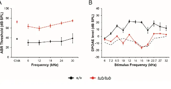
To compare hearing ability of wild-type and tubby mice, I performed ABR and DPOAE experiments. ABR is the widely used method for measuring hearing ability.35,36 ABR is the type of auditory evoked potential detected by electrodes
installed on the scalp. The ABR appears in seven positive wave forms, among these the first five waves (wave I to V) which are analyzed for access hearing ability. ABR thresholds of P21 wild-type and tubby mice were measured to check hearing abilities. As reported in previous studies, elevated ABR threshold values were observed in tubby mice when compared to that of wild-type.13 The result showed
that tubby mice had 40 to 50 dB elevated ABR threshold values than wild-type mice (Figure 1A). These elevated ABR threshold values represent severe hearing loss of
tubby mice. DPOAE is also a widely used method for hearing ability measurement.
DPOAE is reflected sound signals from the cochlea when stimulated by two pure tone frequencies. It is known that cochlear amplification is important for DPOAE. DPOAE measurement can detect the problems in cochlea function.37,38 To further
confirm the results and figure out the cause of hearing loss, DPOAE experiments were conducted using P21 wild-type and tubby mice. DPOAE levels reflect cochlea amplification ability by measuring otoacoustic emissions. If amplification ability of
19
the cochlea is normal, specific frequency (2f1 − f2) of otoacoustic emission
responses will be detected by microphone sensors when two different frequencies (f1, f2) of pure tone stimuli were given. Wild-type mice showed DPOAE
components above the noise floor when sound stimuli were given, however tubby mice did not show any detectable DPOAE response (Figure 1B). These results indicate that hearing loss of tubby mice are originated from disability of cochlea amplification.
20
Figure 1. Auditory phenotypes of wild-type and tubby mice. (A) ABR threshold
of wild-type (+/+, black line) and tubby (tub/tub, red line) mice. ABR threshold values were measured at 6, 12, 18, 24, 30 kHz and Click sound. ABR threshold values are significantly different (P < 0.05). +/+ (n=4), tub/tub (n=12). (B) DPOAE level of wild-type (black line) and tubby (red line) mice. Dashed black line represents noise level. DPOAEs above 7.2 kHz stimulus frequency are significantly different (P < 0.05) +/+ (n=3), tub/tub (n=7).
21
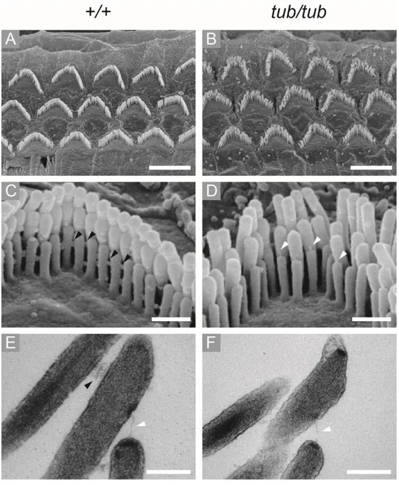
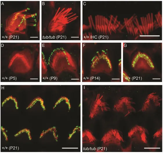
2. Horizontal top connectors and lateral links are disappeared in OHC hair bundles of tubby mice
Previous studies showed that cochlea in tubby mice are progressively degenerate, however the detailed morphology of hair bundles has not yet been reported.2,3 In
hearing tests, I found that the DPOAE responses of the tubby mice are almost lost, which means that the OHCs of tubby mice are not functioning properly. Through SEM image analysis, I found that the OHC hair bundles array is slightly disrupted in tubby mice (Figure 2A, B). I next analyzed stereociliary link formation using high magnification SEM images. High magnification images of wild-type OHC hair bundles showed horizontal top connectors and other types of lateral links (Figure 2C). However, in tubby mice, there are no horizontal top connectors or any other lateral links at OHC hair bundles (Figure 2D). Notably, tip links still existed in
tubby mice (Figure 2D). TEM images showed corresponding results with SEM
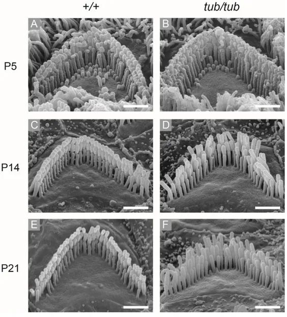
images. In wild type OHC hair bundles, horizontal top connectors and tip links were shown in TEM images (Figure 2E). However, I could not observe any lateral links including horizontal top connectors in TEM images of tubby mice while tip links existed (Figure 2F). These results suggest that hearing loss of tubby mice originated from functional failure of OHCs caused by incomplete stereociliary links. It is known that OHC hair bundles maturation is completed by 3 wk of age.39 At P5,
there were no significant morphological differences between wild-type and tubby mice’s hair bundle of OHCs (Figure 3A, B). However, the OHC hair bundles array
22
was slightly disturbed in tubby mice after P14 (Figure 3C-F). This result suggests a morphological defect of OHC hair bundles in tubby mice appeared after horizontal top connectors generated.
23
Figure 2. OHCs hair bundle morphology of wild-type and tubby mice. SEM and
TEM images of wild-type (A, C, E) and tubby mice (B, D, F). Low magnification SEM images show 3 rows of OHCs, wild-type (A) and tubby mice (B). High magnification SEM images show horizontal top connectors in wild-type OHCs hair
24
bundles (C, black arrowhead). In tubby mice, there are no horizontal top connectors and other lateral links between stereocilia, but tip links are present (D, white arrowhead). TEM images also shows that horizontal top connectors (E, black arrowhead) and tip link (E, white arrowhead) are exist in wild-type OHCs hair bundle. But in tubby mice, only tip-links (F, white arrowhead) are exist. Scale bars : 5 μm (A, B), 0.5 μm (C, D), 0.2 μm (E, F).
25
Figure 3. OHCs hair bundle morphology of wild-type and tubby mice during OHCs hair bundle maturation. At P5, there are no morphological differences
between wild-type and tubby mice OHC hair bundles (A, B). After P14, OHC hair bundles array of wild-type mice is well aligned until P21 (C, E), while OHC hair bundles array is slightly disturbed after P14 in tubby mice (D, F). Scale bars : 1 μm (A-F).
26
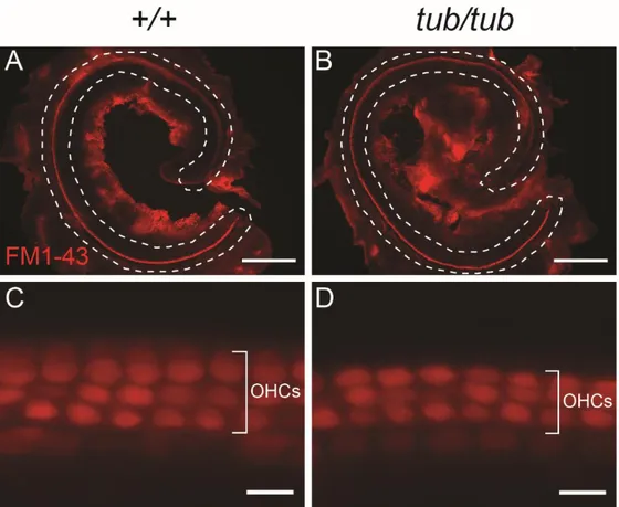
3. MET channels of OHCs are functionally normal in tubby mice
FM1-43 styryl dye is widely used for identifying actively firing neurons and studying vesicular recycling.40-42 In the inner ear, FM1-43 dye is quickly and
specifically enter the sensory hair cells through MET channels.43,44 For testing the
MET channels are properly working in tubby mice, an FM1-43 dye uptake assay was performed using P6 cochlea. The result showed that both OHCs of wild-type and tubby mice are strongly labeled by FM1-43 dye (Figure 4A-D). This result indicates that MET channels are normally open in tubby mice. It is known that functional tip-links is important for the opening of MET channels.45 In the
morphological analysis of OHC hair bundles, I found that tip-links still exist in
tubby mice (Figure 2D, F). The FM1-43 dye uptake assay result also suggests that
27
Figure 4. FM1-43 dye enters OHCs of tubby mice through MET channels. (A, B)
Low magnification images of cochlea of wild-type (A) and tubby mice (B). From apex to base, FM1-43 dye uptake is observed in cochlea OHCs of wild-type and
tubby mice (dashed line). (C, D) High magnification images of cochlear basal
region of wild type (C) and tubby mice (D). FM1-43 dye is uptake to 3 rows of OHCs in both wild-type and tubby mice cochlea. Scale bars : 10 μm (A, B), 0.5 μm (C, D).
28
4. Tub proteins are localized to the stereocilia tip of OHC hair bundles
Previous studies revealed that Tub proteins are expressed in OHCs, IHCs, supporting cells and spiral ganglia neurons, and there are no differences in expression pattern of the Tub protein between wild type and tubby mice.33 To check
the localization of Tub proteins at the cochlea, I conducted whole mount cochlear immunostaining with polyclonal antibody specifically binds to Tub protein. Whole mount cochlear immunostaining of P21 wild-type mice with tubby antibody showed enriched Tub protein localization at the stereocilia tip of OHCs (Figure 5A), however the Tub protein was not observed in the OHC stereocilia of tubby mice (Figure 5B, I). Tub protein localization at the stereocilia tip of IHCs was not observed (Figure 5C). To monitor the changes in Tub protein localization during development of OHCs hair bundle links, cochlea of P5, P9, P14 and P21 wild-type mice were immunostained by tubby antibody. I found that Tub protein was not localized at OHC hair bundles until P5 (Figure 5D). Tub protein was detected at the tallest stereocilia tip of OHC hair bundle at P9 (Figure 5E), and after P14, Tub proteins were localized at all 3 rows of stereocilia tip of OHC hair bundles (Figure 5F, G, H). The time point of localization of Tub protein at the stereocilia tip is similar to that of formation of horizontal top connectors.39
29
Figure 5. Localization of Tub protein in cochlear OHCs. Confocal microscope
images of OHC hair bundles stained for Tub (green) and F-actin (red). (A, B) Tub protein is localized to stereocilia tip of OHC hair bundles (P21) in wild-type mice but not in tubby mice. (C) In hair bundles of IHCs (P21), there are no Tub protein signals. Tub protein is only localized to OHC hair bundles. (D) At P5, there are no Tub proteins in OHC hair bundles. (E) At P9, Tub protein is localized to tip of tallest stereocilia of OHC hair bundles. (F, G) At P14 and P21, Tub protein is
30
localized to tip of all 3 rows of stereocilia tip of OHC hair bundles. (H, I) Low magnification images of OHC hair bundles (P21) in wild-type (H), and tubby mice (I). Scale bars : 2 μm (A, B, D-G), 5 μm (C, H, I).
31
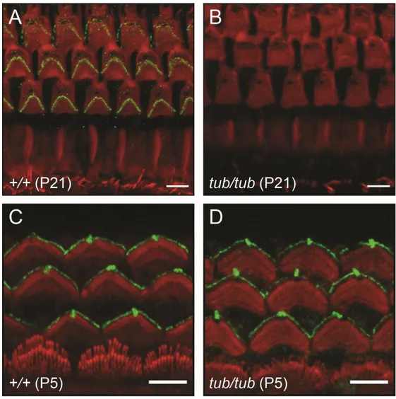
5. The OHC hair bundle localization of stereocilin is disrupted in tubby mice
Stereocilin is a protein specifically expressed in OHCs. This protein is located at the OHC hair bundles, especially at horizontal top connectors and TM attachment crowns.9,46 The features of the OHC hair bundles morphology shown in tubby mice,
such as the less clearly aligned OHC hair bundles and disruption of horizontal top connectors are very similar with the OHC morphological phenotypes of stereocilin null mice.9,46 Furthermore, the OHC hair bundle localization pattern of Tub protein
during P5 to P21 is also similar to that of stereocilin.46 These results strongly
suggest that tubby might be closely related with stereocilin. To confirm the localization of stereocilin at cochlea of tubby mice, I performed a cochlear whole mount immunofluorescence imaging assay with stereocilin antibody. As reported in previous studies, stereocilin was localized to the OHC hair bundles in wild-type mice (Figure 6A). However, in tubby mice, stereocilin was not localized to OHC hair bundles (Figure 6B). Stereocilin is the main component of the horizontal top connectors and is essential for its formation.9,46 This immunostaining result supports
the phenomenon that horizontal top connectors had been disappeared in tubby mice. Previous study shows that stereocilin is located at kinocilium before it appears at OHC hair bundles.46 To test kinociliary localization of stereocilin in tubby mice, I
performed whole mount cochlea immunostaining with P5 cochlea. In wild-type, stereocilin is localized to kinocilium and hair bundles of OHCs (Figure 6C). Interestingly, stereocilin is also localized to kinocilium and OHC hair bundles of
32
tubby mice (Figure 6D). This result suggests that trafficking of stereocilin to
kinocilium may be conducted via a tubby-independent pathway. In addition, this result also suggests that initial localization of stereocilin to the OHC hair bundles do not requires Tub protein. In other words, Tub protein plays an important role in maintaining the OHC hair bundle localization of stereocilin.
33
Figure 6. Disrupted OHCs hair bundle localization of stereocilin in tubby mice.
Confocal microscope images of OHC hair bundles stained for stereocilin (green) and F-actin (red). At P21, stereocilin is localized to OHC hair bundles of wild-type OHCs (A) but not in tubby mice (B). At P5, stereocilin is localized to kinocilium and OHC hair bundles of wild-type (C) and tubby mice (D). Scale bars : 5 μm.
34
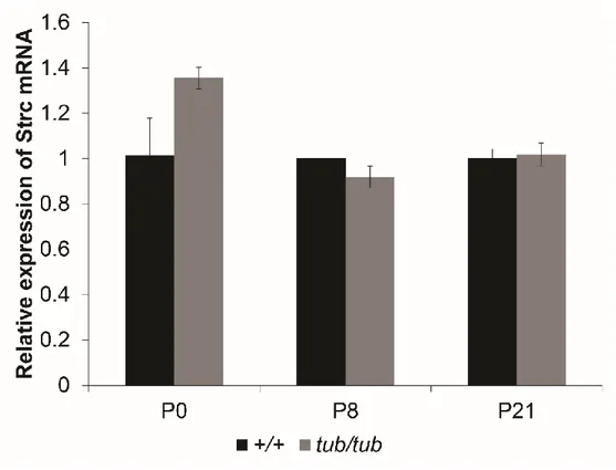
6. Expression of stereocilin mRNA in cochlea of tubby mice is normal
Since Tub protein has a DNA binding domain at carboxy terminal and nuclear localization sequence at the amino terminal, the possibility of its role as transcriptional regulator has been proposed by previous studies.4,7 To test whether
stereocilin gene expression level is reduced in cochlea of tubby mice, expression of stereocilin mRNA was analyzed by semi-quantitative real-time PCR. As a result, there were no significant differences on expression level of stereocilin mRNA between wild-type and tubby mice (Figure 7). This result suggests that stereocilin is expressed normally in cochlea of tubby mice but has defects in stereociliary localization.
35
Figure 7. Gene expression level of stereocilin in cochlea of wild-type and tubby mice. The graph shows relative expression of stereocilin mRNA. Quantitative
real-time PCR was performed. There are no significantly differences in expression level of stereocilin mRNA between wild-type (+/+) and tubby (tub/tub) mice cochlea (P > 0.05).
36
IV. DISCUSSION
In this study, I attempted to figure out the cause of hearing loss in tubby mice. At 3 wk of age, tubby mice showed elevated ABR threshold value at every tested frequency, and DPOAE response almost completely lost. According to previous reports, cochlear degenerative phenotypes like apoptotic cell death had been detected after 6 mo of age of tubby mice.2,3 However, hearing ability of tubby mice
is reduced before the onset of cochlear degeneration. This result suggests that cochlea of tubby mice is functionally disabled. The results of ABR and DPOAE measurement suggest that there is a defect in the cochlear amplification of tubby mice. The OHCs are known to play an important role in cochlear amplification.38,47,48 This suggests that OHCs of tubby mice is functionally defect.
The gross morphology of the OHC hair bundles of tubby mice analyzed by electron microscopy was not seems to make a big difference compared to wild-type. However, the alignment of OHC hair bundles was slightly disturbed in tubby mice. High resolution image analysis showed that the horizontal top connectors were completely lost in the OHC hair bundles of tubby mice. Interestingly, however, tip-links were normally exist in the OHC hair bundles of tubby mice. And these tip links were tightly connected to MET channels, which was confirmed by FM1-43 fluorescent dye experiment. The horizontal top connector serves to connect each stereocilia in the OHC hair bundles, and it is known to be important for the stiffness
37
of OHC hair bundles, and DPOAE response.9,46 The disturbance of OHC hair
bundles array in tubby mice appeared after P10, which is the time point when the horizontal top connectors are formed. This suggests that the morphological phenotypes of the OHC hair bundles in tubby mice is due to the absence of the horizontal top connectors.
Previous study has reported that Tub proteins are present in sensory hair cells, supporting cells, and spiral ganglia neurons in the mice cochlea.33 In this study,
cochlear whole mount immunostaining results showed that the Tub protein was specifically localized in the stereocilia tip of OHCs. Many previous studies have suggested that tubby and other tubby family proteins might be closely related to microtubule cytoskeleton system such as primary cilium.21,24,49,50 Surprisingly,
however, the results of this study showed that the Tub protein is present on the stereocilia which composed of the F-actin structure. Stereociliary tip localization of Tub protein was completely absent in OHC hair bundles of tubby mice. In this study, it is currently not known that what is the role of Tub protein in the OHC stereocilia tip, but it seems clear that the Tub protein is closely related to the actin cytoskeleton.
Stereocilin gene is located at DFNB16 locus, and its mutation cause non-syndromic hearing loss in human.51 Previous studies reported that stereocilin
knockout mice have no DPOAE response, progressive hearing loss, and disruption of horizontal top connectors.9,46 Although many genes affecting stereociliary link
formation are known, stereocilin is the only known gene that affects the horizontal top connector.52 OHC hair bundles of streocilin knockout mice have no horizontal
38
top connectors, but tip link remain, as seen in tubby mice. Based on the similarity of the phenotypes of tubby and stereocilin knockout mice, and the similarity of protein localization of tubby and stereocilin on OHC hair bundles, I expected that tubby is closely related to stereocilin. And the cochlear whole mount immunostaining with anti stereocilin antibody showed that there are no stereocilin in the OHC hair bundles of tubby mice as I expected. This suggests that the morphological phenotypes of tubby mice OHC hair bundle were due to the absence of stereocilin. Stereocilin is known to be also present in kinocilia, although it is not known about the role of stereocilin at kinocilia.46 Kinocilium is microtubule based cilium
structure which disappeared during hair bundle development process.39 Interestingly
stereocilin was present in the kinocilia of tubby mice during the early hair bundle developmental stage. These results suggest that there might be distinct mechanisms of stereocilin trafficking to kinocilia and stereocilia. In addition, at this time point, when the kinocilia remain, stereocilin was transiently present in the OHC hair bundles of tubby mice. This suggests that Tub protein is not required for initial stereociliary trafficking of stereocilin. However, as the OHC hair bundles are gradually maturated, stereocilin disappears from the OHC hair bundles of tubby mice, suggesting that the Tub protein plays an important role in the maintenance of OHC hair bundle localization of stereocilin.
Previous studies have shown that Tub protein is likely to act as a transcriptional regulator.4,7 However, at least in the cochlear OHCs, Tub proteins do not seem to
39
in cochlea of tubby mice was not different from that of wild-type mice. This suggests that, unlike the hypothesis that Tub protein will function as a transcription factor, it may play a totally different role in vivo.
Stereocilia are composed of filamentous actin fiber and distinct stereociliary membrane components.48 Molecular trafficking in stereocilia is known to be carried
out by unconventional myosins. Myosin IIIa and XVa are known to transport the cargo protein to the tip of OHC stereocilia.53-55 It is however uncertain that these
unconventional myosins are essential for stereociliary trafficking of tubby and stereocilin. To figure out stereociliary trafficking mechanism of tubby and stereocilin, further investigation is needed.
Overall results demonstrate that hearing loss phenotype of tubby mice is caused by disrupted stereociliary localization of stereocilin and loss of horizontal top connectors. It is clear that this would be the primary cause of hearing loss mechanism in tubby mice, but there are some reports that sequence variation of microtubule associated protein 1A (Map1a) affect tubby mice hearing ability.10,33,56
These reports suggest possible roles of Tub protein at the ganglia neuron, but functional role of Tub protein at ganglia neuron and the relationship between tubby and Map1a is still elusive, and needed further studies. In human, it has been reported that homozygous mutation in tub gene is associated with obesity and retinal degeneration, but not associated with hearing loss.57 It is currently not known
why hearing defect does not appear in the people with tub gene mutation, but it is possible that human tub was affected by other genes, as in case of Map1a in mice.
40
Tub protein binds to PIP2 through the PIP2 binding domain present in the tubby
domain, and this binding is very specific and strong, even to be used as a PIP2
imaging tool in cellular experiments.8,58 In Drosophila, dINPP5E mutant fly, it is
known that the ciliary localization of drosophila tubby homolog (dTulp) is altered due to the change of the PIP2 composition of the chordotonal cilia membrane,
resulting in hearing defects in this mutant fly. This means that the localization of the Tub protein is dependent on the PIP2 composition of the cell membrane. In addition,
although not cochlear hair cells, it has been reported that PIP2 is concentrated in the
stereocilia of vestibule hair cells.59 Based on these facts, it is likely that the
enrichment of the Tub protein specifically to the stereocilia of OHCs is closely related to the PIP2 composition of the stereocilia membrane. Further studies are
needed to determine whether OHC hair bundles localization of Tub protein is associated with PIP2 composition of stereocilia membrane.
41
V. CONCLUSION
Overall results demonstrate that the hearing loss of tubby mice results from the dysfunction of OHCs due to the loss of the horizontal top connector in the OHC hair bundles. And the loss of the horizontal top connectors observed in tubby mice is caused by stereocilin mislocalization. Stereocilin is present in the OHC hair bundles of tubby mice at P5, but it disappeared from after P9. This suggests that stereocilin does not require a Tub protein for initial trafficking to the OHC hair bundles. Instead, it means that Tub protein is essential for maintain of stereocilin at OHC hair bundles and formation of horizontal top connectors by stereocilin. This study firstly demonstrated that Tub protein is specifically located at OHC hair bundles of cochlea. Tub protein has long been suggested to be closely associated with primary cilia which is microtubule-based cellular organelle. However, the results of this study suggest that Tub protein may be closely associated with actin-based cellular organelles in vivo. The results of this study could also provide a new insight into the molecular mechanism of another phenotypes of tubby mice such as late onset obesity, and retinal degeneration. The remaining question is how Tub protein moves to the stereocilia and what is the molecular function of Tub protein on the stereocilia. Further study is needed to answer these questions.
42
REFERENCES
1. Coleman DL, Eicher EM. Fat (fat) and tubby (tub): two autosomal recessive mutations causing obesity syndromes in the mouse. J Hered 1990;81:424-7. 2. Ohlemiller KK, Hughes RM, Mosinger-Ogilvie J, Speck JD, Grosof DH, Silverman
MS. Cochlear and retinal degeneration in the tubby mouse. Neuroreport 1995;6:845-9.
3. Ohlemiller KK, Hughes RM, Lett JM, Ogilvie JM, Speck JD, Wright JS, et al. Progression of cochlear and retinal degeneration in the tubby (rd5) mouse. Audiol Neurootol 1997;2:175-85.
4. Carroll K, Gomez C, Shapiro L. Tubby proteins: the plot thickens. Nat Rev Mol Cell Biol 2004;5:55-63.
5. Noben-Trauth K, Naggert JK, North MA, Nishina PM. A candidate gene for the mouse mutation tubby. Nature 1996;380:534-8.
6. Stubdal H, Lynch CA, Moriarty A, Fang Q, Chickering T, Deeds JD, et al. Targeted deletion of the tub mouse obesity gene reveals that tubby is a loss-of-function mutation. Mol Cell Biol 2000;20:878-82.
7. Boggon TJ, Shan WS, Santagata S, Myers SC, Shapiro L. Implication of tubby proteins as transcription factors by structure-based functional analysis. Science 1999;286:2119-25.
8. Santagata S, Boggon TJ, Baird CL, Gomez CA, Zhao J, Shan WS, et al. G-protein signaling through tubby proteins. Science 2001;292:2041-50.
9. Verpy E, Weil D, Leibovici M, Goodyear RJ, Hamard G, Houdon C, et al. Stereocilin-deficient mice reveal the origin of cochlear waveform distortions. Nature 2008;456:255-8.
10. Ikeda A, Zheng QY, Zuberi AR, Johnson KR, Naggert JK, Nishina PM. Microtubule-associated protein 1A is a modifier of tubby hearing (moth1). Nat Genet 2002;30:401-5.
11. Hildebrandt F, Benzing T, Katsanis N. Ciliopathies. N Engl J Med 2011;364:1533-43.
12. Girard D, Petrovsky N. Alstrom syndrome: insights into the pathogenesis of metabolic disorders. Nat Rev Endocrinol 2011;7:77-88.
13. Heckenlively JR, Chang B, Erway LC, Peng C, Hawes NL, Hageman GS, et al. Mouse model for Usher syndrome: linkage mapping suggests homology to Usher type I reported at human chromosome 11p15. Proc Natl Acad Sci U S A 1995;92:11100-4.
14. Kleyn PW, Fan W, Kovats SG, Lee JJ, Pulido JC, Wu Y, et al. Identification and characterization of the mouse obesity gene tubby: a member of a novel gene family. Cell 1996;85:281-90.
15. Ikeda S, Shiva N, Ikeda A, Smith RS, Nusinowitz S, Yan G, et al. Retinal degeneration but not obesity is observed in null mutants of the tubby-like protein 1 gene. Hum Mol Genet 2000;9:155-63.
16. Gu S, Lennon A, Li Y, Lorenz B, Fossarello M, North M, et al. Tubby-like protein-1 mutations in autosomal recessive retinitis pigmentosa. Lancet protein-1998;35protein-1:protein-1protein-103-4. 17. Hagstrom SA, North MA, Nishina PL, Berson EL, Dryja TP. Recessive mutations
43
in the gene encoding the tubby-like protein TULP1 in patients with retinitis pigmentosa. Nat Genet 1998;18:174-6.
18. Hagstrom SA, Adamian M, Scimeca M, Pawlyk BS, Yue G, Li T. A role for the Tubby-like protein 1 in rhodopsin transport. Invest Ophthalmol Vis Sci 2001;42:1955-62.
19. North MA, Naggert JK, Yan Y, Noben-Trauth K, Nishina PM. Molecular characterization of TUB, TULP1, and TULP2, members of the novel tubby gene family and their possible relation to ocular diseases. Proc Natl Acad Sci U S A 1997;94:3128-33.
20. Ikeda A, Ikeda S, Gridley T, Nishina PM, Naggert JK. Neural tube defects and neuroepithelial cell death in Tulp3 knockout mice. Hum Mol Genet 2001;10:1325-34.
21. Mukhopadhyay S, Wen X, Chih B, Nelson CD, Lane WS, Scales SJ, et al. TULP3 bridges the IFT-A complex and membrane phosphoinositides to promote trafficking of G protein-coupled receptors into primary cilia. Genes Dev 2010;24:2180-93. 22. Norman RX, Ko HW, Huang V, Eun CM, Abler LL, Zhang Z, et al. Tubby-like
protein 3 (TULP3) regulates patterning in the mouse embryo through inhibition of Hedgehog signaling. Hum Mol Genet 2009;18:1740-54.
23. Cameron DA, Pennimpede T, Petkovich M. Tulp3 is a critical repressor of mouse hedgehog signaling. Dev Dyn 2009;238:1140-9.
24. Mukhopadhyay S, Jackson PK. The tubby family proteins. Genome Biol 2011;12:225.
25. Li QZ, Wang CY, Shi JD, Ruan QG, Eckenrode S, Davoodi-Semiromi A, et al. Molecular cloning and characterization of the mouse and human TUSP gene, a novel member of the tubby superfamily. Gene 2001;273:275-84.
26. van Duyvenvoorde HA, Lui JC, Kant SG, Oostdijk W, Gijsbers AC, Hoffer MJ, et al. Copy number variants in patients with short stature. Eur J Hum Genet 2014;22:602-9.
27. Vieira AR, de Carvalho FM, Johnson L, DeVos L, Swailes AL, Weber ML, et al. Fine Mapping of 6q23.1 Identifies TULP4 as Contributing to Clefts. Cleft Palate Craniofac J 2015;52:128-34.
28. Coyle CA, Strand SC, Good DJ. Reduced activity without hyperphagia contributes to obesity in Tubby mutant mice. Physiol Behav 2008;95:168-75.
29. Backberg M, Madjid N, Ogren SO, Meister B. Down-regulated expression of agouti-related protein (AGRP) mRNA in the hypothalamic arcuate nucleus of hyperphagic and obese tub/tub mice. Brain Res Mol Brain Res 2004;125:129-39. 30. Wang Y, Seburn K, Bechtel L, Lee BY, Szatkiewicz JP, Nishina PM, et al.
Defective carbohydrate metabolism in mice homozygous for the tubby mutation. Physiol Genomics 2006;27:131-40.
31. Guan XM, Yu H, Van der Ploeg LH. Evidence of altered hypothalamic pro-opiomelanocortin/ neuropeptide Y mRNA expression in tubby mice. Brain Res Mol Brain Res 1998;59:273-9.
32. Ikeda S, He W, Ikeda A, Naggert JK, North MA, Nishina PM. Cell-specific expression of tubby gene family members (tub, Tulp1,2, and 3) in the retina. Invest Ophthalmol Vis Sci 1999;40:2706-12.
33. Ikeda A, Zheng QY, Rosenstiel P, Maddatu T, Zuberi AR, Roopenian DC, et al. Genetic modification of hearing in tubby mice: evidence for the existence of a major gene (moth1) which protects tubby mice from hearing loss. Hum Mol Genet 1999;8:1761-7.
44
34. Kong L, Chen GD, Zhou X, McGinnis JF, Li F, Cao W. Molecular mechanisms underlying cochlear degeneration in the tubby mouse and the therapeutic effect of sulforaphane. Neurochem Int 2009;54:172-9.
35. Kotlarz JP, Eby TL, Borton TE. Analysis of the efficiency of retrocochlear screening. Laryngoscope 1992;102:1108-12.
36. Melcher JR, Kiang NY. Generators of the brainstem auditory evoked potential in cat. III: Identified cell populations. Hear Res 1996;93:52-71.
37. Mehrparvar AH, Mirmohammadi SJ, Davari MH, Mostaghaci M, Mollasadeghi A, Bahaloo M, et al. Conventional Audiometry, Extended High-Frequency Audiometry, and DPOAE for Early Diagnosis of NIHL. Iran Red Crescent Med J 2014;16:e9628. 38. Avan P, Buki B, Petit C. Auditory distortions: origins and functions. Physiol Rev
2013;93:1563-619.
39. Goodyear RJ, Marcotti W, Kros CJ, Richardson GP. Development and properties of stereociliary link types in hair cells of the mouse cochlea. J Comp Neurol 2005;485:75-85.
40. Betz WJ, Bewick GS. Optical analysis of synaptic vesicle recycling at the frog neuromuscular junction. Science 1992;255:200-3.
41. Betz WJ, Mao F, Bewick GS. Activity-dependent fluorescent staining and destaining of living vertebrate motor nerve terminals. J Neurosci 1992;12:363-75. 42. Betz WJ, Mao F, Smith CB. Imaging exocytosis and endocytosis. Curr Opin
Neurobiol 1996;6:365-71.
43. Gale JE, Marcotti W, Kennedy HJ, Kros CJ, Richardson GP. FM1-43 dye behaves as a permeant blocker of the hair-cell mechanotransducer channel. J Neurosci 2001;21:7013-25.
44. Meyers JR, MacDonald RB, Duggan A, Lenzi D, Standaert DG, Corwin JT, et al. Lighting up the senses: FM1-43 loading of sensory cells through nonselective ion channels. J Neurosci 2003;23:4054-65.
45. Gillespie PG, Muller U. Mechanotransduction by hair cells: models, molecules, and mechanisms. Cell 2009;139:33-44.
46. Verpy E, Leibovici M, Michalski N, Goodyear RJ, Houdon C, Weil D, et al. Stereocilin connects outer hair cell stereocilia to one another and to the tectorial membrane. J Comp Neurol 2011;519:194-210.
47. Fettiplace R, Hackney CM. The sensory and motor roles of auditory hair cells. Nat Rev Neurosci 2006;7:19-29.
48. Schwander M, Kachar B, Muller U. Review series: The cell biology of hearing. J Cell Biol 2010;190:9-20.
49. Sun X, Haley J, Bulgakov OV, Cai X, McGinnis J, Li T. Tubby is required for trafficking G protein-coupled receptors to neuronal cilia. Cilia 2012;1:21.
50. Mukhopadhyay S, Jackson PK. Cilia, tubby mice, and obesity. Cilia 2013;2:1. 51. Verpy E, Masmoudi S, Zwaenepoel I, Leibovici M, Hutchin TP, Del Castillo I, et al.
Mutations in a new gene encoding a protein of the hair bundle cause non-syndromic deafness at the DFNB16 locus. Nat Genet 2001;29:345-9.
52. Peng AW, Salles FT, Pan B, Ricci AJ. Integrating the biophysical and molecular mechanisms of auditory hair cell mechanotransduction. Nat Commun 2011;2:523. 53. Belyantseva IA, Boger ET, Friedman TB. Myosin XVa localizes to the tips of inner
ear sensory cell stereocilia and is essential for staircase formation of the hair bundle. Proc Natl Acad Sci U S A 2003;100:13958-63.
54. Belyantseva IA, Boger ET, Naz S, Frolenkov GI, Sellers JR, Ahmed ZM, et al. Myosin-XVa is required for tip localization of whirlin and differential elongation of