S H O R T G E N O M E R E P O R T
Open Access
Draft genome sequence of the extremely
halophilic archaeon Haladaptatus cibarius
type strain D43
T
isolated from fermented
seafood
Hae-Won Lee
1,2†, Dae-Won Kim
3†, Mi-Hwa Lee
4, Byung-Yong Kim
5, Yong-Joon Cho
5, Kyung June Yim
1,
Hye Seon Song
1, Jin-Kyu Rhee
6, Myung-Ji Seo
7, Hak-Jong Choi
2, Jong-Soon Choi
1, Dong-Gi Lee
1,
Changmann Yoon
1, Young-Do Nam
4*and Seong Woon Roh
1*Abstract
An extremely halophilic archaeon, Haladaptatus cibarius D43
T, was isolated from traditional Korean salt-rich
fermented seafood. Strain D43
Tshows the highest 16S rRNA gene sequence similarity (98.7 %) with Haladaptatus
litoreus RO1-28
T, is Gram-negative staining, motile, and extremely halophilic. Despite potential industrial applications
of extremely halophilic archaea, their genome characteristics remain obscure. Here, we describe the whole genome
sequence and annotated features of strain D43
T. The 3,926,724 bp genome includes 4,092 protein-coding and 57
RNA genes (including 6 rRNA and 49 tRNA genes) with an average G + C content of 57.76 %.
Keywords: Extremely halophilic archaea, Haladaptatus cibarius, Genome sequence, Salt-fermented seafood, Glycine
betaine, Trehalose
Introduction
The extremely halophilic archaea, called haloarchaea,
possess the small retinal protein halorhodopsin [1–3]
and currently consists of more than 47 genera that live
in hypersaline environments [4, 5]. Three members of
the genus Haladaptatus—H. paucihalophilus [6], H.
litoreus [7], and H. cibarius [8]—were isolated from a
low-salt, sulfide-rich spring; marine solar saltern; and
salt-fermented seafood, respectively. Haladaptatus
com-prises Gram-negative staining, non-motile haloarchaea
that have polar lipids including phosphatidylglycerol,
phosphatidylglycerol phosphate methyl ester, and
phos-phatidylglycerol sulfate [6]. The genomic analysis
re-vealed that H. paucihalophilus survives in low salinity
conditions because of trehalose synthesis with OtsAB
pathway and trehalose glycosyl-transferring synthase
pathway, and glycine betaine uptake [9]. However, other
members in the genus Haladaptatus have not been
ana-lyzed at the genome level.
H. cibarius was isolated from the traditional Korean
salt-fermented seafood, which is made with shellfish [8].
D43
T(= DSM 19505
T= JCM 15962
T) is a representative
strain and designated as the type strain of the species. It
can grow in 10%–30% (w/v) NaCl (optimum, 15%), with
Mg
2+required for growth. In addition, cells are not lysed
in distilled water. The genome sequences of this genus
are expected to provide fundamental information for
the halotolerant features and biotechnological
applica-tions of the haloarchaea. Here, we describe the first
whole genome sequence of H. cibarius along with its
annotated features, and summarize the taxonomic
classification.
* Correspondence:[email protected];[email protected]
†Equal contributors 4
Research Group of Gut Microbiome, Korea Food Research Institute, Sungnam 463-746, Republic of Korea
1
Biological Disaster Analysis Group, Korea Basic Science Institute, Daejeon 305-806, Republic of Korea
Full list of author information is available at the end of the article
© 2015 Lee et al.Open Access This article is distributed under the terms of the Creative Commons Attribution 4.0 International License (http://creativecommons.org/licenses/by/4.0/), which permits unrestricted use, distribution, and reproduction in any medium, provided you give appropriate credit to the original author(s) and the source, provide a link to the Creative Commons license, and indicate if changes were made. The Creative Commons Public Domain Dedication waiver (http://creativecommons.org/publicdomain/zero/1.0/) applies to the data made available in this article, unless otherwise stated.
Organism information
Classification and features
The taxonomic position for H. cibarius D43
Twas
iden-tified with type strains obtained from the EzTaxon-e
server [10]. The 16S rRNA sequences of D43
Tand
closely related strains were aligned using the ClustalW
multiple sequence alignment program [11] and were
subsequently used for the phylogenetic analysis.
Phylogen-etic trees were constructed using the neighbor-joining
[12], maximum-parsimony [13], and maximum likelihood
[14] algorithms with bootstrap values of 1,000 using
MEGA version 5 molecular evolutionary genetics analysis
program [15]. Strain D43
Tclustered with type strains of
Haladaptatus species (Fig. 1), exhibiting 16S rRNA gene
sequence similarities of 98.7% and 95.1% between strain
D43
T(EF660747) and the type strain of H. litoreus and H.
paucihalophilus, respectively. Classification and general
features of H. cibarius D43
Tare shown in Table 1.
Strain D43
Tis a Gram-negative staining, coccus or
coccobacillus, motile archaeon approximately 1.0
μm in
diameter (Fig. 2). Catalase and oxidase tests yielded
posi-tive results, but reduction of nitrate to nitrite under
aer-obic conditions was negative. Cells contained the polar
lipids phosphatidylglycerol, phosphatidylglycerol
phos-phate methyl ester, and two unidentified glycolipids.
Strain D43
Thydrolyzed gelatin and Tween 80, utilized
formate and acetate as carbon sources, and produced
acid from sucrose and D-glucose. The strain was
sensi-tive to anisomycin, aphidicolin, chloramphenicol, and
ri-fampicin, and was resistant to ampicillin, erythromycin,
kanamycin, streptomycin, and polymycin B.
Genome sequencing and annotation
Genome project history
The genome project and sequence of the H. cibarius
D43
Tgenome were deposited in the Genomes OnLine
Database [16] (project ID: Gp0086819) and GenBank
(accession number: JDTH00000000), respectively. The
BioProject number was PRJNA236630. Sequencing and
annotation were performed by Chun Lab Inc. (Seoul,
Korea) and Integrated Microbial Genomes Expert
Re-view (IMG-ER) [17].
Growth conditions and genomic DNA preparation
H. cibarius D43
Tgrew optimally on halophilic medium
[6] supplemented with 15% (w/v) NaCl and 20 mM Mg
2+adjusted to pH 7.0, producing colonies with a pink color
after incubation at 37°C as previously described [8].
Genomic DNA was extracted and purified using a
G-spin DNA extraction kit (iNtRON Biotechnology
Inc., Sungnam, Korea), according to the manufacturer
’s
instructions.
Haladaptatus cibarius D43T(EF660747)
Haladaptatus cibarius D43T(JDTH00000000; Gene ID:2582599458)
Haladaptatus cibarius D43T(JDTH00000000; Gene ID:2582596273)
Haladaptatus cibarius D43T(JDTH00000000; Gene ID:2582599457)
Haladaptatus litoreus RO1-28T(EU887285)
Haladaptatus litoreus RO1-22 (FJ773394)
Haloarchaeon Nie 14-2 (AB291225) Haloarchaeon Nie 14-1 (AB291220)
Haladaptatus paucihalophilus strain GY252 copy 1 (DQ867122)
Haloarchaeon 30AH clone 1 (AY292392) Haloarchaeon W1 clone 1 (AY292394)
Haladaptatus paucihalophilus strain DX253Tcopy 1 (DQ344973)
Haladaptatus paucihalophilus GY252 copy 2 (DQ867123)
Haloarchaeon 30AH clone 2 (AY292393)
Haladaptatus paucihalophilus DX253Tcopy 2 (DQ344974)
Natrialba asiatica Aidin-3 (AB046875) Natrialba chahannaoensis C112T(AJ004806)
Natronococcus occultus NCIMB 2192T(Z28378)
Haloterrigena thermotolerans PR5T(AF115478)
Natronorubrum texcoconense B4T(HQ456225)
Haloterrigena turkmenica VKM B-1734T(AB004878)
Natronobacterium gregoryi NCIMB 2189T(D87970)
Halalkalicoccus tibetensis DS12T(AF435112)
Halococcus dombrowskii H4T(AJ420376)
Methanosarcina semesiae MD1T(AJ012742)
100/99/100 99/98/99 98/82/96 98/93/94 9387/92 79/-/-77/80/79 99/86/-99/91/94 0.05
Fig. 1 Phylogenetic tree constructed using the neighbor-joining method based on 16S rRNA gene sequences, showing the taxonomic position of strain D43Tin genus Haladaptatus. Bootstrap values (>70%) at nodes are shown as percentages calculated using the neighbor-joining/maximum
parsimony/maximum likelihood probabilities based on 1000 replicates. Filled circles indicate identical branches generated using three algorithms. Methanosarcina semesiae MD1Twas used as an outgroup. Bar, 0.05 substitutions per nucleotide position
Fig. 2 Scanning electron micrographs of H. cibarius D43Tobtained by SUPRA 55VP (Carl Zeiss, Jena, Germany). Scale bars represent 200 nm
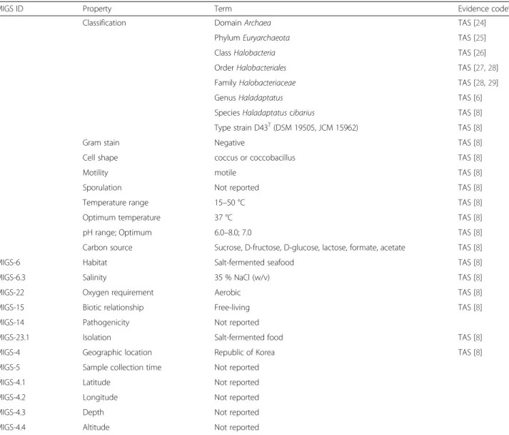
Table 1 Classification and general features of Haladaptatus cibarius D43
T[18]
MIGS ID Property Term Evidence codea
Classification Domain Archaea TAS [24]
Phylum Euryarchaeota TAS [25]
Class Halobacteria TAS [26]
Order Halobacteriales TAS [27,28]
Family Halobacteriaceae TAS [28,29]
Genus Haladaptatus TAS [6]
Species Haladaptatus cibarius TAS [8]
Type strain D43T(DSM 19505, JCM 15962) TAS [8]
Gram stain Negative TAS [8]
Cell shape coccus or coccobacillus TAS [8]
Motility motile TAS [8]
Sporulation Not reported TAS [8]
Temperature range 15–50 °C TAS [8]
Optimum temperature 37 °C TAS [8]
pH range; Optimum 6.0–8.0; 7.0 TAS [8]
Carbon source Sucrose, D-fructose, D-glucose, lactose, formate, acetate TAS [8]
MIGS-6 Habitat Salt-fermented seafood TAS [8]
MIGS-6.3 Salinity 35 % NaCl (w/v) TAS [8]
MIGS-22 Oxygen requirement Aerobic TAS [8]
MIGS-15 Biotic relationship Free-living TAS [8]
MIGS-14 Pathogenicity Not reported
MIGS-23.1 Isolation Salt-fermented food TAS [8]
MIGS-4 Geographic location Republic of Korea TAS [8]
MIGS-5 Sample collection time Not reported
MIGS-4.1 Latitude Not reported
MIGS-4.2 Longitude Not reported
MIGS-4.3 Depth Not reported
MIGS-4.4 Altitude Not reported
a
Genome sequencing and assembly
Genomic sequences of H. cibarius D43
Twere generated
from a total of 9,237,360 quality-filtered reads
(710.3-fold coverage) by combining 5,074,634 reads (374.9-(710.3-fold
coverage) obtained from Mi-Seq 300 bp paired-end
li-brary (Illumina, San Diego, CA, USA), 4,112,798 reads
(292.1-fold coverage) obtained from an Ion Torrent
Per-sonal Genome Machine 318v2 chip (Life Technologies,
Carlsbad, CA, USA), and 49,928 reads (43.3-fold coverage)
obtained from PacBio RS 10 kb library (Pacific
Biosci-ences, Menlo Park, USA). Illumina and PGM data were
assembled de novo with CLC Genomics Workbench 6.5.1
(CLC bio, Boston, MA, USA) and PacBio data were
as-sembled using the HGAP2 algorithm in SMRT Analysis
2.1 (Pacific Biosciences). Resultant contigs were assembled
with CodonCode Aligner 3.7 (CodonCode Corporation,
Centerville, MA, USA). Sequences were assembled to 13
scaffolds with an N50 contig size of 985,075 bp; the genome
sequencing project information and its associated MIGS
version 2.0 compliance levels [18] are shown in Table 2.
Genome annotation
The open reading frames of the assembled genome were
predicted and annotated using IMG-ER [17], NCBI COG
[19], Pfam [20], and EzTaxon-e [10] databases. The rRNA
and tRNA genes were identified using RNAmmer 1.2 [21]
and tRNA scan-SE 1.23 [22], respectively.
Genome properties
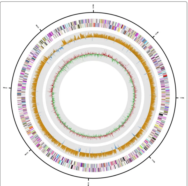
The draft genome sequence for H. cibarius D43
Tcon-tained 3,926,724 bp, with 13 scaffolds. The G + C content
was 57.76 % (Fig. 3 and Table 3), and 4,092 protein-coding
genes were predicted along with 57 RNA genes, including
six rRNA (two 5S, three 16S, and one 23S rRNA), 49
tRNA, and two additional RNA genes. There were 2,676
protein-coding genes with predicted functions: 773 were
enzymes, 98 encoded signal peptides, and 1,049 encoded
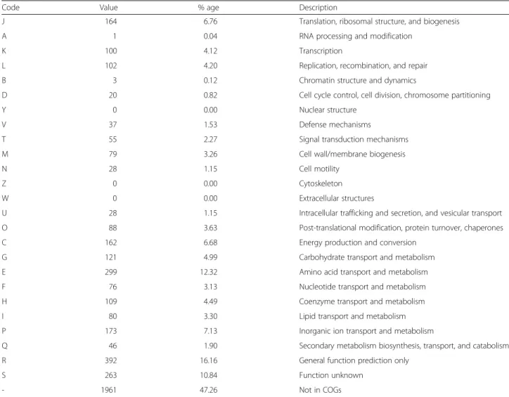
transmembrane proteins. The distribution of genes in
the COG functional categories is shown in Table 4. A
large number of genes were associated with the COG
functional categories of cell wall biogenesis (79, 3.3 %);
transcription (100, 4.1 %); and transport and
metabol-ism of amino acids (299, 12.3 %), carbohydrates (121,
5.0 %), and lipids (80, 3.3 %). Further analysis with
dbCAN
[23],
a
database
for
annotation
of
carbohydrate-active enzymes, showed that the genome
contains genes encoding various enzymes for the
break-down and biosynthesis of carbohydrates such as
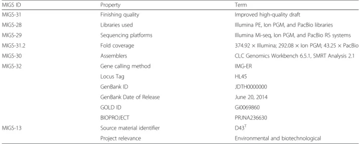
Table 2 Project information
MIGS ID Property Term
MIGS-31 Finishing quality Improved high-quality draft
MIGS-28 Libraries used Illumina PE, Ion PGM, and PacBio libraries
MIGS-29 Sequencing platforms Illumina Mi-seq, Ion PGM, and PacBio RS systems
MIGS-31.2 Fold coverage 374.92 × Illumina; 292.08 × Ion PGM; 43.25 × PacBio
MIGS-30 Assemblers CLC Genomics Workbench 6.5.1, SMRT Analysis 2.1
MIGS-32 Gene calling method IMG-ER
Locus Tag HL45
GenBank ID JDTH0000000
GenBank Date of Release June 20, 2014
GOLD ID Gi0069860
BIOPROJECT PRJNA236630
MIGS-13 Source material identifier D43T
Project relevance Environmental and biotechnological
Table 3 Genome statistics
Attribute Value % of Total
Genome size (bp) 3,926,724 100.00 DNA coding (bp) 3,378,684 86.04 DNA G + C (bp) 2,267,915 57.76 DNA scaffolds 13 100.00 Total genes 4,149 100.00 Protein-coding genes 4,092 98.63 RNA genes 57 1.37
Genes in internal clusters 3,135 75.56
Genes with function prediction 2,676 64.50
Genes assigned to COGs 2,188 52.74
Genes assigned Pfam domains 2,699 65.05
Genes with signal peptides 98 2.36
Genes with transmembrane helices 1049 25.28
chitinase
(GH18),
chitosanase
(GH5),
pullulanase
(GH13), trehalose synthase (GT4 and 20), cellulose
syn-thase (GT2), and alginate lyase (PL6).
Insights from the genome sequence
The genome analysis of H. cibarius D43
Trevealed genes
involved in glycine betaine synthesis—including betaine
aldehyde dehydrogenase, glycine betaine demethylase, and
choline-glycine betaine transporter gene—that allow H.
cibarius to maintain osmotic balance in hypersaline
environments. In addtion, trehalose-related genes of
trehalose-6-phosphate synthase, trehalose-6-phosphatase,
trehalose-6-phosphate synthase and trehalose-6-phosphate
hydrolase, and trehalose-utilization protein genes were
an-alyzed in the genome sequences of H. cibarius D43
T. The
genes related with trehalose synthesis in the genome show
the possibility of trehalose production that is important in
food industry.
Fig. 3 Graphical map of the H. cibarius D43Tpseudochromosome. From outside to center: RNA genes (red, tRNA and blue, rRNA) and genes on the antisense and sense strands (colored according to COG categories). Inner circle shows the GC skew, with yellow and blue indicating positive and negative values, respectively. GC content is indicated in red and green. The genome map was visualized using CLgenomics 1.06 (Chun Lab Inc.)
Conclusions
The draft genome sequences of the extremely halophilic
archaeon isolated from the salt-fermented seafood were
analyzed. Genes related with glycine betaine and trehalose
for the survival in extreme environments were identified.
The extremely halophilic archaeon could be a valuable
re-source for biotechnological applications because
hypersa-line conditions minimize the risk of contamination by
other microorganisms. Further characterization of
halo-philic enzymes of the haloarchaea based on the genomic
analyses can provide more detailed information on enzyme
structures and potential industrial applications.
Abbreviations
PGM:Personal genome machine; IMG-ER: Integrated microbial genomes expert review; ORF: Open reading frame.
Competing interests
The authors declare that they have no competing interests.
Authors’ contributions
KJY and HSS carried out the microbial cultivation and DNA isolation. HWL, DWK, SWR, BYK, YJC and KJY participated in the sequence analyses. HWL, DWK and SWR drafted the manuscript. MJS, JKR, DGL and CY helped to draft the manuscript. SWR and YDN conceived of the study and participated in its design. HJC and JSC participated in its design and coordination. All authors read and approved the final manuscript.
Acknowledgements
This research was supported by the Basic Science Research Program through the National Research Foundation of Korea (2012R1A1A2040922) and a Korea Basic Science Institute NAP grant (T34780).
Author details
1
Biological Disaster Analysis Group, Korea Basic Science Institute, Daejeon 305-806, Republic of Korea.2World Institute of Kimchi, Gwangju 503-360,
Republic of Korea.3Systems Biology Team, Center for Immunity and Pathology, Korea National Institute of Health, Cheongju 361-951, Republic of Korea.4Research Group of Gut Microbiome, Korea Food Research Institute, Sungnam 463-746, Republic of Korea.5ChunLab Inc., Seoul National
University, Seoul 151-742, Republic of Korea.6Department of Food Science and Engineering, Ewha Womans University, Seoul 120-750, Republic of Korea.
7
Division of Bioengineering, Incheon National University, Incheon 406-772, Republic of Korea.
Table 4 Number of genes associated with general COG functional categories
Code Value % age Description
J 164 6.76 Translation, ribosomal structure, and biogenesis
A 1 0.04 RNA processing and modification
K 100 4.12 Transcription
L 102 4.20 Replication, recombination, and repair
B 3 0.12 Chromatin structure and dynamics
D 20 0.82 Cell cycle control, cell division, chromosome partitioning
Y 0 0.00 Nuclear structure
V 37 1.53 Defense mechanisms
T 55 2.27 Signal transduction mechanisms
M 79 3.26 Cell wall/membrane biogenesis
N 28 1.15 Cell motility
Z 0 0.00 Cytoskeleton
W 0 0.00 Extracellular structures
U 28 1.15 Intracellular trafficking and secretion, and vesicular transport
O 88 3.63 Post-translational modification, protein turnover, chaperones
C 162 6.68 Energy production and conversion
G 121 4.99 Carbohydrate transport and metabolism
E 299 12.32 Amino acid transport and metabolism
F 76 3.13 Nucleotide transport and metabolism
H 109 4.49 Coenzyme transport and metabolism
I 80 3.30 Lipid transport and metabolism
P 173 7.13 Inorganic ion transport and metabolism
Q 46 1.90 Secondary metabolism biosynthesis, transport, and catabolism
R 392 16.16 General function prediction only
S 263 10.84 Function unknown
- 1961 47.26 Not in COGs
Received: 10 September 2014 Accepted: 27 July 2015
References
1. Lanyi JK. Halorhodopsin: a light-driven chloride ion pump. Annu Rev Biophys Biophys Chem. 1986;15:11–28.
2. Lanyi JK. Halorhodopsin, a light-driven electrogenic chloride-transport system. Physiol Rev. 1990;70:319–30.
3. Oesterhelt D, Tittor J, Bamberg E. A unifying concept for ion translocation by retinal proteins. J Bioenerg Biomembr. 1992;24:181–91.
4. Oren A. The order Halobacteriales. In: Dworkin M, Falkow S, Rosenberg E, Shleifer KH, Stakebrandt E, editors. The prokaryotes. Volume 3. 3rd ed. New York: Springer; 2006. p. 113–64.
5. Oren A, Ventosa A. International Committee on Systematics of Prokaryotes Subcommittee on the taxonomy of Halobacteriaceae and Subcommittee on the taxonomy of Halomonadaceae: minutes of the joint open meeting, 31 July 2014, Montreal, Canada. Int J Syst Evol Microbiol. 2014;64:3915–8. 6. Savage KN, Krumholz LR, Oren A, Elshahed MS. Haladaptatus paucihalophilus
gen. nov., sp. nov., a halophilic archaeon isolated from a low-salt, sulfide-rich spring. Int J Syst Evol Microbiol. 2007;57:19–24.
7. Cui HL, Sun FF, Gao X, Dong Y, Xu XW, Zhou YG, et al. Haladaptatus litoreus sp. nov., an extremely halophilic archaeon from a marine solar saltern, and emended description of the genus Haladaptatus. Int J Syst Evol Microbiol. 2010;60:1085–9.
8. Roh SW, Lee ML, Bae JW. Haladaptatus cibarius sp. nov., an extremely halophilic archaeon from seafood, and emended description of the genus Haladaptatus. Int J Syst Evol Microbiol. 2010;60:1187–90.
9. Youssef NH, Savage-Ashlock KN, McCully AL, Luedtke B, Shaw EI, Hoff WD, et al. Trehalose/2-sulfotrehalose biosynthesis and glycine-betaine uptake are widely spread mechanisms for osmoadaptation in the Halobacteriales. ISME J. 2014;8:636–49.
10. Kim OS, Cho YJ, Lee K, Yoon SH, Kim M, Na H, et al. Introducing EzTaxon-e: a prokaryotic 16S rRNA gene sequence database with phylotypes that represent uncultured species. Int J Syst Evol Microbiol. 2012;62:716–21. 11. Thompson JD, Higgins DG, Gibson TJ. CLUSTAL W: improving the sensitivity
of progressive multiple sequence alignment through sequence weighting, position-specific gap penalties and weight matrix choice. Nucleic Acids Res. 1994;22:4673–80.
12. Saitou N, Nei M. The neighbor-joining method: a new method for reconstructing phylogenetic trees. Mol Biol Evol. 1987;4:406–25. 13. Kluge AG, Farris JS. Quantitative phyletics and the evolution of anurans.
Syst Biol. 1969;18:1–32.
14. Felsenstein J. Evolutionary trees from DNA sequences: a maximum likelihood approach. J Mol Evol. 1981;17:368–76.
15. Tamura K, Peterson D, Peterson N, Stecher G, Nei M, Kumar S. MEGA5: molecular evolutionary genetics analysis using maximum likelihood, evolutionary distance, and maximum parsimony methods. Mol Biol Evol. 2011;28:2731–9.
16. Liolios K, Chen IM, Mavromatis K, Tavernarakis N, Hugenholtz P, Markowitz VM, et al. The Genomes On Line Database (GOLD) in 2009: status of genomic and metagenomic projects and their associated metadata. Nucleic Acids Res. 2010;38:D346–354.
17. Markowitz VM, Mavromatis K, Ivanova NN, Chen IM, Chu K, Kyrpides NC. IMG ER: a system for microbial genome annotation expert review and curation. Bioinformatics. 2009;25:2271–8.
18. Field D, Garrity G, Gray T, Morrison N, Selengut J, Sterk P, et al. The minimum information about a genome sequence (MIGS) specification. Nat Biotechnol. 2008;26:541–7.
19. Tatusov RL, Galperin MY, Natale DA, Koonin EV. The COG database: a tool for genome-scale analysis of protein functions and evolution. Nucleic Acids Res. 2000;28:33–6.
20. Finn RD, Bateman A, Clements J, Coggill P, Eberhardt RY, Eddy SR, et al. Pfam: the protein families database. Nucleic Acids Res. 2014;42:D222–230. 21. Lagesen K, Hallin P, Rodland EA, Staerfeldt HH, Rognes T, Ussery DW.
RNAmmer: consistent and rapid annotation of ribosomal RNA genes. Nucleic Acids Res. 2007;35:3100–8.
22. Lowe TM, Eddy SR. tRNAscan-SE: a program for improved detection of transfer RNA genes in genomic sequence. Nucleic Acids Res. 1997;25:955–64. 23. Yin Y, Mao X, Yang J, Chen X, Mao F, Xu Y. dbCAN: a web resource for
automated carbohydrate-active enzyme annotation. Nucleic Acids Res. 2012;40:W445–451.
24. Woese CR, Kandler O, Wheelis ML. Towards a natural system of organisms: proposal for the domains Archaea, Bacteria, and Eucarya. Proc Natl Acad Sci U S A. 1990;87:4576–9.
25. Garrity GM, Holt JG. Phylum AII. Euryarchaeota phy. nov. In: Boone DR, Castenholz RW, Garrity GM, editors. Bergey’s Manual of Systematic Bacteriology. Volume 2. 2nd ed. New York: Springer; 2001. p. 211–355. 26. Grant WD, Kamekura M, McGenity TJ, Ventosa A. Class III. Halobacteria class
nov. In: Boone DR, Castenholz RW, Garrity GM, editors. Bergey’s Manual of Systematic Bacteriology. Volume 2. 2nd ed. New York: Springer; 2001. p. 294. 27. Grant WD, Kamekura M, McGenity TJ, Ventosa A. Order I. Halobacteriales. In: Boone DR, Castenholz RW, Garrity GM, editors. Bergey’s Manual of Systematic Bacteriology. Volume 2. 2nd ed. New York: Springer; 2001. p. 299–301. 28. Gupta RS, Naushad S, Baker S. Phylogenomic analyses and molecular
signatures for the class Halobacteria and its two major clades: a proposal for division of the class Halobacteria into an emended order Halobacteriales and two new orders, Haloferacales ord. nov. and Natrialbales ord. nov. Int J Syst Evol Microbiol. 2015;65:1050–69.
29. Grant WD, Kamekura M, McGenity TJ, Ventosa A. Family I. Halobacteriaceae. In: Boone DR, Castenholz RW, Garrity GM, editors. Bergey’s Manual of Systematic Bacteriology. Volume 2. 2nd ed. New York: Springer; 2001. p. 299–301. 30. Ashburner M, Ball CA, Blake JA, Botstein D, Butler H, Cherry JM, et al. Gene
ontology: tool for the unification of biology. The Gene Ontology Consortium. Nat Genet. 2000;25:25–9.
Submit your next manuscript to BioMed Central
and take full advantage of:
• Convenient online submission
• Thorough peer review
• No space constraints or color figure charges
• Immediate publication on acceptance
• Inclusion in PubMed, CAS, Scopus and Google Scholar
• Research which is freely available for redistribution
Submit your manuscript at www.biomedcentral.com/submit