Rigid External Distraction System을 이용한
구순구개열 상악열성장의 치료 후 회귀현상
도형식, 송영일, 장환용, 이진용, 장현석, 임재석, 권종진, 이의석*
고려대학교 구강악안면외과ABSTRACT
Relapse after Treatment of Maxillary Hypoplasia with Cleft Lip and
Palate by Rigid External Distraction System
Hyoung-Sik DO, Young-Il Song, Hwan-Yong Jang, Jin-Yong Lee,
Hyun-Seok Jang, Jae-Suk Rim, Jong-Jin Kwon, Eui-Seok Lee*
Department of Oral and Maxillofacial Surgery, Korea University
Distraction osteogenesis is useful treatment which the gradual separation of cut bone edges results in the generation of new bone. It is effective treatment for correcting maxillofacial deformities. Patients with cleft lip and palate usually have maxillary hypoplasia due to scarring of lip and palate. To correct these deformities, we chose to use a 2-jaw orthognathic surgery or distraction osteogenesis. But despite improvements in surgical techniques for maxillofacial deformities, postoperative stability still leaves the question of when relapse may occur. This case report describes the Relapse after treatment of maxillary hypoplasia with cleft lip and palate by Rigid External distraction system over a 2-year treatment and follow-up period. In addition, we reviewed related articles about the influence of the occlusal stability on postoperative stability in patients with cleft lip and palate correction with Distraction osteogenesis.
Key words: Distraction osteogenesis, Cleft lip and palate, Orthognathic surgery, Relapse, Stability
Ⅰ. 서론
골신장술은 골절단술 후 골편을 점진적으로 신장 시킴으로써 신생골 형성을 유도하는 골 재생 방법 이다1). 구강악안면외과 영역에서는 1973년 Snyder 등2)이 개의 하악골에 골신장술을 적용하여 하악골 신장을 실험하였고, 1992년 McCarthy 등3)은 반안 면왜소증(hemifacial microsomia) 환자에서 하악 골 골신장 장치를 사용하여 하악골을 신장시켰다. 골신장술은 두개안면기형의 치료에 많이 적용되고 있다. 구순구개열 환자는 선천적인 중안면부 열성장과 조기의 구개열 수술로 인한 반흔의 영향 등으로 상 악의 수평적, 횡적 열성장을 보인다4,5). 구순구개열 환자들은 어렸을 때부터의 잦은 수술로 생긴 술후 반흔 등에 의해서 상악열성장이 발생할 가능성이높기 때문에 상,하악골의 불균형이 발생하거나 골 격성 Class III 교합 양상을 보이는 경우가 많다4,6). 심하지 않은 상악 열성장 환자에서는 상악골 골절 단술을 통한 악교정 수술을 시행할 수 있으나, 심한 상악 열성장 환자에 있어서는 전방 이동량에 제한 이 있으므로 골신장술이 추천된다7,8). 상악골을 전 방 이동시키기 위한 골신장기에는 구강내 장치뿐 만 아니라 Rigid External Distraction (RED) System (RED II system, KLS Martin, Germany) 과 같은 구강외 장치도 있다. 골신장기를 이용한 골 신장술은 상악골의 이동 방향을 조절할 수 있으며, 좌/우측에 다른 양의 견인을 할 수 있을 뿐만 아니 라, 연조직 또한 신장시킬 수 있다9,10). 하지만 골신 장 수술 후, 장기적으로 관찰 하였을 때 신장된 골 편이 회귀되는 보고가 있으며, 이를 방지하기 위하 여 많은 양의 신장(over-correction)을 권장하는 연구들이 있다11,12). 본 연구에서는 구순구개열을 동반한 상악골 열성 장 환자에게 구강외 신장 장치(RED System)를 이 용한 골신장술을 시행한 증례를 보고한다. 수술 후 1년 동안의 회귀 현상에 대하여 기술하고 문헌을 고 찰하고자 한다.
Ⅱ. 증례보고
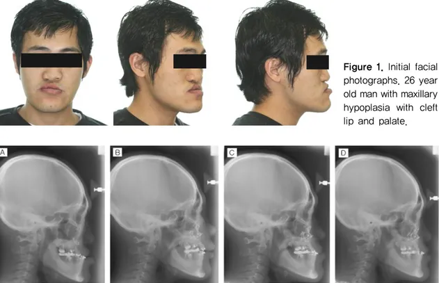
2008년 편측성 구순구개열(Cleft hard palate with unilateral cleft lip )을 가진 26세 남자 환자 가 상악열성장을 주소로 고려대학교병원 구강악안 면외과에 내원하였다. 환자는 생후 4개월에 구순성 형술을, 4세에 경구개성형술을 받은 병력이 있다. 내원 당시 환자의 상악열성장은 상악과 코, 그 주변 조직, 안와하부와 협골부에서 나타나고 있었다. 좌 측에 비해 작은 우측 비공, 그리고 비익기저부가 넓 고 비대칭인 소견이 관찰되었다(Figure 1). 측모두 부규격 방사선 사진상으로는 비중격의 우측편위, 우측상방으로 편향된 교합평면, 우측으로 심하게 변위되어 하악면 비대칭을 동반한 상악골 열성장이 확인되었다(Figure 2A). 구내 소견으로는 수평피개 도(overjet)의 양은 -9 mm 였고 수직피개도(overbite) 의 양은 2.5 mm였다(Figure 3A). 다수 치아(#11, 12, 13, 15, 16, 36, 37)가 결손되어 있었고, 하악좌 측제2소구치는 하악좌측제1대구치 결손 부위로 전 위되어 있었다. 상하 제1대구치의 관계에서 III급 부정교합 관계를 보였다. 다수 치아(#28, 47, 48)는 심한 우식으로 발치하였다. 27세 때 하악정중부에 채취한 자가골을 이용하여 상악 전치부에 Alveolar cleft repair를 시행하였다. 중안면 열성장 및 전치 부 반대교합을 해소하기 위하여 교정 치료만으로는 치료에 한계가 예상되고, 상악골의 전방 이동시에 연조직의 신장도 필요하다고 판단되어 골신장 수술 을 시행하기로 하였다. 추후 필요시 하악시상지분 할술 및 이부성형술을 시행하여 비대칭을 교정하기 로 결정하였다. 수술은 비기관 삽관을 이용한 전신 마취 하에 Le Fort I형 골절단술을 시행하였다. 상악골 전방부의 골절단을 시행할 때 상악골 혈액공급을 유지하기 위해 구개측 점막을 보존하였다. 상악골의 하방 골 절(Down-fracture)을 시행하고, 상악 골편의 완전 한 동요를 확인한 다음, 상악 구외신장기를 상악골 에 연결하기 위하여 협골 받침대 부위의 골절단선 하방과 전비극 부위에 각각 2개씩, 총 4개의 metal plate를 고정하였다. 견고 구외신장기의 고정장치 부분(halo portion) 을 환자의 두개골에 맞도록 조절하였다. 이때 trial pin을 이용하여 견고 구외신장기와 수평 막대
Figure 1. Initial facial photographs. 26 year old man with maxillary hypoplasia with cleft lip and palate.
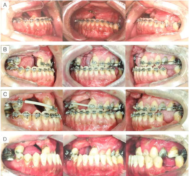
Figure 2. Intraoral photographs. Preoperative photo (A), Postoperative photo (B), 6-month postdistraction photo (C), 1-year postdistraction photo (D).
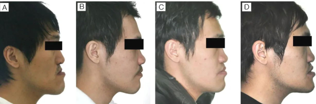
(horizontal bar)를 위치시키고, 이개(auricle)의 최 상점에서 3cm위의 높이에서 FH 평면과 평행하게 놓이도록 조절한 다음, 양측 두정골에 각각 3개 씩, 6개의 나사(scalp screw)를 이용하여 고정하였다. 견고 구외신장기의 수직막대(vertical bar)를 상악 골의 비대칭을 조정하기 위해 안면정중선을 기준으 로 좌측으로 위치시켜 수술계획과 일치하도록 확인 하였다. 골절단술 후 7일간의 잠복기를 두었다. 상 악골의 신장은 술 후 8일부터 시작하여 매일 아침과 저녁에 각각 0.5 mm 씩 하루 1 mm 의 신장속도로 11일동안 총 11 mm 신장시켰다. 3주간의 골경화기 를 가진 다음 두부 나사(scalp screw)와 윤상의 고 정장치를 제거하면서 상악골의 위치 안정성을 확보 하기 위해 소형금속판과 나사로 골편을 고정하였다 (Figure 4). 골신장 후 중안모와 상순의 돌출도가 증가하여 개선되었고, 상악 열성장으로 인한 III급 부정교합 이 정상교합으로 개선된 것을 관찰할 수 있었다 (Figure 5B). 술 후 방사선 사진에서는 신장된 익돌 상악접합부에서 방사선불투과상이 관찰되어 신생 골이 형성되고 있음을 확인할 수 있었다(Figure 2B). 측모두부규격방사선계측사진 중첩결과, 술 전 에 비해 상악이 전상방 견인되고 코와 상순의 돌출 도가 개선된 것을 관찰할 수 있었다(Figure 6). SNA는 74.0에서 78.3으로 증가하였고, SNB는 85.1에서 81.1로 감소하여 수술 전보다 개선된 계측 치를 확인할 수 있었다. SN 평면을 기준으로 상악 의 교합평면이 반시계방향으로 회전하였고, 상하악
Figure 3. Intraoral photographs. Preoperative photo (A), Postoperative photo (B), 6-month postdistraction photo (C), 1-year postdistraction photo (D).
전치부에서는 정상 교합 관계가 관찰되었다. 수평 피개도의 양은 2.0 mm, 수직피개도의 양은 0.6 mm로 전치부의 교합 관계가 정상이 되면서 하악이 시계방향으로 회전하였다(Figure 3B). Bjork’s Sum 값은 388.0에서 392.6으로 증가하였고 안모 의 길이는 증가되는 효과를 얻었다. 11 mm를 신장 시켰으나 N-perp-A 값이 6.4 mm만 증가한 이유 는 상악골의 비대칭을 조정하기 위해서 전진시킨 방향이 좌측을 향했기 때문이라고 분석하였다 (Table 1). 6개월 후 재평가를 시행하였다. 수술 전보다는 코와 상순이 돌출되어 있었으나, 골신장을 시행한 직후와 비교하였을 때는 중안모와 코, 상순의 돌출 도가 회귀된 모습을 관찰할 수 있었다(Figure 5C).
Figure 4. Latera facial photographs of the Rigid External Distraction (RED) System (RED II system, KLS Martin, Germany).
Figure 5. Lateral facial photographs. Preoperative photo (A), Postoperative photo (B), 6-month postdistraction photo (C), 1-year postdistraction photo (D).
Figure 6. The superimposition of the lateral cephalometric tracings at the preoperation (black line), postoperation (blue line) and 1-year postdistraction (red line).
Pre Distraction Osteogenesis
(Post OP 1 month) (Post OP 6 month) (Post OP 1 year)
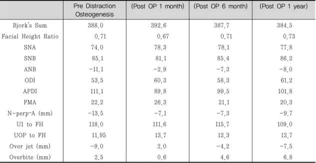
Bjork’s Sum 388.0 392.6 387.7 384.5
Facial Height Ratio 0.71 0.67 0.71 0.73
SNA 74.0 78.3 78.1 77.8 SNB 85.1 81.1 85.4 86.2 ANB -11.1 -2.9 -7.3 -8.0 ODI 53.5 60.3 58.3 61.2 APDI 111.1 89.8 99.5 101.8 FMA 22.2 26.3 21.1 20.3 N-perp–A (mm) -13.5 -7.1 -7.3 -9.7 U1 to FH 118.0 111.6 115.7 109.0 UOP to FH 11.95 13.7 12.3 12.7 Over jet (mm) -9.0 2.0 -4.2 -7.5 Overbite (mm) 2.5 0.6 4.6 6.8
Table 1. Values of the variables in the presurgical stages, immediate postsurgical stage, 6-month postdistraction and 1-year postdistraction.
수술 직후 회복되었던 상하악골의 관계가 상악이 후방으로 이동하여, III급 부정교합 관계로 회귀되 는 경향을 보였다(Figure 2C). SNA는 78.3에서 78.1으로 유지되었고, SNB는 81.1에서 85.4로 증 가하였으며, ANB는 -2.9에서 -7.3로 변화되었음 을 확인할 수 있었다. 수평피개도의 양은 -4.2 mm, 수직피개도의 양은 4.6 mm로 전치부의 교합 관계는 III급 부정교합 관계를 보였다(Figure 3C). 하악은 반시계방향으로 회전하여 Bjork’s Sum 값은 392.6에서 387.7로 감소하였다(Table 1). 1년 후 평가를 한번 더 시행하였다. 연조직 평가 에서 코와 상순의 돌출도는 6개월 전과 유사한 모습 을 관찰할 수 있었다(Figure 5D). 6개월 전과 비교 하였을 때, 수평피개도의 양은 -7.5 mm, 수직피개 도의 양은 6.8 mm로 교합이 술 전 보다 더 교합이 더 깊어진 양상을 보였다(Figure 3D). 방사선 사진 상으로 III급 부정교합 관계를 관찰할 수 있었다 (Figure 2D). SNB는 85.4에서 86.2로 증가, ANB는 -7.3에서 -8.0로 증가되었다. 수직피개도가 깊어 지면서 하악은 반시계방향으로 회전하였고, Bjork’s Sum은 387.7에서 384.5로 감소하였다(Table 1).
Ⅲ. 결론 및 고찰
골신장술을 통한 구순구개열 환자의 열성장 교정 술은 점진적인 상악의 전방이동으로 인해, 연조직 의 적응이 일어나고, 많은 양의 전방이동이 가능하 다는 장점이 있으며, 성장기의 환자에서도 시술이 가능하다9,10,13). 하악골 후퇴술을 동시에 시행해야 하는 악교정 수술에 비하여 골신장술은 수술 중 수 혈 가능성이 적고, 예측가능성이 높은 술식으로 알 려져 있다7,14). RED system을 이용한 골신장술은 상악의 수직, 수평이동량을 조절할 수 있는 장점이있다9,15). 그러나 악교정 수술과는 달리, 정확한 상 악의 위치를 설정하기 어렵고, 상하악 간의 안정된 교합을 얻기 어려우며, 정중선 불일치에도 주의해 야 한다. 구순구개열 환자에게 골신장술을 시행시 에는 정상인에 비해 회귀, 치유 지연, 치아 상실, 연 조직과 골의 괴사 그리고 감염 등과 같은 문제가 일 어날 수 있다16). 신장 도중 상악은 구순의 반흔으로 인해 시계방향으로 회전하는 경향을 가지게 되어 상악의 수직 신장을 보이기도 하므로 신장방향 조 절 시 고려를 요한다17-20). 골신장술은 골신장의 방 향과 양을 계속적으로 관찰해야 하며, 개교합 등의 원하지 않은 방향으로의 골신장 가능성, 술 후 재귀 현상 등이 단점으로 지적되어 왔다. 또한 견인기간 동안 혐오스러운 외부 장치 부착으로 심리적인 부 담감을 주거나, 사회생활이 불편하다21).
Polley 등13)은 구순구개열 환자들에게 RED system 을 이용한 골신장술을 시행한 결과 상악골의 전방변화 가 악교정 수술로 이동가능한 한계보다(6 mm) 더 큰 이동량인 평균 12.7 mm 얻었다고 보고하였다. 본 증 례에서도 골신장기를 이용하여 10 mm 이상의 전방 이동을 시행하는데 특별한 어려움이 없었다. Cedars 등14,22)에 의하면 교합면 수준에서는 전진 양상의 훌륭한 안정이 관찰되었고, 그의 견인 방법 으로 중안면전진술 후 안와 수준에서는 약간의 회 귀가 관찰되었다고 하였다. 이 결과는 교합의 안정 성이 뒷받침되었기 때문에 견인된 상태로 유지가 된 거라 분석해 볼 수 있다. 본 증례에서 수술 후 소 형금속판과 나사 고정을 하였음에도 불구하고 상악 골의 회귀현상을 관찰하였다. 이와 같은 결과는 환 자의 구강내 구치부에 결손치가 많아 교합 안정성 이 떨어지면서 생긴 것이라 평가하였다. Singh 등 23)은 회귀될 것을 고려하려 30% 정도의 과신장을 하는게 좋다고 하였다. Chen 등24)은 술 후 신장된 상악골의 위치 안정성과 재귀를 고려하여 목표양의 최대 53% 까지 과신장을 시행하는 것을 제안하였 고, 골신장 양이 충분하지 않은 경우 부가적인 악교 정 수술을 시행하는 것이 필요하다고 하였다. Marianetti 등25)은 결손치가 많은 환자에게 술 전 보철치료를 통하여 교합관계의 안전성을 얻은 후 악교정 수술을 시행하여 장기적인 교합 안정성 을 얻은 증례를 보고한 바 있다. Yosano A 등26)은 악교정 수술 후의 안정성은 소형금속판의 고정보다 상하악의 교합평면이 더 중요하고 큰 영향을 미친 다고 주장하였다. 이는 본 증례에서는 소형금속판 을 이용하여 견고 고정을 시행하였으나 상하악 간 의 안정된 교합 관계를 얻지 못하여 회귀된 현상과 연관성이 있다고 생각한다. 추후 보철 치료를 병행하면서 교합관계의 변화 양상에 대하여 지속적으로 관찰을 시행하고, 2차적 인 악교정 수술을 고려할 예정이다.
Ⅳ. 결론
본 증례보고에서 상악골 열성장을 보이는 구순구 개열 환자에 골신장기를 이용하여 상악골 전진술을 시행한 후 1년 동안 경과관찰하였다. 수술 직후 상악 이 전방 견인되고 코 상순의 돌출도가 개선되면서 III 급 부정교합이 개선된 모습을 보였다. 그러나 6개월, 1년 경과관찰 결과 상악골을 소형금속판으로 고정하 였음에도 불구하고 회귀현상을 관찰할 수 있었다. 이 는 환자의 구치부 결손치가 많아 교합 불안정성이 발 생하여 상악 골편의 안정성을 유지하지 못하고, 후방 으로 재귀된 것이라고 평가하였다. 이번 증례를 통해 서 골신장술을 시행할 환자에게도 술 전, 술 후의 교 합 안정성이 중요하다는 것을 경험하였다.참고문헌
1. Campisi P, et al. Expression of bone mor-phogenetic proteins during mandibular distraction osteogenesis. Plast Reconstr Surg 2003;111:201-8;discussion 209-210. 2. Snyder CC, et al. Mandibular lengthening
by gradual distraction. Preliminary report. Plast Reconstr Surg 1973;51:506-508. 3. McCarthy JG, et al. Lengthening the human
mandible by gradual distraction. Plast Reconstr Surg 1992;89:1-8; discussion 9-10. 4. Ross RB. The clinical implications of facial
growth in cleft lip and palate. Cleft Palate J 1970;7:37-47.
5. Tateishi C, Moriyama K, Takano-Yamamoto T. Dentocraniofacial morphology of 12 Japanese subjects with unilateral cleft lip and palate with a severe Class III maloc-clusion: a cephalometric study at the pre-treatment stage of surgical orthodontic treatment. Cleft Palate Craniofac J 2001; 38:597-605.
6. Gilley FP. A cephalometric analysis of the developmental pattern and facial morphol-ogy in cleft palate. Dent Res Grad Study 1947;48:13-15.
7. Figueroa AA, Polley JW, Figueroa AL. Introduction of a new removable adjustable intraoral maxillary distraction system for correction of maxillary hypoplasia. J Craniofac Surg 2009;20Suppl 2:1776-1786. 8. Figueroa AA, Polley JW, Ko EW. Maxillary
distraction for the management of cleft maxillary hypoplasia with a rigid external
distraction system. Semin Orthod 1999;5: 46-51.
9. Polley JW, Figueroa AA. Rigid external dis-traction: its application in cleft maxillary deformities. Plast Reconstr Surg, 1998;102:
1360-72;discussion 1373-1374.
10. Polley JW, Figueroa AA. Maxillary dis-traction osteogenesis with rigid external distraction. Atlas Oral Maxillofac Surg Clin North Am 1999;7:15-28.
11. Joss CU, et al. Skeletal and dental stability of segmental distraction of the anterior mandibular alveolar process. A 5.5-year follow-up. Int J Oral Maxillofac Surg 2012. 12. Joss CU, et al. Skeletal and dental stability
of segmental distraction of the anterior mandibular alveolar process. A 2-year fol-low-up. Int J Oral Maxillofac Surg 2012;41:
553-559.
13. Polley JW, Figueroa AA. Management of se-vere maxillary deficiency in childhood and adolescence through distraction osteo-genesis with an external, adjustable, rigid distraction device. J Craniofac Surg 1997; 8:181-185;discussion 186.
14. Cedars MG, et al. Advancement of the mid-face using distraction techniques. Plast Reconstr Surg, 1999;103:429-441.
15. Figueroa AA, Polley JW. Management of se-vere cleft maxillary deficiency with dis-traction osteogenesis: procedure and results. Am J Orthod Dentofacial Orthop, 1999;115:1-12.
16. Willmar K. On Le Fort I osteotomy; A fol-low-up study of 106 operated patients with
maxillo-facial deformity. Scand J Plast Reconstr Surg 1974;12:suppl 12:1-68. 17. Harada K, Sato M, Omura K. Long-term
maxillomandibular skeletal and dental changes in children with cleft lip and palate after maxillary distraction. Oral Surg Oral Med Oral Pathol Oral Radiol Endod 2006;102:292-299.
18. Harada K, Sato M, Omura K. Long-term skeletal and dental changes in patients with cleft lip and palate after maxillary dis-traction: a report of three cases treated with a rigid external distraction device. Cranio 2005;23:152- 157.
19. Harada K, et al. Maxillary distraction os-teogenesis for cleft lip and palate children using an external, adjustable, rigid dis-traction device: a report of 2 cases. J Oral Maxillofac Surg 2001;59:1492- 1496. 20. Cheung LK, Chua HD. A meta-analysis of
cleft maxillary osteotomy and distraction osteogenesis. Int J Oral Maxillofac Surg 2006;35:14-24.
21. Klein C, Howaldt HP. Mandibular micro-gnathism as a sequela of early childhood capitulum fractures and their treatment
using distraction osteogenesis. Fortschr Kiefer Gesichtschir 1996;41:147-151. 22. Toth BA, et al. Distraction osteogenesis and
its application to the midface and bony orbit in craniosynostosis syndromes. J Craniofac Surg 1998;9:100-13; discussion 119-122. 23. Singh SP, et al. Treatment outcome and
long-term stability of skeletal changes fol-lowing maxillary distraction in adult sub-jects of cleft lip and palate. Contemp Clin Dent 2012;3:188-192.
24. Chen PK, et al. Maxillary distraction os-teogenesis in the adolescent cleft patient: three-dimensional computed tomography analysis of linear and volumetric changes over five years. Cleft Palate Craniofac J 2011;48:445-454.
25. Marianetti TM, et al. Case report of oligo-dontia: long term stability of orthognatic surgery and prosthetic rehabilitation. Minerva Stomatol 2011;60:139-147.
26. Yosano A, et al. Influence of mandibular fixation method on stability of the maxil-lary occlusal plane after occlusal plane alteration. Bull Tokyo Dent Coll 2009; 50:71-82.
교신 저자
Professor Eui-seok Lee
Department of Oral and Maxillofacial Surgery, Guro Hospital, Korea University Tel: +82-2-2626-1523 / Fax: +82-303-0482-7575 / E-mail: [email protected]