저작자표시-비영리-변경금지 2.0 대한민국 이용자는 아래의 조건을 따르는 경우에 한하여 자유롭게 l 이 저작물을 복제, 배포, 전송, 전시, 공연 및 방송할 수 있습니다. 다음과 같은 조건을 따라야 합니다: l 귀하는, 이 저작물의 재이용이나 배포의 경우, 이 저작물에 적용된 이용허락조건 을 명확하게 나타내어야 합니다. l 저작권자로부터 별도의 허가를 받으면 이러한 조건들은 적용되지 않습니다. 저작권법에 따른 이용자의 권리는 위의 내용에 의하여 영향을 받지 않습니다. 이것은 이용허락규약(Legal Code)을 이해하기 쉽게 요약한 것입니다. Disclaimer 저작자표시. 귀하는 원저작자를 표시하여야 합니다. 비영리. 귀하는 이 저작물을 영리 목적으로 이용할 수 없습니다. 변경금지. 귀하는 이 저작물을 개작, 변형 또는 가공할 수 없습니다.
Clinical Outcome in the Surgical Intervention
of Congenital Muscular Torticollis
by
JooHyoungKim
Major in Medicine
Department of Medical Sciences
The Graduate School, Ajou University
Clinical Outcome in the Surgical Intervention
of Congenital Muscular Torticollis
by
JooHyoungKim
A Dissertation Submitted to The Graduate School of
Ajou University in Partial Fulfillment of the Requirements
for the Degree of Ph.D.
of Medicine
Supervised by
MyongChul Park, M.D., Ph.D.
Major in Medicine
Department of Medical Sciences
The Graduate School, Ajou University
This certifies that the dissertation
ofJooHyoungKim is approved.
SUPERVISORY COMMITTEE
MyongChul Park
Dong Ha Park
Goo Hyun Baek
Goo-Hyun Mun
Il Jae Lee
The Graduate School, Ajou University
June, 21st, 2016
-ABSTRACT-
Clinical Outcome in the Surgical Intervention of
Congenital Muscular Torticollis
A number of studies have shown that facial asymmetry improves in congenital
muscular torticollis (CMT) patients after surgical release. This study confirmed the improvement in facial asymmetry, and analyzed factors that affect the change of facial asymmetry in CMT patients after surgical release by using objective and quantitative methods.Facial asymmetry was analyzed in 60 CMT patients who underwent surgical release before 10 years of age. Horizontal and lower facial asymmetry angles (HFAA and LFAA) in the clinical photograph were used to measure facial asymmetry. Postoperative improvements in HFAA and LFAA were evaluated in each age group, after grouping the patients by age. Patients were divided into 2 groups according to the postoperative head tilt and functional deficit. Postoperative improvements in HFAA and LFAA were compared between 2 groups. The relationships between postoperative improvements in HFAA and LFAA and independent variables (age, follow-up period, preoperative HFAA or LFAA, postoperative head tilt, and postoperative functional deficit) were analyzed. Mean age at operation was 34.8 months (range, 6-120 mo). Horizontal facial asymmetry angle was improved significantly postoperatively in groups <5 years of age. Lower facial asymmetry angle was improved significantly postoperatively in all age groups. No significant difference was found in the postoperative improvements in HFAA and LFAA between 2 groups according to the postoperative head tilt and functional deficit. In the correlation analysis,
postoperative improvements in HFAA and LFAA were proportional to the follow-up period (r = 0.256, P = 0.048) and preoperative HFAA or LFAA (r = 0.600, P < 0.001). Facial asymmetry in CMT patients can be improved in part if surgical release is performed before 10 years of age and the possibility of improvement may be different according to the area of the face. After surgical release, facial asymmetry will improve over a long period of time, and patients with more severe facial asymmetry have a better remodeling potential to achieve facial symmetry.
TABLE OF CONTENTS
ABSTRACT ··· i
TABLE OF CONTENTS ··· iiii
LIST OF FIGURES ··· iv
LIST OF TABLES ··· v
ABBREVIATION ··· vii
I. INTRODUCTION ··· 1
II. MATERIALS AND METHODS ··· 3
A. Method of Assessment ··· 4 B. Assess of Results ··· 8 III. RESULTS ··· 10 IV. DISCUSSION··· 21 REFERENCES ··· 27 국문요약 ··· 31
LIST OF FIGURES
Fig. 1. Standardized anteroposterior photographs before and after operation ··· 6 Fig. 2. Scatter plot between improvement of facial asymmetry index and follow-up period
after surgical release in congenital muscular torticollis patients ··· 14 Fig. 3. Scatter plot between improvement of facial asymmetry index and preoperative facial
LIST OF TABLES
Table 1. Patient Characteristics ··· 11 Table 2. Comparisons Between Preoperative and Postoperative Facial Asymmetry Index ·· 13 Table 3. Comparison of Improvement in Facial Asymmetry Index According to the
Postoperative Head Tilt Angle ··· 17 Table 4. Comparison of Improvement in Facial Asymmetry Index According to the
Postoperative Functional Deficit ··· 18 Table 5. Correlation Analyses Between Improvement in Facial Asymmetry Index, and Age
at Operation, Follow-Up Period, Preoperative Facial Asymmetry Index,
ABBREVIATION
CMT: congenital muscular torticollis SCM: sternocleidomastoid
PROM: passive range of motions
HFAA: horizontal facial asymmetry angle
LFAA: lower facial asymmetry angle
HTA: head tilt angle
1
I. INTRODUCTION
Congenital muscular torticollis (CMT) is caused by an idiopathic fibromatous contracture of the sternocleidomastoid (SCM) muscle that restricts movement of the neck. Contracture of the SCM muscle causes the head to turn toward the affected side and the chin to point to the opposite side. The prolonged unilateral contracture of the SCM muscle may induce
craniofacial asymmetry.1–4 The characteristic appearance associated with torticollis includes deformational plagiocephaly with flattening of the contralateral side of the occiput,
ipsilateral recessed orbit and zygoma, reduction in ramal height on the affected side, inferior orbital dystopia on the affected side, deviation of the nasal tip and chin point, commissural canting toward the affected side, and inferiorly and posteriorly positioned ipsilateral ear.1,4– 6
Surgical release is indicated in CMT patients who do not improve after physical therapy or botulinum toxin injections or whose SCM muscle is severely fibrotic.7,8A number of studies have shown that the craniofacial asymmetry in CMT patients improves after surgical release by variable operative methods. It was, however, difficult to confirm the objective and quantitative improvement in craniofacial asymmetry because the severity of craniofacial asymmetry was evaluated on the basis of subjective and categorical values in these studies.8–14
Several studies quantitatively evaluated the craniofacial asymmetry in CMT patients using cephalometry 1,3,5,13,15,16 or three-dimensional computed tomography imaging,4 and
analyzed the quantitative change in craniofacial asymmetry after surgical release 3; however, little is still known about the factors that affect the change of craniofacial asymmetry in CMT patients after surgical release.
In our previous study, we quantitatively measured the severity of preoperative craniofacial asymmetry in untreated CMT patients and showed that cranial asymmetry is already
determined in those <6 months of age, although facial asymmetry is progressive if the release of contracted SCM muscle is delayed.2Hence, this study evaluated the postoperative change in facial asymmetry except cranial asymmetry, and identified the factors that affect the postoperative change of facial asymmetry in CMT patients by using objective and quantitative methods.
3
II. MATERIALS AND METHODS
Three hundred nineteen CMT patients underwent complete tight fibrous band release and resection of the SCM muscle 7 at the Department of Plastic and Reconstructive Surgery of Ajou University Hospital from February 2007 to December 2012. Clinical photographs and medical records of the following patients were reviewed: patients who underwent surgical release at <10 years of age, and patients who revisited the outpatient clinic after >1 year postoperatively. Seventy-two patients were followed up for >1 year postoperatively. Eleven patients were >10 years of age at the time of operation. One patient whose clinical photograph was missing was excluded from this study. Sixty patients were evaluated in our study. The principles outlined in the Declaration of Helsinki were followed in our study.
Medical record review was performed to assess age at operation, sex, and passive range of motions (PROMs) for rotation and lateral flexion of the neck. A standardized anteroposterior photograph was taken with a digital camera wherein rotation of upper body was restricted and head was maintained naturally. Facial asymmetry and head tilt was evaluated on the photograph.
Surgical release of CMT was performed at the age of >=6 months only if the patients simultaneously met the following 2 criteria: functional limitation of the neck motion was evident because of shortening of the unilateral SCM, and the parents or the patients realized that there was no more benefit from the stretching exercises or that implementation of stretching exercises was not practically possible because of the patient's resistance to perform stretching exercises.
Postoperatively, a soft neck collar was immediately applied in the neutral neck position and was worn for 3 weeks. After the surgical wound had stabilized, an intensive physiotherapy program was undertaken to acquire full PROM of the neck by performing active
strengthening exercises for 3 to 4 months.
A. Method of Assessment
Measurement of PROMs for rotation and lateral flexion of the neck was performed using the arthrodial protractor, with the patient in the supine position, both shoulders stabilized, and head and neck supported by the examiner over the edge of the examination couch, so that the neck was free to rotate and bend laterally.17 When a limitation during neck movement was noted, further movement was stopped and the PROM was recorded. The difference between the affected side and the normal side was recorded as the rotational or flexional deficit. The sum of rotational and flexional deficits was represented by the functional deficit
Facial asymmetry index (horizontal facial asymmetry angle [HFAA] and lower facial asymmetry angle [LFAA]) was used to measure the degree of facial asymmetry on
standardized anteroposterior photographs (Fig. 1). Horizontal facial asymmetry angle is the sharp angle between the horizontal line through the bilateral exocanthions and the line through the bilateral mouth corners.16,18,19 Lower facial asymmetry angle is the angle between the line which connects nasion projection on the interexocanthal plane and gnathion, and the ideal facial midline.18 The ideal facial midline was defined as the line which is
5
perpendicular to interexocanthal plane because the glabella, subnasale, Cupid's bow,
maxillary interincisive line, and gnathion cannot be aligned perfectly in cases of asymmetry. Head tilt angle (HTA) was used to measure the amount of head tilt on standardized
anteroposterior photographs (Fig. 1). Head tilt angle is the sharp angle between the interacromial line and the ideal facial midline.
Measurement of HFAA, LFAA, and HTA was performed twice with Adobe Photoshop CS3 (Adobe Systems, San Jose, CA), and the mean value of these measurements was recorded.
Fig. 1. Standardized anteroposterior photographs before and after operation. (Above) Operation was performed at the age of 10 months and the postoperative photograph was taken 25 months after the operation. Preoperative horizontal facial
asymmetry angle (HFAA) = 4.1[degrees], preoperative lower facial asymmetry angle (LFAA) = 0.1[degrees]; postoperative HFAA = 2.0[degrees], postoperative LFAA = 0.3[degrees]. (Below) Operation was performed at the age of 20 months and the postoperative photograph was taken 48 months after the operation. Preoperative HFAA = 3.6[degrees], preoperative
7
LFAA = 1.9[degrees]; postoperative HFAA = 2.1[degrees], postoperative LFAA = 1.1[degrees]. HFAA: the sharp angle between the horizontal line through the bilateral exocanthions (red line) and the line through the bilateral mouth corners (yellow line). LFAA: the angle between the line which connects nasion projection on the interexocanthal plane and gnathion (blue line), and the ideal facial midline (green line). Head tilt angle: the sharp angle between the interacromial line (orange line) and the ideal facial midline.
B. Assessment of Results
The authors assumed that the age at operation, follow-up period, and preoperative severity of facial asymmetry might affect the postoperative change of facial asymmetry in CMT patients. In addition, residual limited range of motion (ROM) of the neck and residual head tilt after the operation can prevent improvement of facial asymmetry in CMT patients. To verify the above assumptions, the following tests were carried out.
First, the sample of patients was divided into 4 subgroups according to the age at the time of operation: Age Group 1, surgically treated before the age of 1 year; Age Group 2, from 1 to 3 years; Age Group 3, from 3 to 5 years; Age Group 4, from 5 to 10 years. Preoperative and postoperative facial asymmetry indexes were compared using the 2-tailed paired t-test. The analysis was performed separately for each age group.
Second, the patients were divided into 2 groups according to the postoperative HTA: HTA Group 1, <=5°; HTA Group 2, >5° and <=10°. Five patients with postoperative HTA >10° were excluded because we considered the operation as an incomplete release. The
improvements in facial asymmetry index were compared between the 2 groups using the 2-tailed independent t-test.
Third, the patients were divided into 2 groups according to the postoperative rotational and flexional deficits: Functional Group 1, all of the postoperative rotational and flexional
deficits <=5°; Functional Group 2, all of the postoperative rotational and flexional deficits >5° and <=10°. Five patients with postoperative rotational or flexional deficit >10° were
9
outcomes according to the Cheng and Tang scoring system 8. The improvements in facial asymmetry index were compared between the 2 groups using the 2-tailed independent t-test.
Parametric tests were used because all variables followed a normal distribution in the Shapiro-Wilk test (P > 0.05).
Finally, correlation and regression analyses were performed to identify the relationship between postoperative improvement in facial asymmetry index, and age at operation, follow-up period, preoperative facial asymmetry indexes, postoperative HTA, postoperative
functional deficit. Regression analysis was also used to predict the effect of the above variables on improvement in facial asymmetry index.
III. RESULTS
The study population composed of 60 patients (37 males and 23 females). Congenital muscular torticollis occurred on the right side in 38 cases and on the left side in 22 cases. Mean age at operation was 34.8 months (range, 6–120 mo). Mean follow-up period was 21.7 months (range, 12–53 mo). Among the 60 patients, 19 patients were included in Age Group 1, 19 in Age Group 2, 10 in Age Group 3, and 12 in Age Group 4 (Table 1).
11
In our study, the mean of HFAA and LFAA was improved significantly (P < 0.001) after surgical release, although facial asymmetry still persisted to some degree at the latest follow-up (Table 2) (Fig. 1) and facial asymmetry progressed a little in some patients (Fig. 2 and Fig. 3). The mean of HFAA was improved significantly postoperatively in Age Groups 1, 2, and 3 (Table 2). The mean of LFAA was improved significantly postoperatively in all age groups (Table 2).
13
Table 2. Comparisons Between Preoperative and Postoperative Facial Asymmetry Index
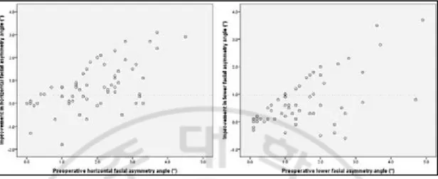
Fig. 2. Scatter plot between improvement of facial asymmetry index and follow-up period after surgical release in congenital muscular torticollis patients. In the correlation analysis, improvement in HFAA (Pearson correlation coefficient r = 0.256, P = 0.048) and LFAA (r = 0.461, P < 0.001) after surgical release was proportional to the follow-up period.
15
Fig. 3. Scatter plot between improvement of facial asymmetry index and preoperative facial asymmetry index after surgical release in congenital muscular torticollis patients. In the correlation analysis, improvement in HFAA after surgical release was proportional to the preoperative HFAA (r = 0.600, P r = 0.676, P < 0.001).
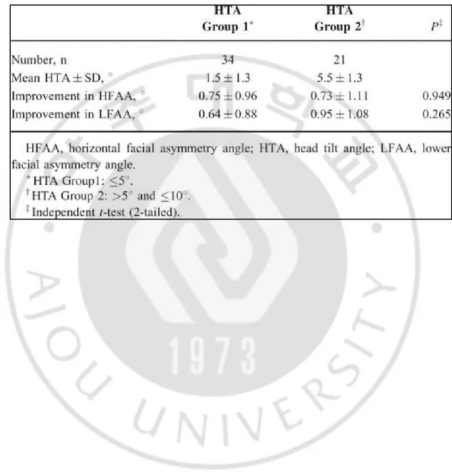
No significant difference was found in the improvements in HFAA and LFAA
postoperatively between the head tilt groups 1 and 2 (Table 3) and between the functional groups 1 and 2 (Table 4). Obviously, no significant difference was found in the confounding factors, including age at operation, follow-up period, preoperative facial asymmetry index, and postoperative functional deficit or postoperative HTA in the head tilt groups and the functional groups.
17
Table 3. Comparison of Improvement in Facial Asymmetry Index According to the Postoperative Head Tilt Angle
Table 4. Comparison of Improvement in Facial Asymmetry Index According to the Postoperative Functional Deficit*
19
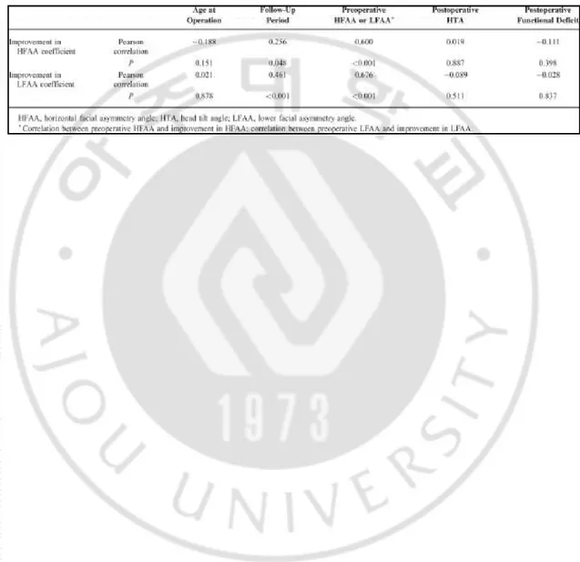
In the correlation analysis, the improvements in HFAA and LFAA after surgical release were proportional to the follow-up period (Table 5) (Fig. 2), and preoperative HFAA or LFAA significantly (Table 5) (Fig. 3). The improvements in HFAA and LFAA, however, were not related significantly to the age at operation, postoperative HTA, and postoperative functional deficit (Table 5). In the regression analysis, the following equation was derived:
Improvement in HFAA = -0.724 + 0.019 × follow-up period + 0.554 × preoperative HFAA
Table 5. Correlation Analyses Between Improvement in Facial Asymmetry Index, and Age at Operation, Follow-Up Period, Preoperative Facial Asymmetry Index, Postoperative Head Tilt Angle, Postoperative Functional Deficit
21
IV. DISCUSSION
There have been several studies that measured facial asymmetry in CMT patients and analyzed the change in facial asymmetry quantitatively after surgical release of the contracted SCM muscle. In many studies, the cephalometric radiograph was used to
quantitatively evaluate facial asymmetry in CMT patients.1,3,5,13,15,16 Lee et al 3 analyzed the objective and quantitative change of craniofacial deformity in CMT patients after
surgical release, using the cephalometric radiograph and the appropriate statistical method. These studies are, however, still insufficient to thoroughly understand the change of facial asymmetry in CMT patients after surgical release. Kim et al 16 did not evaluate the postoperative change in facial asymmetry because the follow-up periods were too short to show the postoperative change in facial asymmetry. Arslan et al 13 quantitatively measured the postoperative change of facial asymmetry in CMT patients, but they failed to show a statistically significant change. Furthermore, patients in that study were >6 years, and the number of patients was 12. Moreover, other factors that may affect the postoperative change in facial asymmetry were seldom investigated except age at operation. To compensate for such limitations, we quantitatively measured facial asymmetry in CMT patients and
investigated the variable factors that may affect the change in facial asymmetry after surgical release, including age at operation, follow-up period, preoperative facial asymmetry,
postoperative head tilt, and postoperative functional deficit.
Anthropometric angles on a frontal digital photograph were used to measure facial asymmetry. Although cephalometric measurement has high reproducibility and is more
objective than measurement using a distal photograph, it was impossible to examine patients <1 years of age holding their head in a standard position with use of a cephalostat.
Anthropometric measurements were also used to evaluate facial asymmetry in many studies as a quantitative and objective method.2,18–20 A digital photograph can show facial soft tissue asymmetry and be taken repeatedly without the risk of radiation exposure. Severity of preoperative facial asymmetry, however, may be measured lower in our photograph than in a cephalometric standard position because patient's affected face is rotated to an unaffected side in our photograph.
Many studies reported about the relation between age and postoperative improvement of craniofacial asymmetry in CMT patients. Maslon et al 21 showed that facial asymmetry is more often persistent in children operated after 3 years of age. Some authors stated that complete restoration of craniofacial asymmetry can be expected in surgically treated patients <5 to 6 years of age.22–24 Based on an average follow-up period of 15 years in 55 patients, Wirth et al 9 showed that even severe facial asymmetry may resolve completely within a few years if the patient is operated before 5 years of age. Postoperative changes in craniofacial asymmetry, however, were not evaluated quantitatively in these studies. On the basis of quantitative data, Lee et al 3 recently showed that craniofacial deformity was better improved when surgical release was performed before 5 years of age in CMT patients.
In our study, horizontal facial asymmetry was improved significantly in age groups <5 years of age and lower facial asymmetry was improved significantly in all age groups after surgical release (Fig. 1) (Table 2). This result may be affected by distinguishing facial
23
The orbits, for example, have a maximum growth rate from 1 to 2 years of age and have usually attained their full adult size by the age of 7 years. The palate and maxilla have achieved two-thirds of adult size by the age of 6 years. And the mandible has later maximum growth rate between the age of 8 and 14 years than other facial areas.25 Especially, the improvement in horizontal facial asymmetry in patients <1 year of age was greater than that in older age groups, although the follow-up period was shorter (Table 2). This result may be affected by the remodeling potential of orbits. Raposo et al 26 showed that surgical
correction of hypertelorbitism in patients <8 years old leads to relapse in a 30-year
longitudinal study because of continued periorbital growth. Based on facial growth pattern and their study, horizontal facial asymmetry of patients >5 years of age in our study may be improved if they are followed up until completely grown. The correlation analysis did not show a significant negative linear correlation between age at operation and improvement in facial asymmetry index after surgical release (Table 5). Consequently, we noticed that facial asymmetry in CMT patients can be improved in part if surgical release is performed before 10 years of age and the possibility of improvement may be different according to the area of the face, although improvement in facial asymmetry is not inversely proportional to age at operation. The authors, however, think that it is better to perform the operation at a younger age than at an older age if patients need surgical release and their facial asymmetry is
prominent because facial asymmetry in the untreated CMT patients is progressive. Definitely, additional measuring which evaluate asymmetry in different area of face has to be done to understand thoroughly improvement in facial asymmetry in CMT patients after surgical release.
It is considered that the improvement of facial asymmetry in CMT patients may be prevented by residual head tilt and residual limited ROM of the neck after surgical release. In our study, there was, however, no significant difference in the improvement in facial asymmetry after surgical release according to the postoperative HTA or the postoperative functional deficit (Tables 3 and 4). Postoperative HTA and postoperative rotational and flexional deficits, which are <=10°, are evaluated as excellent or good outcomes of the CMT operation.8,12 Correction of the head tilt and regaining the ROM of the neck is the primary goal of CMT surgery. If the primary goal of CMT surgery is achieved by obtaining excellent or good outcomes, improvement in facial asymmetry will not be prevented by residual head tilt and residual limited ROM of the neck.
Improvement of facial asymmetry in CMT patients was proportional to the follow-up period after surgical release (Table 5) (Fig. 2). In other words, facial asymmetry in CMT patients may change postoperatively until the facial skeleton grows completely; therefore, facial asymmetry should be traced until the facial skeleton grows completely. Indeed, the remodeling potential of the facial bone is greater and more long-lasting than that of the cranial bone and the growth rate differ according to the area of the face.25 Facial skeletal growth continues until the age of 18 years.11 Furthermore, a case report described that facial asymmetry was resolved almost completely postoperatively in a 10-year-old girl with CMT who was followed up for 14 years.27
There was a significant positive correlation between the preoperative severity of facial asymmetry and the improvement in facial asymmetry after surgical release (Table 5) (Fig. 3). Furthermore, in the regression analysis, improvement in facial asymmetry was more affected
25
by preoperative facial asymmetry than other factors. This result indicated that the facial bone with more severe asymmetry has a higher remodeling potential to attain the symmetric status, if the SCM contracture is released. Indeed, this result is quite interesting. Up till now, there have been no studies showing same results as those in our report or demonstrating the effect of preoperative severity of facial asymmetry on improvement of facial asymmetry in CMT patients after surgical release. Indeed, this result might be possible because all of the patients in our study were growing and their mean age was only 34.8 months.
There are several limitations in our study. To understand the change of facial asymmetry in CMT patients after surgical release, it is essential to follow up the CMT patients until they achieve the completely grown status. We evaluated, however, the improvement of facial asymmetry in patients who were still growing. In addition, only 72 patients, among the patients, who underwent surgical release were followed up in our study. The authors tried to follow up all patients, but a number of patients did not reply to our request to visit again because they were living at distant areas, or did not notice any problems about their operation. Although this creates a selection bias, this does not contradict our results which facial asymmetry in CMT patients is improved quantitatively after surgical release because patients who were not followed up have commonly the better surgical results. Obviously, further studies are needed to confirm the results of our study. We will attempt to follow up more number of patients until they are completely grown. Despite these limitations, this study can provide help in understanding the postoperative change of facial asymmetry in CMT patients
In conclusion, we noticed that facial asymmetry in CMT patients can be improved in part if surgical release is performed before 10 years of age, and the possibility of improvement may be different according to the area of the face. The postoperative follow-up period and the severity of preoperative facial asymmetry are also important for determining the
improvement in facial asymmetry postoperatively. In CMT patients, facial asymmetry will improve over a long period of time, and patients with more severe facial asymmetry have more remodeling potential to achieve facial symmetry.
27
REFERENCES
1. Hollier L, Kim J, Grayson BH, et al. Congenital muscular torticollis and the associated craniofacial changes. Plast Reconstr Surg 105:827-835, 2000
2. Seo SJ, Yim SY, Lee IJ, et al. Is craniofacial asymmetry progressive in untreated congenital muscular torticollis? Plast Reconstr Surg 132:407-413, 2013
3. Lee JK, Moon HJ, Park MS, et al. Change of craniofacial deformity after
sternocleidomastoid muscle release in pediatric patients with congenital muscular torticollis. J Bone Joint Surg Am 94:e93, 2012
4. Yu CC, Wong FH, Lo LJ, et al. Craniofacial deformity in patients with uncorrected congenital muscular torticollis: an assessment from three-dimensional computed tomography imaging. Plast Reconstr Surg 113:24-33, 2004
5. Ferguson JW. Cephalometric interpretation and assessment of facial asymmetry secondary to congenital torticollis. The significance of cranial base reference lines.
Int J Oral Maxillofac Surg 22:7-10, 1993
6. Ferguson JW. Surgical correction of the facial deformities secondary to untreated congenital muscular torticollis. J Craniomaxillofac Surg 21:137-142, 1993
7. Lee IJ, Lim SY, Song HS, et al. Complete tight fibrous band release and resection in congenital muscular torticollis. J Plast Reconstr Aesthet Surg 63:947-953, 2010 8. Cheng JC, Tang SP. Outcome of surgical treatment of congenital muscular torticollis.
9. Wirth CJ, Hagena FW, Wuelker N, et al. Biterminal tenotomy for the treatment of congenital muscular torticollis. Long-term results. J Bone Joint Surg Am 74:427-434, 1992
10. Stassen LF, Kerawala CJ. New surgical technique for the correction of congenital muscular torticollis (wry neck). Br J Oral Maxillofac Surg 38:142-147, 2000 11. Bharadwaj VK. Sternomastoid myoplasty: surgical correction of congenital
torticollis. J Otolaryngol 26:44-48, 1997
12. Shim JS, Jang HP. Operative treatment of congenital torticollis. J Bone Joint Surg
Br 90:934-939, 2008
13. Arslan H, Gunduz S, Subasi M, et al. Frontal cephalometric analysis in the evaluation of facial asymmetry in torticollis, and outcomes of bipolar release in patients over 6 years of age. Arch Orthop Trauma Surg 122:489-493, 2002
14. Shim JS, Noh KC, Park SJ. Treatment of congenital muscular torticollis in patients older than 8 years. J Pediatr Orthop 24:683-688, 2004
15. Lee DY, Song BW, Cho TJ, et al. Craniofacial asymmetry in congenital muscular torticollis patients: a study using cephalometry. J Korean Orthop Assoc 42:24-31, 2007
16. Kim HT, Kang KJ, Yoo CI. Head tilt and facial asymmetry in congential muscular torticollis. J Korean Orthop Assoc 38:217-222, 2003
17. Cheng JC, Wong MW, Tang SP, et al. Clinical determinants of the outcome of manual stretching in the treatment of congenital muscular torticollis in infants. A
29
prospective study of eight hundred and twenty-one cases. J Bone Joint Surg Am 83-A:679-687, 2001
18. Yu CC, Bergeron L, Lin CH, et al. Single-splint technique in orthognathic surgery: intraoperative checkpoints to control facial symmetry. Plast Reconstr Surg 124:879-886, 2009
19. Song WC, Koh KS, Kim SH, et al. Horizontal angular asymmetry of the face in korean young adults with reference to the eye and mouth. J Oral Maxillofac Surg 65:2164-2168, 2007
20. Kim HT, Kang JH, Yoo CI. Head tilt and facial asymmetry in congenital muscular torticollis. J Korean Orthop Assoc 38:217-222, 2003
21. Maslon A, Lebiedzinski R, Domzalski M, et al. [Facial asymmetry in children with congenital muscular torticollis after surgical treatment]. Chir Narzadow Ruchu
Ortop Pol 74:31-34, 2009
22. Ling CM. The influence of age on the results of open sternomastoid tenotomy in muscular torticollis. Clin Orthop Relat Res 142-148,1976
23. Ippolito E, Tudisco C, Massobrio M. Long-term results of open sternocleidomastoid tenotomy for idiopathic muscular torticollis. J Bone Joint Surg Am 67:30-38, 1985 24. Lee EH, Kang YK, Bose K. Surgical correction of muscular torticollis in the older
child. J Pediatr Orthop 6:585-589, 1986
25. Dufresne CR, Manson PN. Pediatric facial injuries. In: Mathes SJ, Hentz VR, eds.
26. Raposo-Amaral CE, Raposo-Amaral CM, Raposo-Amaral CA, et al. Age at surgery significantly impacts the amount of orbital relapse following hypertelorbitism correction: a 30-year longitudinal study. Plast Reconstr Surg 127:1620-1630, 2011 27. Chate RA. Facial scoliosis from sternocleidomastoid torticollis: long-term
31 -국문요약-