대한소화기학회지 2009;53:187-193 □ REVIEW □ 연락처: 김원호, 120-752, 서울시 서대문구 신촌동 134 연세대학교 의과대학 내과학교실 Tel: (02) 2228-1950, Fax: (02) 393-6884 E-mail: kimwonho@yuhs.ac * 본 연구는 보건복지부 보건의료기술진흥사업의 지원에 의하여 이루어진 것임(과제고유번호: A080588).
Correspondence to: Won Ho Kim, M.D.
Department of Internal Medicine, Yonsei University College of Medicine, 134, Sinchon-dong, Seodaemun-gu, Seoul 120- 752, Korea Tel: +82-2-2228-1950, Fax: +82-2-393-6884 E-mail: kimwonho@yuhs.ac
베체트장염 진단 가이드라인
연세대학교 의과대학 내과학교실*, 아주대학교 의과대학 내과학교실†, 가톨릭대학교 의과대학 내과학교실‡, 서울대학교 의과대학 내과학교실§천재희*ㆍ신성재
†ㆍ김상우
‡ㆍ이강문
‡ㆍ김주성
§ㆍ김원호*ㆍ대한장연구학회 IBD 연구회
Diagnosis of Intestinal Behçet’s Disease
Jae Hee Cheon, M.D.*, Sung Jae Shin, M.D.†, Sang Woo Kim, M.D.‡,
Kang Moon Lee, M.D.‡, Joo Sung Kim, M.D.§, Won Ho Kim, M.D.*, and
IBD Study Group of the Korean Association of the Study of Intestinal Diseases Department of Internal Medicine, Yonsei University College of Medicine*, Seoul, Ajou University
College of Medicine†, Suwon, The Catholic University of Korea College of Medicine‡, Seoul National University College of Medicine§, Seoul, Korea
Due to similar manifestations of intestinal Behçet’s disease (BD) to those of other colitis such as Crohn’s disease
or intestinal tuberculosis, it is still challenging for gastroenterologist to accurately diagnose intestinal BD in
pa-tients with ileo-colonic ulcers. Moreover, no reliable diagnostic criteria for intestinal BD have been developed yet.
Therefore, IBD Study Group of KASID was formulated to establish the guideline for the diagnosis of intestinal
BD using a modified Delphi process. The novel diagnostic criteria for intestinal BD were developed based on
two aspects; colonoscopic findings and extra-intestinal systemic manifestations, in which patients were categorized
into 4 groups including definite, probable, suspected, and non-diagnostic for intestinal BD. Furthermore, Disease
Activity Index for intestinal BD was developed through a Korean multicenter study. These diagnostic and disease
activity guidelines will contribute to understand intestinal BD. (Korean J Gastroenterol 2009;53:187-193)
Key Words: Intestinal Behçet’s disease; Diagnostic criteria
서 론
베체트병은 1937년 터키 피부과 의사인 Behçet가 재발 구강 궤양, 외음부 궤양 및 안구 염증의 3대 증상을 특징으로 하는 만성 재발 질환으로 처음 기술한 전신 질환으로 그 밖에도 피부, 관절, 혈관계, 신경계 및 위장관 등을 침범할 수 있다.1 그러나 베체트병은 아직 정확한 질병 원인이나 병태생리가 밝혀져 있지 않고 특이 치료가 없으며 기존의 류마티스 질환 이나 염증성 장질환에 사용되고 있는 약제를 경험적으로 사 용하고 있는 실정이다. 베체트병이 장관을 침범할 경우 치료 에 대한 반응이 나쁘며 천공 등의 합병증이 흔하므로 베체트 장염은 베체트병의 중요한 유병 및 사망 원인이 된다.2 하지 만 지금까지 제안된 베체트병 진단 기준에서 예후에 중요한 영향을 주는 베체트장염은 포함되어 있지 않고 있는 것은 이 에 대한 관심과 연구가 부족하다는 것을 반영한다. 베체트장 염을 정확하게 진단하기 위해서는 진단 가이드라인이 필요하 나, 아직까지 체계화된 미국 및 유럽의 진단 가이드라인이 확 립되어 있지 않다. 이는 베체트장염의 진단기준이 명확하지188 대한소화기학회지: 제53권 제3호, 2009
현재 널리 쓰이는 베체트병의 임상 진단 기준은 국제 베체트병 연구그룹[International Study Group for Behçet’s Disease (ISGBD), 1990]과 1987년에 일본 베체트병연구회 에서 제안한 진단 기준이다. ISGBD 진단 기준은 간단하 고 명료하다는 장점이 있으나, 베체트장염이 진단 기준 에는 포함되어 있지 않다. 일본의 진단 기준은 증상 발현 정도에 따라 여러 단계의 진단 아형을 두고 있는 복잡한 구조를 보이나 다양한 임상 형태를 반영할 수 있다는 장 점이 있다.
Table 1. Diagnostic Criteria (International Study Group for
Behçet’s Disease, 1990) Recurrent oral ulceration
Minor aphthous
Major aphthous or herpetiform ulceration observed by a physician or reported reliably by patient
Recurrent at least three times in one 12-month period Plus two of the following:
Recurrent genital ulceration
Recurrent genital aphthous ulceration or scarring, especially males, observed by physician or reliably reported by patient
Eye lesions
a. Anterior uveitis b. Posterior uveitis
c. Cells in vitreous on slit lamp examination or d. Retinal vasculitis observed by qualified physician
(ophthalmologist) Skin lesions
a. Erythema-nodosum-like lesions observed by physician or reliably reported by patient
b. Pseudofolliculitis c. Papulopustular lesions or
d. Acneiform nodules consistent with Behçet’s Disease observed by a physician and in post-adolescent patients not receiving corticosteroids
Positive pathergy test
An erythematous papule, >2 mm, at the prick site 48 h after the application of a sterile needle, 20-22 gauge, which obliquely penetrated avascular skin to a depth of 5 mm, read by physician at 48 h 않고, 치료제 및 치료 반응과 예후를 파악할 수 있는 객관적 인 지표 등이 없으며, 또한, 우리나라를 포함한 동아시아와는 달리 서구에서는 베체트장염이 드물기 때문이다. 다만, 최근 들어 일본에서 2007년에 전문가들에 의해 베체트장염의 진단 및 치료에 대한 합의가 처음으로 제안되었으나, 이 합의안 역 시 아직 검증되지 않았고, 우리나라의 의료 환경 및 환자 특 성을 고려할 때 그대로 적용하기에는 다소 어려운 실정이다. 따라서, 우리나라 실정에 맞는 베체트장염 진단 가이드라인 설정이 필요하며, 이를 통해 치료의 지연으로 인한 문제를 예 방하고 질병에 관한 통일된 용어를 사용함으로써 임상의사와 연구자들 서로간 의사 소통을 원활하게 하며, 특히 일선에서 진료를 담당하는 의사가 베체트장염을 진단하는 데 방향을 제시할 수 있을 것이다. 이를 위하여 본 연구회는 현재까지 국내외에서 발표된 다양한 연구 자료들을 검토하였고, 특히, 국내에서 베체트장염을 진단하는 데 고려하여야 할 문제점도 반영하였다. 사안에 따라 근거 자료가 불충분할 경우는 대한 장연구학회 산하 IBD 연구회 전문가들의 토론을 통해 권고 안을 제시하였다. 다만, 궤양성 대장염이나 크론병과는 달리 아직까지 베체트장염의 경우 질병에 대한 의학적인 연구 및 자료가 부족하고, 체계화된 기존의 가이드라인이 없는 관계 로 주로 국내 자료를 이용하여 가이드라인을 제정하였다. 따 라서, 가이드라인 제정에 의학 근거 등급(evidence level) 및 추천 정도(recommendation grade)는 제시하지 못하였으며, 앞 으로 추가 연구를 통하여 이를 달성할 수 있으리라 생각한다. 이번 가이드라인은 국내에서 처음으로 제정되는 것이므로, 향 후 새로운 진단 방법이 개발되거나, 새로운 연구 결과가 발표 되는 경우 그에 따라 수정 및 보완이 필요하며, 특히 전 세계 적으로 볼 때 가장 많은 환자군을 갖고 있는 우리나라의 경우 앞으로 많은 연구가 이루어질 것으로 기대한다.
본 론
1. 정의(Definitions) 및 분류(Classification) 1) 전신 베체트병의 진단 및 분류 베체트병은 구강 궤양, 외음부 궤양, 포도막염, 피부병변 을 특징으로 하며 악화와 호전을 반복하는 만성 전신 염증 성 질환이다.3,4 현재 널리 사용되고 있는 임상 진단 기준은 1990년 국제 베체트병 연구그룹[International Study Group for Behçet’s Disease (ISGBD), 1990] (Table 1)과 1987년에 일본 베 체트병 연구회에서 제안한 진단 기준이다(Table 2).5,6 ISGBD 진단 기준은 간단하고 명료하다는 장점이 있으나, 베체트병 증상이 반드시 동시에 나타나는 것이 아니고 시간 차이를 두며 하나씩 나타날 수 있으며 구강 궤양이 반드시 포함되 어야 하는 불합리한 점이 있다. 또한, 베체트장염이 진단 기 준에 포함되어 있지 않으며, 우리나라의 경우 pathergy test 의 양성률이 서구사회에 비해 10-40% 이하로 비교적 낮은 편으로 진단 기준 만족률 또한 낮다.7-10 반면, 일본의 진단 기준은 증상 발현 정도에 따라 여러 단계의 진단 아형을 두 고 있는 복잡한 구조를 보이나 다양한 임상 형태를 반영할 수 있다는 장점이 있다.천재희 외 5인. 베체트장염 진단기준 189 일반적으로 베체트장염은 전신 베체트병이 있으며 전 형적인 장 궤양이 증명되면 진단이 가능하다. 하지만 전 신 베체트병의 진단 기준을 만족시키지 못하나, 베체트 장염이 의심되는 환자에서 시간 경과에 따라 전신 증상 의 발현이 뒤늦게 나타날 수 있음을 고려할 때, 베체트병 진단 기준에 합당하고 전형적인 장 궤양 소견을 가진 경 우 베체트장염 확정형(definite type), 전신 베체트병 진단 기준에 합당하나 비전형적인 장 궤양이 있을 경우 혹은 전신 베체트병 증상이 있으나 진단 기준에 합당하지 않 으면서 전형적인 장 궤양이 있을 때는 유력형(probable type), 전형적인 장 궤양이 있으나 전신 증상이 전혀 없는 경우는 의심형(suspected type)으로 분류하기로 한다.
Table 3. Guideline Statements for Diagnosis of Intestinal
Behçet’s Disease (Japan)11
Diagnosis of intestinal Behçet’s disease can be made if A. There is a typical oval-shaped large ulcer in the
terminal ileum, OR
B. There are ulcerations or inflammation in the small or large intestine,
AND clinical findings meet the diagnostic criteria of Behçet’s disease
Table 2. Diagnostic Criteria (Behçet’s Disease Research
Com-mittee of Japan, 1987) 1. Major
(1) Recurrent aphthous ulceration of the oral mucous membrane
(2) Skin lesions: Erythema nodosum
Subcutaneous thrombophlebitis Folliculitis, acne-like lesions Cutaneous hyperirritability (3) Eye lesions:
Iridocyclitis
Chorioretinitis, retinouveitis
Definite history of chorioretinitis or retinouveitis (4) Genital ulcers
2. Minor
(1) Arthritis without deformity and ankylosis
(2) Gastrointestinal lesions characterized by ileocecal ulcers (3) Epididymitis
(4) Vascular lesions
(5) Central nervous system symptoms Diagnosis
(1) Complete type: 4 major features (2) Incomplete type:
a. 3 major features b. 2 major+2 minor
c. Typical ocular symptom+1 major or 2 minor features (3) Suspected type: a. 2 major features b. 1 major+2 minor 2) 베체트장염 진단 및 분류 (1) 진단 기준 일반적으로 베체트장염이라고 진단을 내리기 위해서는 상기 ISGBD 혹은 일본 진단 기준을 만족하는 환자에서 실 제적인 장관의 병변이 있어야 하며 이러한 병변은 다른 원 인에 의한 것이 아니어야 하고 일시적인 급성 병변이 아니 어야 한다. 최근 일본에서도 베체트장염 진단 기준을 제안 하였는데 그 기준에서는 반드시 전신 베체트병이 있어야 베 체트장염으로 진단하도록 하였다(Table 3).11 반면, 실제 임상에서는 소화기내과 의사의 판단에 따라 전신 베체트병 기준을 만족하지 못하더라도 베체트장염으 로 진단하고 경과 관찰하는 경우가 더욱 흔하며 이에 대해 명확하고 일관된 진단 기준이 필요하다. 이에 대한 근거로 상기 진단 기준을 만족시키지 못하면서 전형적인 베체트장 염을 보이는 경우를 단순 궤양(simple ulcer)이라고 부르기도 하며,12 이러한 환자의 경우 시간이 경과함에 따라 진단 기 준을 만족시키는 경우가 많으므로 주의 깊은 추적 관찰이 필요하다.12-14 또한 전신 증상의 동반 여부가 전형적인 베체 트장염에 일치하는 궤양을 갖고 있는 환자의 임상 경과에 영향을 미치지 못하며 두 군의 임상 양상이 다르지 않다는 연구 결과가 있다.15 따라서 베체트장염의 진단은 일반적인 임상 양상, 내시경 소견, 조직 검사 소견 등을 종합적으로 고려하여 판단하여야 한다. 장연구학회 IBD 연구회에서 베체트장염 전문가들이 문헌 고찰과 임상 경험, 변형된 Delphi 합의 과정을 거쳐 제안하 는 가이드라인은 다음과 같다. 즉 전신 증상이 베체트병에 합당하고 전형적인 내시경 소견을 보이는 말단회장 또는 대 장 궤양이 있으면 베체트장염으로 확정 진단한다(확정형, definite type). 전신 베체트병이 있으면서 전형적이지 않은 궤양이 있을 때, 구강 궤양이나 전신 베체트병 증상이 있으 나 진단 기준에 합당하지 않는 경우 전형적인 장 궤양이 있 을 때는 유력형(probable type)으로 진단한 후 베체트장염에 준하여 치료하면서 추적 관찰한다. 전형적인 장 궤양이 있으 나 전신 증상이 없는 경우에는 의심형(suspected type)으로 하 여 역시 경과 관찰한다. 전신 베체트병의 진단 기준은 시간 변화에 따른 단계별 분류가 가능한 일본 베체트병 연구회 진단 기준을 사용한다.
190 The Korean Journal of Gastroenterology: Vol. 53, No. 3, 2009
아직까지 정립된 베체트장염 고유의 질병 활성도 측정 기준은 없다. 대한장연구학회에서는 Disease Activity Index for Intestinal Behçet’s Disease를 다기관 연구를 통 해 제시하고자 하며 임상에서의 적용 가능성과 이에 대 한 검증이 필요한 상태이다. 베체트장염은 서양이나 지중해 연안 국가에 비하여 우리 나라와 일본에 흔하다. 베체트병이 진단되고 나서 평균 적으로 4-6년 경과한 30대 중반에 많이 진단되며, 남자에 서 다소 많다. 베체트장염의 진단은 일반적인 임상 양상, 내시경 소 견, 조직 검사 소견 등을 종합적으로 고려하여 판단하여 야 한다. 가장 중요한 소견은 내시경을 통한 전형적인 베 체트 궤양을 육안으로 확인하는 것이다. 일반 혈액 검사에서는 백혈구 증가, ESR 또는 CRP의 상승, 철분 결핍 빈혈, 혈소판 증가, 그리고 저알부민 혈증 이 동반될 수 있으며, 궤양성 대장염이나 크론병에서 발견 되는 질병 특이적인 혈청 표지자는 알려져 있지 않다.
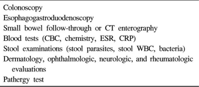
Table 4. Tests for the Diagnosis of Intestinal Behçet’s Disease
Colonoscopy
Esophagogastroduodenoscopy
Small bowel follow-through or CT enterography Blood tests (CBC, chemistry, ESR, CRP)
Stool examinations (stool parasites, stool WBC, bacteria) Dermatology, ophthalmologic, neurologic, and rheumatologic
evaluations Pathergy test
CT, computed tomography; CBC, complete blood cell; ESR, erythrocyte sedimentation rate; CRP, C-reactive protein; WBC, white blood cell.
(2) 질병 활성도
현재까지 베체트장염에서 확립된 질병의 활성도 측정 기준 은 없었으며 그동안 임상의사의 주관적인 경험으로 판단하거 나 크론병 질병활성도 지수 등이 사용되어 왔다. 따라서 대한 장연구학회에서 Disease Activity Index for Intestinal Behçet’s Disease를 만들기 위한 다기관 연구가 진행되었다. 그 결과 고유의 질병활성도 측정 기준을 만들었으며 이에 대한 임상 적용 가능성에 대한 검증이 필요한 상태이다.16 상기 항목에 대한 점수를 더하여 관해기(<20), 경도(20-44), 중등도(45-74), 중증(75 이상)으로 분류한다. 2. 역학(Epidemiology) 베체트병은 주로 지중해 및 중동으로부터 우리나라를 비 롯한 동아시아에 이르는 지역에서 호발되고 있으며 20-30대 의 젊은 환자들이 많다.17 소화기내과 의사가 관심을 갖는 베체트병의 위장관 침범 빈도는 연구자마다 결과가 다른데, 이는 베체트병의 어떤 진단 기준을 채택하였는지에 따라 모 집단의 범위가 달라질 수 있고, 장관에 병변이 확인된 경우 에 한해 진단을 내려야 하나 일부 연구는 위장관 증상만으 로 진단하는 오류를 범하고 있고, 위장관 증상이 있는 경우 에 한해 장검사를 시행할 경우 무증상 베체트장염 환자를 발견하지 못해 유병률이 낮게 평가될 수 있기 때문이다. 따 라서, 정확한 유병률을 구하기 위해서는 정확한 진단기준에 의해 확진된 모든 베체트병 환자들을 대상으로 장검사를 시 행하여야 할 것이다.2 베체트장염은 지역에 따라 큰 편차를 보이는데, 터키를 포함한 지중해 연안 국가들은 유병률이 가장 높은 지역이지만 중동이나 서양 환자들에서는 0-5%로 드물며, 주로 우리나라를 비롯한 동아시아에서는 10-30% 정 도로 비교적 흔하게 발생하는 것으로 알려져 있다.18-20 베체트장염은 베체트병 진단 후 평균 4-6년 내외에 많이 진단 되며, 진단 당시의 평균 연령은 36-38세로 알려져 있고, 남녀비는 전신 베체트병과는 달리 1.1-2.0:1로 남자에서 다소 높다.2,10,15,21,22 3. 진단 대장내시경 소견 및 조직 검사에서 베체트장염이 진단되 거나 의심되면 Table 4와 같은 검사를 진행한다. 식도, 위 침범 유무를 파악하기 위해 상부위장관 내시경을 시행할 수 있으며, 소장 침범 유무를 알기 위해 소장조영술이나 CT 소 장조영술을 시행한다. 대변 검사는 세균 및 기생충에 의한 감염성 장염과 감별 진단을 위해 시행한다. 전신 베체트병 침범 정도를 파악하기 위해 피부과, 안과, 신경과, 류마티스 내과의 협의 진료가 필요하다. 1) 임상양상 베체트장염의 증상으로서 가장 흔히 나타나는 것은 복통 이며 약 90%에서 발생한다고 알려져 있다.23,24 특히, 우하복 부의 통증 및 반발통이 나타날 수 있으며 심할 경우 종괴가 만져지기도 한다. 그 이외에 흔히 보이는 증상으로 혈변 및 흑색변, 설사, 누공, 그리고 전신 증상으로 발열 등이 있다. 그 이외에 베체트병의 전신 증상이 동반되었는지 확인하면 진단에 도움이 된다. 2) 검사실 소견 일반 혈액 검사에서는 백혈구 증가, ESR 또는 CRP의 상 승, 철분 결핍 빈혈, 혈소판 증가, 그리고 저알부민 혈증이
Cheon JH, et al. Diagnosis of Intestinal Behçet’s Disease 191 전형적인 베체트장염의 대장 내시경 소견은 주로 회맹 부에 국한된 소수의 큰 둥근 혹은 난원형의 깊은 궤양으 로 궤양의 가장자리는 융기되어 있고 주변 정상 점막과 의 경계는 명확하며 궤양의 바닥은 두터운 백태로 덮혀 있는 경우가 많다. 비전형적인 경우로 아프타 궤양, 지도 상 궤양, 다발 분절형 및 미만 궤양 등으로 나타날 수 있 다. 전형적인 베체트 장염의 조직학적인 특징은 혈관염과 그 주변으로의 림프구 침윤이나, 많은 경우에서 이러한 소견을 보이지 않으므로 임상 특성과 내시경 소견을 고 려하여 진단을 내려야 한다.
Table 5. Endoscpic Findings of Intestinal Behçet’s Disease
Typical findings
1. Single or a few large ulcer in ileo-cecal area 2. Round or oval shape deep ulceration 3. Discrete and elevated margin 4. Ulcer base covered with exudates Atypical findings
1. Aphthoid or geographic ulcers 2. Multi-segmental or diffuse distribution 동반될 수 있다. 그러나, 다른 염증성 장질환인 궤양성 대장
염이나 크론병에서 관찰되는 pANCA (perinuclear anti-neutrophilic cytoplasmic antibody) 혹은 ASCA (anti-saccharo-myces cerevisiae antibody)와 같은 혈청 지표의 의의는 베체 트장염의 경우 알려져 있지 않다.25-28 최근 베체트장염 환자 중 약 44% 정도에서 ASCA 양성이었으며 이들에서 음성인 환자들보다 누적 수술률이 높았고,29 항alpha-enolase 항체는 베체트병 환자 중 베체트장염이 동반되어 있을 경우 약 65%에서 양성을 보여 이런 혈청 검사의 베체트장염 진단 유용성에 대해 추후 귀추가 주목된다.30 3) 내시경검사 소견 전형적인 베체트장염의 대장 내시경 소견은 주로 회맹부 에 존재하는 소수의 큰 둥근 혹은 난원형의 깊은 궤양으로 궤양의 가장자리는 융기되어 있고 주변 정상 점막과의 경계 는 명확하며 궤양의 바닥은 두터운 백태로 덮혀 있는 경우 가 많다(Table 5). 그 외에도 비전형적인 경우로 아프타 궤 양, 지도상 궤양, 다발 분절형 및 미만 궤양 등으로 나타날 수 있다.13,31,32 한 연구 결과 전형적인 내시경 소견, 즉 궤양 의 모양과 분포, 개수를 종합할 때 크론병과의 감별이 대부 분에서 가능하였다.32 전형적인 베체트장염 소견이 있을 때 전신 베체트병 진단 기준의 만족 여부에 따른 임상 양상이 나 내시경 소견의 차이가 없으며,13 궤양의 모양이 예후에 관련되어 있다는 연구 결과가 있다.31 이는 베체트장염 진단 에 전신 증상 유무가 처음부터 필수 조건이 아닐 가능성을 제시한다. 또한 처음 베체트장염에 전형적인 궤양이 있을 때 전신 증상이 없어도 추적 관찰 중 상당수의 환자들에서 전신 증상이 장 침범 이후에 나타나므로 처음부터 전신 증 상을 만족시켜야 베체트장염을 진단하게 되면 많은 수의 환 자들에서 진단이 늦어지게 된다.11,15 4) 영상의학검사 소견 베체트장염의 소장조영술 소견은 주로 회장 말단 부위에 단독 혹은 다발성의 뚜렷한 궤양이 관찰되며 점막 주름 비 대, 형태의 변형(contour deformity), 장관의 협착 소견이 관찰 된다. 대장조영술에서는 심부 관통 궤양이나 다발 궤양, 염 증 종괴, 누공 및 가성 용종 등이 관찰될 수 있다.33-35 5) 조직검사 소견 조직학적으로 작은 혹은 중간크기 혈관의 염증(vasculitis involving small and medium sized vessels)이 관찰되며 혈관 주변으로 림프구의 침윤(lymphocyte infiltration in the peri-vascular space) 소견을 보인다. 또한 육아종(granuloma) 형성 은 드문 것으로 알려져 있다. 그러나, 이런 전형적인 소견을 보이는 경우는 50% 이하이며 생검 조직에서 비특이적인 염 증 소견만을 보이는 경우가 많기 때문에 전형적인 조직검사 소견을 보이지 않더라도 임상 특성을 고려하여 판단하여야 한다.36-38 6) 감별진단 베체트장염과 감별을 요하는 질환으로 아래와 같은 장질 환이 있다. (1) 크론병 (2) 대장 결핵 (3) 약인 대장염 (4) CMV 대장염 (5) 아메바 장염 및 살모넬라 장염 (6) 대장암 이 중 특히 크론병과의 감별이 중요한데 크론병이 분절 혹은 미만성의 분포를 보이는 경우가 많으나 베체트장염은 대부분 회맹 부위에 국소 분포하며, 궤양의 모양이 베체트 장염의 경우 둥근 난원형이 많으나 크론병의 경우는 부정형 및 종주형이 많다. 또한, 궤양의 깊이에 있어서도 베체트장 염에서 크론병에 비하여 깊고 경계 부위가 뚜렷하다.32,39
결 론
베체트장염 진단에서 가장 중요한 것은 전신 베체트병 증192 대한소화기학회지: 제53권 제3호, 2009 상과 전형적인 내시경 소견이다. 이를 토대로 대한장연구학 회에서는 베체트장염을 진단하는 가이드라인을 제시하였 다. 앞으로 이 가이드라인에 대한 검증 작업뿐만 아니라 베 체트장염에 대한 병태생리, 진단 방법, 질병 특이 혈청 지 표, 질병활성도 측정, 특이 치료제 및 치료 반응률, 예후 등 에 관한 추가 연구가 필요하리라 생각한다.
참고문헌
1. O'Duffy JD. Behçet's disease. Curr Opin Rheumatol 1994; 6:39-43.
2. Yang SK. Intestinal Behçet’s disease. Intest Res 2005;3:1-10. 3. Behçet H. Uber rezidivierende apthuse durch ein virus ver-usachte Geswure am Mund, am Auge und an den Genitalien. Dermatol Wochenschr 1937;105:1152-1157.
4. Sakane T, Takeno M, Suzuki N, Inaba G. Behçet's disease. N Engl J Med 1999;341:1284-1291.
5. International Study Group for Behçet's disease. Criteria for diagnosis of Behçet's disease. Lancet 1990;335:1078-1080. 6. Mizushima Y, Inaba G, Mimura Y, Ono S. Diagnostic criteria
for Behçet's disease in 1987, and guidelines for treating Behçet's disease. Saishin-Igaku 1988;43:382-391.
7. Friedman-Birnbaum R, Bergman R, Aizen E. Sensitivity and specificity of pathergy test results in Israeli patients with Behçet's disease. Cutis 1990;45:261-264.
8. Yoon MS, Lee SH, Bang D, Lee S. Cutaneous manifestations of Behçet's syndrome. Yonsei Med 1987;28:291-296. 9. Chang HK, Cheon KS. The clinical significance of a pathergy
reaction in patients with Behçet's disease. J Korean Med Sci 2002;17:371-374.
10. Bang D, Lee JH, Lee ES, et al. Epidemiologic and clinical survey of Behçet's disease in Korea: the first multicenter study. J Korean Med Sci 2001;16:615-618.
11. Kobayashi K, Ueno F, Bito S, et al. Development of con-sensus statements for the diagnosis and management of in-testinal Behçet's disease using a modified Delphi approach. J Gastroenterol 2007;42:737-745.
12. Muto T. Historical review of so called “simple ulcer” of the intestine. Stomach Intestine 1979;14:739-748.
13. Jung HC, Rhee PL, Song IS, et al. Temporal changes in the clinical type or diagnosis of Behçet's colitis in patients with aphthoid or punched-out colonic ulcerations. J Korean Med Sci 1991;6:313-318.
14. Kim JE, Han DS, Cho HS, et al. Clinical features of in-testinal Behçet's disease according to disease subtypes. Intest Res 2007;5:26-32.
15. Lee CR, Kim WH, Cho YS, et al. Colonoscopic findings in intestinal Behçet's disease. Inflamm Bowel Dis 2001;7:243- 249.
16. Cheon JH. Development of a novel disease activity index for intestinal Behçet’s disease. The 2nd Korea-Japan IBD Sym-posium.
17. Suzuki Kurokawa M, Suzuki N. Behçet's disease. Clin Exp Med 2004;4:10-20.
18. Bayraktar Y, Ozaslan E, Van Thiel DH. Gastrointestinal man-ifestations of Behçet's disease. J Clin Gastroenterol 2000;30: 144-154.
19. Lakhanpal S, Tani K, Lie JT, Katoh K, Ishigatsubo Y, Ohokubo T. Pathologic features of Behçet's syndrome: a re-view of Japanese autopsy registry data. Hum Pathol 1985;16: 790-795.
20. Kim HJ, Bang D, Lee SH, et al. Behçet's syndrome in Korea: a look at the clinical picture. Yonsei Med J 1988;29:72-78. 21. Bang DS, Oh SH, Lee KH, Lee ES, Lee SN. Influence of
sex on patients with Behçet's disease in Korea. J Korean Med Sci 2003;18:231-235.
22. Kim DK, Yang SK, Byeon JS, et al. Clinical manifestations and course of intestinal Behçet’s disease: an analysis in rela-tion to disease subtype. Intes Res 2005;3:48-54.
23. Shin SJ, Kim BC, Park SY, Kim TI, Kim WH. Systemic manifestations of Behçet's disease in diagnosis of intestinal Behçet's disease. Gut 2006;55(suppl):A120.
24. Choi IJ, Kim JS, Cha SD, et al. Long-term clinical course and prognostic factors in intestinal Behçet's disease. Dis Colon Rectum 2000;43:692-700.
25. Cambridge G, Rampton DS, Stevens TR, McCarthy DA, Kamm M, Leaker B. Anti-neutrophil antibodies in inflam-matory bowel disease: prevalence and diagnostic role. Gut 1992;33:668-674.
26. Quinton JF, Sendid B, Reumaux D, et al. Anti-Saccharomyces cerevisiae mannan antibodies combined with antineutrophil cytoplasmic autoantibodies in inflammatory bowel disease: prevalence and diagnostic role. Gut 1998;42:788-791. 27. Duerr RH, Targan SR, Landers CJ, et al. Anti-neutrophil
cy-toplasmic antibodies in ulcerative colitis: comparison with other colitides/diarrhoeal illnesses. Gastroenterology 1992;100: 1590-1596.
28. Peeters M, Joossens S, Vermeire S, Vlietinck R, Bossuyt X, Rutgeerts P. Diagnostic value of anti-Saccharomyces cer-evisiae and antineutrophil cytoplasmic autoantibodies in in-flammatory bowel disease. Am J Gastroenterol 2001;96:730- 734.
cer-천재희 외 5인. 베체트장염 진단기준 193
evisiae antibody in intestinal Behçet’s disease patients: rela-tion to clinical course. Dis Colon Rectum 2006;49:1849-1859. 30. Shin SJ, Kim BC, Park S, et al. Anti α-enolase antibody in
intestinal Behçet’s disease patients: useful serologic marker and relation to clinical course. J Gastroenterol Hepatol 2005; 20(S):143A.
31. Kim JS, Lim SH, Choi IJ, et al. Prediction of the clinical course of Behçet's colitis according to macroscopic classi-fication by colonoscopy. Endoscopy 2000;32:635-640. 32. Kim TI, Kim WH, Lee JH, et al. Differential diagnosis of
in-testinal Behçet’s disease and Crohn’s disease by colonoscopic findings. J Gastroenterol Hepatol 2002;16(S):144A.
33. Chung SY, Ha HK, Kim JH, et al. Radiologic findings of Behçet's syndrome involving the gastrointestinal tract. Radio-graphics 2001;21:911-926.
34. Kim JH, Choi BI, Han JK, Choo SW, Han MC. Colitis in Behçet’s disease: characteristics on double-contrast barium
en-ema examination in 20 patients. Abdom Imaging 1994;19: 132-136.
35. You JK, Kim MJ, Park S, Chung JJ, Kim WH. Intestinal Behçet's disease: breath-hold MR imaging. Abdom Imaging 2001;26:309-314.
36. Lee RG. The colitis of Behçet's syndrome. Am J Surg Pathol 1986;10:888-893.
37. Bradbury AW, Milne AA, Murie JA. Surgical aspects of Behçet's disease. Br J Surg 1994;81:1712-1721.
38. Kasahara Y, Tanaka S, Nihino M, Umemura H, Shiraha S, Kuyama T. Intestinal involvement in Behçet’s disease: review of 136 surgical cases in the Japanese literature. Dis Colon Rectum 1981;24:103-106.
39. Kim KJ, Yang SK, Myung SJ, et al. Diagnostic value of co-lonoscopic findings in the differential diagnosis between Crohn's disease and intestinal Behçet's disease. Gastrointest Endosc 2002;55:AB263.