246 Copyright © 2018 Korean Neurological Association
JCN
Open AccessParaneoplastic Neuromyelitis Optica Associated
with Lung Adenocarcinoma in a Young Woman
Dear Editor,
Neuromyelitis optica spectrum disorder (NMOSD) is an autoimmune inflammatory disor-der of the central nervous system that preferentially affects the optic nerve, spinal cord, and area postrema.1 The serum anti-aquaporin-4 antibody (anti-AQP-4 antibody) is specific for NMOSD. There are only a few reports of NMOSD presenting in the context of paraneoplastic neurological syndromes. Here we report a young woman who developed recurrent NMOSD in the setting of a lung adenocarcinoma.
A previously healthy 37-year-old woman presented with a palpable mass on the right side of the neck. The patient was diagnosed with metastatic adenocarcinoma after a biopsy of the mass, which was in a neck lymph node. Chest computed tomography and whole-body positron-emission tomography/computed tomography (PET-CT) revealed that the main mass was in her lung and that multiple metastases were present, classifying it as stage 4 (Fig. 1A). An EGFR mutation (deletion of exon 19) was also detected in the biopsied tissue.
During the diagnostic workup she developed progressive weakness of all extremities. When she visited our clinic, a neurological examination revealed weakness of Medical Re-search Council grade 3 to 4 in the upper extremities and grade 1 in the lower extremities, with hypoesthesia below the neck. She did not have an oral or genital ulcer, sicca syndrome, abnormal rash, or other manifestations implicating autoimmune disorders. Spine magnetic resonance imaging (MRI) showed a T2-weighted hyperintense lesion from level C2 to level T7 with gadolinium enhancement (Fig. 1B and C). Brain MRI revealed no abnormality. Ce-rebrospinal fluid analysis showed pleocytosis (60 cells/µL, 98% mononuclear cells) and an elevated protein level (138.2 mg/dL) without malignant cells or an oligoclonal band. Anti-AQP-4 antibody (indirect immunofluorescence test; Euroimmun, Lübeck, Germany) was de-tected in the serum. She was negative for antinuclear antibody. Based on these results she was diagnosed with NMOSD and was treated with a daily intravenous infusion of 1 g of methyl-prednisolone for 5 days, followed by a tapering course of oral methyl-prednisolone from an initial 45 mg daily [1 mg/kg body weight (47 kg)] to 10 mg daily at 3 months after the onset of myelitis. The lung adenocarcinoma was treated concurrently with gefitinib.
Three months after presenting with myelitis she experienced a painful visual disturbance in the left eye. An examination revealed bilateral papilledema and a visual field defect of the lower quadrant in the left eye. The visual acuity of her left eye was finger counting at 50 cm. Orbit MRI revealed an enlarged bilateral optic nerve with segmental enhancement of the left optic nerve (Fig. 1D and E). Optic neuritis was diagnosed. At this time her cancer was stable with gefitinib treatment. She was treated with another steroid pulse therapy (daily intrave-nous infusion of 1 g of methylprednisolone for 5 days), followed by a tapering course of oral prednisolone as for the previous myelitis. In addition, azathioprine was added at 150 mg daily [2.5 mg/kg body weight (60 kg)]. At 8 months after presenting with optic neuritis she was tak-ing 5 mg of prednisolone and 150 mg of azathioprine daily. At 1 month after presenttak-ing with Kyoung Won Baika
Se Hoon Kimb
Ha Young Shina a Departments of Neurology and b Pathology, Yonsei University
College of Medicine, Seoul, Korea
pISSN 1738-6586 / eISSN 2005-5013 / J Clin Neurol 2018;14(2):246-247 / https://doi.org/10.3988/jcn.2018.14.2.246
Received August 30, 2017 Revised December 22, 2017 Accepted December 22, 2017 Correspondence Ha Young Shin, MD Department of Neurology,
Yonsei University College of Medicine, 50-1 Yonsei-ro, Seodaemun-gu, Seoul 03722, Korea
Tel +82-2-2228-1600 Fax +82-2-393-0705 E-mail hayshin@yuhs.ac
cc This is an Open Access article distributed under the terms of the Creative Commons Attribution
Non-Com-mercial License (http://creativecommons.org/licenses/by-nc/4.0) which permits unrestricted non-comNon-Com-mercial use, distribution, and reproduction in any medium, provided the original work is properly cited.
www.thejcn.com 247
Baik KW et al.
JCN
optic neuritis her visual acuity recovered to 0.7. At the latest fol-low up, which was performed 8 months after presenting with optic neuritis, she remained relapse-free with an EDSS score of 3.5.
Immunohistochemistry of formalin-fixed paraffin-embed-ded slides of the patient’s neck lymph nodes showed positivity for membranous AQP-4 immunoreactivity in the cluster of the metastatic carcinoma [anti-AQP4 antibody (A5971, Sig-ma-Aldrich, St. Louis, MO, USA)] (Fig. 1F).
Paraneoplastic NMOSD has been reported recently,2 with some studies showing AQP-4 expression in the actual tumor cells.3,4 AQP-4 may have role in tumor progression, invasion, and metastasis.5 However, the pathophysiology underlying paraneoplastic NMOSD remains to be elucidated.
Combined treatment of anticancer therapy and an immu-nosuppressive agent usually results in paraneoplastic NMOSD that is episodic.3,6 One recurrent case occurred under the cir-cumstance of cancer relapse.7 However, in our patient the NMOSD recurred in spite of combined treatment and rela-tively stable cancer. Therefore, the clinician should keep the possibility of recurrence in mind, and might consider additive immunosuppressive treatment upon the recurrence of para-neoplastic NMOSD.
Conflicts of Interest
The authors have no financial conflicts of interest.
Acknowledgements
This study was supported by a faculty research grant of Yonsei University College of Medicine (6-2016-0110).
REFERENCES
1. Wingerchuk DM, Banwell B, Bennett JL, Cabre P, Carroll W, Chitnis T, et al. International consensus diagnostic criteria for neuromyelitis optica spectrum disorders. Neurology 2015;85:177-189.
2. De Santis G, Caniatti L, De Vito A, De Gennaro R, Granieri E, Tola MR. A possible paraneoplastic neuromyelitis optica associated with lung cancer. Neurol Sci 2009;30:397-400.
3. Figueroa M, Guo Y, Tselis A, Pittock SJ, Lennon VA, Lucchinetti CF, et al. Paraneoplastic neuromyelitis optica spectrum disorder associat-ed with metastatic carcinoid expressing aquaporin-4. JAMA Neurol 2014;71:495-498.
4. Iorio R, Rindi G, Erra C, Damato V, Ferilli M, Sabatelli M. Neuromye-litis optica spectrum disorder as a paraneoplastic manifestation of lung adenocarcinoma expressing aquaporin-4. Mult Scler 2015;21:791-794. 5. Warth A, Muley T, Meister M, Herpel E, Pathil A, Hoffmann H, et al.
Loss of aquaporin-4 expression and putative function in non-small cell lung cancer. BMC Cancer 2011;11:161.
6. Frasquet M, Bataller L, Torres-Vega E, Durán-Moreno M, García-Ver-dugo JM, Sevilla T, et al. Longitudinally extensive transverse myelitis with AQP4 antibodies revealing ovarian teratoma. J Neuroimmunol 2013;263:145-147.
7. Mueller S, Dubal DB, Josephson SA. A case of paraneoplastic my-elopathy associated with the neuromyelitis optica antibody. Nat Clin Pract Neurol 2008;4:284-288.
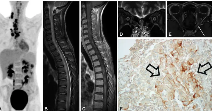
Fig. 1. Findings of imaging and immunohistochemistry studies. A: Positron-emission tomography/computed tomography showed increased FDG up-take in the lung and multiple lymph-node metastases. B: Sagittal T2-weighted image of the spine revealing a hyperintense lesion from level C2 to level T7. C: Sagittal T1-weighted image of the spine after infusing gadolinium showing multifocal enhancement. D: Coronal T2-weighted image of the orbit revealing bilateral optic nerve enlargement. E: Sagittal T1-weighted image of the spine after infusing gadolinium showing segmental enhancement of the left optic nerve (arrow). F: Immunohistochemistry of the patient’s neck lymph nodes showed positive membranous anti-aquaporin-4 immunore-activity (open arrows) (×400).
A B C
D E