2004;10:8105-8113.
Clin Cancer Res
Ki Taek Nam, Ki-Baik Hahm, Sang-Yeon Oh, et al.
Development in a Mouse Model
-Associated Gastric Cancer
Helicobacter pylori
Prevents
The Selective Cyclooxygenase-2 Inhibitor Nimesulide
Updated version
http://clincancerres.aacrjournals.org/content/10/23/8105
Access the most recent version of this article at:
Cited Articles
http://clincancerres.aacrjournals.org/content/10/23/8105.full.html#ref-list-1
This article cites by 40 articles, 17 of which you can access for free at:
Citing articles
http://clincancerres.aacrjournals.org/content/10/23/8105.full.html#related-urls
This article has been cited by 8 HighWire-hosted articles. Access the articles at:
E-mail alerts
Sign up to receive free email-alerts
related to this article or journal.
Subscriptions
Reprints and
.
[email protected]
Department at
To order reprints of this article or to subscribe to the journal, contact the AACR Publications
Permissions
.
[email protected]
Department at
The Selective Cyclooxygenase-2 Inhibitor Nimesulide Prevents
Helicobacter pylori-Associated Gastric Cancer Development
in a Mouse Model
Ki Taek Nam,
1Ki-Baik Hahm,
2Sang-Yeon Oh,
1Marie Yeo,
2Sang-Uk Han,
2Byeongwoo Ahn,
3Young-Bae Kim,
2Jin Seok Kang,
1Dong Deuk Jang,
1Ki-Hwa Yang,
1and
Dae-Yong Kim
41Department of General Toxicology, National Institute of
Toxicological Research, Korea Food and Drug Administration, Seoul; 2Genome Center for Gastroenterology, Ajou University School of Medicine, Suwon;3Department of Veterinary Pathology, College of Veterinary Medicine, Chungbuk National University, Cheongju; and 4Department of Veterinary Pathology, College of Veterinary Medicine and School of Agricultural Biotechnology, Seoul National University, Seoul, Korea
ABSTRACT
Purpose: Helicobacter pylori infection can lead to gastric
cancer, and cyclooxygenase-2 (COX-2) is overexpressed in the stomach during H. pylori infection. Therefore, we inves-tigated whether nonsteroidal anti-inflammatory drugs might protect against this form of cancer. Specifically, we examined the chemopreventive effect of the COX-2 inhibitor nimesulide on H. pylori-associated gastric carcinogenesis in mice.
Experimental Design: C57BL/6 mice were treated with
the carcinogen N-methyl-N-nitrosourea (MNU) and/or H.
pylori. To determine the effect of COX-2 inhibition,
nime-sulide was mixed with feed pellets and administered for the duration of the experiment. All of the mice were sacrificed 50 weeks after the start of the experiment. Histopathology, immunohistochemistry, and Western blotting for COX-2, Bax and Bcl-2 were performed in stomach tissues. In vitro experiments with the human gastric cancer cell line AGS were also performed to identify mechanisms underlying can-cer chemoprevention by nimesulide.
Results: Gastric tumors developed in 68.8% of mice
that were given both MNU and H. pylori, whereas less than 10% developed gastric tumors when given either MNU or H.
pylori alone. These findings indicate that H. pylori promotes
carcinogen-induced gastric tumorigenesis. In mice treated with both MNU and H. pylori, nimesulide administration substantially reduced H. pylori-associated gastric tumori-genesis, whereas substantial inductions of apoptosis were observed. In vitro studies demonstrated that nimesulide and
H. pylori when combined acted synergistically to induce
more apoptosis than either alone.
Conclusions: Our data show that nimesulide prevents H. pylori-associated gastric carcinogenesis, and suggest that
COX-2 may be a target for chemoprevention of gastric cancer.
INTRODUCTION
Helicobacter pylori causes chronic active gastritis and
pep-tic ulcer disease, and is linked with gastric adenocarcinomas, including gastric mucosa-associated lymphoid tissue lymphoma (1). On the basis of epidemiologic data, WHO/IARC classified
H. pylori as a group 1 carcinogen (2). Generally, gastric
ade-nocarcinoma develops through a multistep process from normal gastric mucosa to chronic active gastritis, to gastric atrophy and intestinal metaplasia, and finally to dysplasia and neoplasia (3), and it has been postulated that H. pylori plays a causative role in the early phases of this malignant progression (4, 5). How-ever, debate still exists as to whether H. pylori is really a carcinogen or a cancer promoter, and whether eradication of
H. pylori is beneficial to people free of gastric tumors (6 –9).
A possible explanation for the link between H. pylori infection and gastric carcinogenesis is that H. pylori infection raised cyclooxygenase-2 (COX-2) mRNA/protein levels, and stimulated release of prostaglandin E2 in H. pylori-associated premalignant and malignant gastric lesions (10 –16). There is strong evidence that COX-2 is causally involved in gastrointes-tinal cancer (17–22). In addition, growth of colon polyps was retarded or blocked by either administration of nonsteroidal anti-inflammatory drugs (NSAIDs; refs. 23–25) or targeted de-letion of the COX-2 gene (26). Because induction of COX-2 expression has been shown to play an important role in neo-plastic transformation in the large intestine, it is our hypothesis that COX-2 is involved in H. pylori-associated gastric cancer development, and that NSAID administration can prevent or retard this process.
It has been shown that H. pylori increased release of prostaglandin E2and COX-2, which were overexpressed in most
metaplastic and adenomatous tissues, as well as in gastric ade-nocarcinoma (27). Despite these observations, it remains un-known as to whether COX-2 may be a target for chemopreven-tion of H. pylori-associated gastric carcinogenesis. In the
Received 5/7/04; revised 8/30/04; accepted 9/9/04.
Grant support: Supported by a grant of the Korean Health 21 R&D
Project, Ministry of Health & Welfare, Republic of Korea (02-PJ1-PG3-20802-0014, 01-PJ10-PG6-01GN14-0007) and by Brain Korea 21 Pro-ject; and supported in part by Korea Food and Drug Administration. The costs of publication of this article were defrayed in part by the payment of page charges. This article must therefore be hereby marked
advertisement in accordance with 18 U.S.C. Section 1734 solely to
indicate this fact.
Requests for reprints: Dae-Yong Kim, Department of Veterinary
Pa-thology, College of Veterinary Medicine and School of Agricultural Biotechnology, Seoul National University, San 56-1, Shillim-dong, Kwanak-gu, Seoul 151-742, Korea. Phone: 880-1249; Fax: 82-2-388-6451; E-mail: [email protected].
present study, we used an H. pylori-associated gastric cancer mouse model (28, 29) to investigate the preventive effects of the selective COX-2 inhibitor nimesulide, and we studied the mech-anisms underlying nimesulide-induced chemoprevention with AGS human gastric cancer cell line.
MATERIALS AND METHODS
Mice. Male C57BL/6 mice were obtained from the Jack-son Laboratory (Bar Harbor, ME) at 5 weeks of age. The mice were maintained in an accredited Korea FDA animal facility in accordance with the AAALAC International Animal Care pol-icies (Accredited Unit-Korea Food and Drug Administration: Unit Number-000996). All of the mice were given a standard pellet chow diet (CRF-1, Oriental Yeast Co. Ltd., Tokyo, Japan)
ad libitum and were maintained in specific pathogen-free
con-ditions.
Chemicals and Bacteria. N-methyl-N-nitrosourea
(MNU; Sigma Chemical Co., St. Louis, MO) solutions were freshly prepared twice a week by dissolving 200 ppm MNU in distilled water. When indicated, mice were given the 200-ppm MNU solution ad libitum in light-shielded bottles in place of drinking water. Mouse-adapted H. pylori (SS1) were inoculated on Brucella agar plates (Becton Dickinson, Cockeysville, MD) containing 10% heat-inactivated fetal bovine serum and Skirrow medium (Difco, Detroit, MI). They were kept at 37°C under microaerobic conditions with GasPak jars (Difco) and Campy-Paks (Becton Dickinson). After 24 hours of fasting, a 0.1-mL suspension of H. pylori containing 1 ⫻ 109 colony-forming
units (CFU)/mL was administered by intragastric intubation.
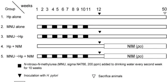
Study Design. The experimental design is illustrated in Fig. 1. Mice were randomized into five groups. Animals of groups 2, 3, and 5 were given MNU. One week after completion of MNU administration, mice in groups 3 and 5 were inoculated with H. pylori three times every other day. Mice in groups 1 and 4 were inoculated with H. pylori three times every other day (no MNU treatment). Animals of groups 4 and 5 were then given a CRF-1 diet (Oriental Yeast Co. Ltd.) containing 200 ppm nime-sulide (Choongwae Pharmaceutical Research Institute, Suwon, Korea). Animals of groups 1, 2, and 3 were maintained on basal
CRF-1 diets throughout the experiment. Mice were sacrificed 38 weeks after infection, making a total of 50 weeks of treatment.
Histopathologic Examination. Immediately after sacri-fice, mouse stomachs were opened along the greater curvature. The number, as well as the long diameter, of tumors in the stomach was measured. A record was kept of the size and number of tumors counted, with a diagnosis made after the final histopathologic examination. One half of the excised stomachs, including neoplastic nodules, were fixed in neutral-buffered 10% formalin and were cut into approximately six strips, which were processed by standard methods, embedded in paraffin, sectioned at 4 m, and stained with hematoxylin and eosin (H&E). The remaining portions were quickly frozen in liquid nitrogen and stored at⫺70°C until analysis. Histologic classi-fication was based on histopathologic and cytologic criteria proposed by Leininger and Jokinen (30). After histopathologic classification was done, tumor incidence and multiplicity was calculated.
Identification of H. pylori in Gastric Mucosa. To con-firm H. pylori infection, we transferred samples (⬃3-mm2) of
stomach mucosa from the greater curvature containing both fundic and pyloric glands to 1.0 mL of sterile 0.1 mol/L PBS; these were homogenized and plated on selective trypticase soy agar/5% sheep blood plates containing vancomycin (20 mg/ mL), nalidixic acid (10 mg/mL), bacitracin (30 mg/mL) and amphotericin B (2 mg/mL) from Sigma Chemical Co., and grown for 3 to 5 days. Colonies were identified by characteristic Gram’s stain morphology, and by urease, catalase, and oxidase activity. Another 3-mm2 sample from the antrum was placed
into the gel of a rapid urease test kit (CLO test, Ballard Medical Products, Draper, VT) and was left for 6 hours at room temper-ature to test for urease activity. The presence of H. pylori in the gastric pit was further confirmed by Warthin–Starry staining.
Immunohistochemistry for COX-2.
Immunohisto-chemical identification of COX-2 expression was performed on replicate sections of stomach tissues. The sections were mounted on silanized slides (Dako, Glostrup, Denmark) and were dewaxed and rehydrated; endogenous peroxidase activity was quenched with hydrogen peroxide. After washing in
dou-Fig. 1 The study design.
Ani-mals were randomized into five groups according to treatment: group 1, H. pylori alone (Hp
alone); group 2, MNU alone;
group 3, five weeks’ MNU ad-ministration followed by H.
lori infection; group 4, H. py-lori infection followed by continuous nimesulide via diet [NIM (po)]; and group 5, five weeks’ MNU administration followed by H. pylori infection and nimesulide treatment [NIM
(po)]. The mice were sacrificed
38 weeks after H. pylori in-fection.
ble-distilled water, sections were subjected to microwave anti-gen retrieval in 0.01 mol/L citric acid. The slides were incubated at 4°C overnight with rabbit anti-COX-2 polyclonal antibody (1:2,000; Cayman Chemical, Ann Arbor, MI). Immunoreaction complexes were detected with the avidin-biotin affinity system (Santa Cruz Biotechnology, Santa Cruz, CA) and were visual-ized with 3,3⬘-diaminobenzidine tetrahydrochloride (Zymed Laboratories Inc., San Francisco, CA) as the chromogen. The sections were counterstained with Mayer’s hematoxylin and examined under a light microscope.
Western Blotting. The frozen stomach tissues were ho-mogenized in radioimmunoprecipitation assay (RIPA) buffer [10 mmol/L Tris (pH 7.6), 1 mmol/L EDTA (pH 8.0), 100 mmol/L NaCl, 1g/mL aprotinin, and 100 g/mL phenylmeth-ylsulfonyl fluoride (all from Sigma Chemical Co.)]. Protein concentration was measured with the Bio-Rad Protein Assay kit (Bio-Rad Laboratories, Hercules, CA). Extracted proteins (40 g/lane) were resolved by SDS-PAGE and were transferred to polyvinylidene difluoride membranes. Membranes were incu-bated overnight at 4°C with rabbit COX-2 polyclonal anti-body (Cayman Chemical), rabbit anti-Bax polyclonal antianti-body (Santa Cruz Biotechnology), mouse anti-Bcl-2 monoclonal an-tibody (Santa Cruz Biotechnology). The membranes were then incubated for 45 minutes with secondary antibody (Santa Cruz Biotechnology). After incubation with the secondary antibody, blots were washed three times with PBS/0.1% Tween 20 and developed with a commercial chemiluminescence detection kit (Amersham Biosciences, Buckinghamshire, United Kingdom). Expression levels of protein were quantified with a Bio-Rad Imaging Densitometer system Model GS690 (Bio-Rad Labora-tories) and the ratio of Bax to Bcl-2 was calculated.
The general procedure for Western immunoblot analyses of cultured AGS cells with antibodies against caspase-3 (Santa Cruz Biotechnology), (poly)ADP-ribose polymerase (PARP; Zymed Laboratories Inc.) were similar to the procedures de-scribed above. Cultured cells were washed twice with cold PBS on ice and harvested by scraping with a rubber scraper. Cells were sedimented by centrifugation at 4°C and resuspended in cell extraction buffer (50 mM/L PIPES/NaOH, 2 mM/L EDTA, 0.1% CHAPS, 5 mM/L DTT, 20g/mL leupeptin, 10 g/mL pepstatin, 10g/mL aprotinin, 1 M/L phenylmethylsulfo-nyl fluoride).
Terminal Deoxynucleotidyl Transferase-Mediated Nick End Labeling Assay. Apoptosis was visualized with terminal deoxynucleotidyl transferase (TdT) FragEL DNA fragmentation detection kit (Oncogene Research Products, Cambridge, MA). The staining procedures were modified based on the manufac-turer’s recommendations. Briefly, after routine deparaffiniza-tion, rehydradeparaffiniza-tion, and washing in 1⫻ PBS (pH 7.4), tissues were digested with proteinase K (20 mg/mL in 1⫻ PBS) for 20 minutes at room temperature and were washed. After incubation in equilibration buffer for 10 minutes, sections were treated with terminal deoxynucleotidyltransferase (TdT) enzyme at 37°C for 1 hour.
Determination of Apoptotic Index. All of the slides
were scored blindly three times without knowledge of the his-tologic findings. To determine the apoptotic index (AI) in each group, we first scanned Tdt-nick end labeling (TUNEL)-immu-nostained sections under low power magnification (⫻100) to
locate the apoptotic hot spots (areas with maximal TUNEL-positive cells). The AI at⫻400 field was then scored by count-ing the number of TUNEL-positive cells. At least five hot spots in a section were selected to determine the average count. Data were expressed as a mean percentage of total cell numbers.
Cell Culture and Cell Viability Assay. The human gas-tric cancer cell line AGS was purchased from American Type Culture Collection (ATCC strain, Manassas, VA) and was main-tained in RPMI 1640 (Life Technologies, Inc., Gaithersburg, MD) supplemented with 10% fetal calf serum (HyClone, Logan, UT) in humidified environment at 37°C in 5% CO2. To
deter-mine the cell growth rate, we seeded AGS cells into 24-well plate at 2⫻ 105cells/mL in triplicate, and we pretreated them
with 5 mol/L nimesulide or celecoxib (Pfizer Pharma, New York, NY) in 0.1% DMSO (Fisher Scientific, Pittsburgh, PA) for 24 hours; we then used H. pylori inoculation (2 ⫻ 106
colony-forming units (CFU)/mL) for 24 hours. H. pylori filtrate was prepared by homogenizing the bacteria in distilled water, pelleting the bacteria by centrifugation, and then filtering through a 0.2m pore size filter (Gelman Sciences, Ann Arbor, MI). Nimesulide and celecoxib were added when the cells had 70 to 80% confluency. Cell numbers and their viability were determined by trypan blue exclusion assay.
Apoptotic Quantitation by Flow Cytometric Analysis.
Apoptotic cells were quantified by staining with FITC-conju-gated Annexin V (Clontech, Palo Alto, CA). Cells (1 ⫻ 106)
were collected at 72 hours for flow cytometric measurement and were stained with FITC-conjugated annexin V and propidium iodide as recommended by the manufacturer, and were then analyzed by flow cytometry with a FACScan (Becton Dickinson Facsort flow cytometer) with an argon laser set to excite at 488 nm. Propidium iodide (40g/100 L PBS) was added to 1 ⫻ 106cells suspended in 800L of PBS, together with 100 L of
RNase A (1g/mL), and was incubated at 37°C for 30 minutes before flow cytometry analysis of 2⫻ 104cells. Red
fluores-cence of propidium-bound DNA was measured with a 630 nm-long bandpass filter.
Statistical Analyses. The data were analyzed with the JMP software package (version 4.0; SAS Institute, Cary, NC) on an IBM computer. Stomach tumor incidence data were analyzed with a2test. Other data were compared with the Dunnett t test
after ANOVA analysis. For all comparisons, P values less than 5% (P⬍ 0.05) were considered to be statistically significant.
RESULTS
Bacterial Colonization. At week 50, stomachs were re-moved, and all of the mice except those in group 2 (MNU alone) showed positive H. pylori colonization as determined by direct bacterial culture and rapid urease tests. The mean (SEM) number of CFU recovered from mice inoculated orally with H. pylori SS1 was 1.41⫾ (0.35) ⫻ 105/mg gastric tissue. Warthin–Starry
staining showed numerous spiral bacteria in mucosal epithelium along the length of the gastric pits in both the antrum and the body of all of the animals inoculated with H. pylori.
H. Pylori Infection Promoted Carcinogen-Induced
Gas-tric Tumorigenesis. Mice were randomized into five groups according to treatment: group 1, H. pylori alone; group 2, MNU alone; group 3, five weeks’ MNU administration followed by H.
pylori infection; group 4, H. pylori infection followed by
con-tinuous nimesulide via diet; and group 5, five weeks’ MNU administration followed by H. pylori infection and nimesulide treatment (see Fig. 1). The mice were sacrificed 38 weeks after
H. pylori infection. The mean body weight of each group was
similar throughout all of the experiments. The incidence of tumors at sacrifice was 0% (0/13) in group 1, 10% (1/10) in group 2, and 68.8% (11/16) in group 3. Group 3 showed a significantly higher incidence of gastric tumor compared with groups 1 and 2 (P⬍ 0.0001 for both comparisons), signifying that the mice were somewhat resistant to carcinogen-stimulated gastric tumorigenesis, and that H. pylori infection promoted, rather than initiated, gastric tumorigenesis. In addition to in-creased tumor incidence, tumor multiplicity was higher in group 3 than in group 2 (2.62⫾ 0.36 versus 0.10 ⫾ 0.10: P ⬍ 0.01; Table 1). Tumors were mostly formed in the pyloric mucosa adjacent to the fundic region. Macroscopically, the majority of tumor masses were polypoid, like type I stomach cancers in humans (Fig. 2A), and some were sessile.
Nimesulide Significantly Suppressed H. pylori-Associ-ated Gastric Carcinogenesis. The incidence of overall stom-ach tumors was lower in group 5 (MNU3H. pylori⫹
nimesu-lide) compared with group 3 [MNU3H. pylori; 27.8% (5/18)
versus 68.8% (11/16); P⬍ 0.01]. In addition, the multiplicity of
gastric tumors in group 5 was lower than that in group 3 (0.44⫾ 0.12 versus 2.62⫾ 0.36; P ⬍ 0.05; Table 1). Although there was no significant difference in the incidence of gastric ade-noma between groups 3 and 5 (Table 2), the incidence of gastric adenocarcinoma was markedly reduced in group 5 compared with group 3 [5.6% (1/18) versus 43.8% (7/16); P⬍ 0.01; Table 2). These data indicate nimesulide inhibited either the develop-ment of H. pylori-associated gastric tumorigenesis or the proc-ess of gastric carcinogenesis.
Histologic Features of H. pylori-Associated Gastric Cancer. In group 1, the fundic and pyloric mucosa revealed chronic gastritis characterized by moderate-to-severe infiltration of lymphocytes, plasma cells, and a few neutrophils with foveolar hyperplasia (Fig. 3A); and in group 4, mild infiltration of inflam-matory cells as well as epithelial hyperplasia was observed, but less than in group 1. The adenoma frequently noted in groups 2, 3, and 5 revealed irregular small compact glandular growths composed of pencil-like and hyperchromatic nuclei, and a few mitotic figures. Occasionally, superficial erosions covered with pinkish amorphous hemorrhagic debris were found on the surface of adenomas in group 3. However, there was no definite stromal invasion (Fig. 3B). Gastric adenocarcinoma, frequently found in group 3, showed an irregular glandular proliferation with solid pattern and angular structure indicative of early stromal invasion. The adenocarcinoma showed an atypical irregular glandular hyperplasia with cribriform appearance, which was composed of hyperchromatic atypical tu-mor cells (Fig. 2C and Fig. 3C). Small irregular atypical glands were observed infiltrating into the muscularis mucosa and submu-cosa (Fig. 3D).
Attenuated Expression of Gastric COX-2 in
Nimesu-lide-Treated Mice. Immunohistochemistry revealed that
COX-2 protein was expressed primarily in stromal cells of the laminar propria in gastric adenocarcinoma, and was character-ized by a strong, intense cytoplasmic expression pattern (Fig. 4Aa and Ac). COX-2 was very strongly localized, not only in the area of gastritis but also in the stromal cells adjacent to erosion, displaying a spotty cytoplasmic staining pattern, and was limited to a small number of epithelial cells in gastric adenomas (Fig.
Fig. 2 Macroscopic (A) and microscopic (B and C) appearance of gastric cancer. A, multiple polypoid tumors developed in the stomach after MNU
and H. pylori infection. B, the polypoid mass shows an intramucosal adenocarcinoma with foveolar epithelial hyperplasia in the antrum and pylorus (⫻100). C, the adenocarcinoma shows an atypical irregular glandular hyperplasia with cribriform appearance, which is composed of hyperchromatic atypical tumor cells (⫻200).
Table 1 Incidence and multiplicity of glandular stomach tumors in mice
Group Effective no. of mice* No. of tumor-bearing mice (% incidence) Tumor multiplicity† 1. Hp alone 13 0 0 2. MNU alone 10 1 (10.0) 0.10⫾ 0.10 3. MNU3Hp 16 11 (68.8)‡ 2.62⫾ 0.36§ 4. Hp⫹ NIM 15 0 0 5. MNU3Hp⫹ NIM 18 5 (27.8)㛳 0.44⫾ 0.12¶ Abbreviation: Hp, H. pylori.
* Living mice with H. pylori at the time of sacrifice, except in group 2 (MNU alone).
†Mean⫾ SEM.
‡P⬍ 0.0001 versus group 2. §P⬍ 0.01 versus group 2. 㛳P⬍ 0.01 versus group 3. ¶P⬍ 0.05 versus group 3.
4Ab). The overall intensity of COX-2 expression was lower in nimesulide-treated groups (groups 4 and 5; Fig. 4B and Ad) than in other groups (groups 1, 2, and 3; Fig. 4B and Ac). COX-2 levels in gastric mucosa homogenates were examined by West-ern blotting, and we found that group 3 mice had the highest level of homogenate COX-2, which was about 1.5-fold higher than that in group 2 (Fig. 4B). The amount of COX-2 protein in group 5 (MNU3H. pylori ⫹ nimesulide) homogenates was significantly lower than that in group 3 (MNU3H. pylori). The data indicate COX-2 expression in H. pylori-associated gastric cancer was significantly inhibited by nimesulide treatment.
A Ratio of Bax to Bcl-2 Was Increased in Nimesulide-Treated Mice. The mean expressions of proapoptotic Bax and antiapoptotic Bcl-2 were measured in mixed homogenates of stomach tissues according to each group. Compared with group 2 (MNU alone), the mean expression of Bax in group 1 (H.
pylori alone) was markedly increased, and the mean expression
of Bcl-2 was rather decreased after H. pylori infection than in group 2, suggesting that significant apoptotic-prone tendency was induced after H. pylori infection. Interestingly, the expres-sion of Bcl-2 in group 3 (MNU3H. pylori) was significantly increased, but the expression of Bax was attenuated, resulting in significant decreased in the mean ratio of Bax/Bcl-2 intensity. Significant increases in the expression of Bax were observed in groups treated with nimesulide (groups 4 and 5), signifying that the apoptotic actions of nimesulide might be the reason why carcinogenesis was ameliorated in animal groups treated with nimesulide (Fig. 4C).
Both COX-2 Inhibitor and H. pylori Acted Synergisti-cally to Increase Apoptosis: A Possible Chemopreventive
Mechanism of Nimesulide. On the basis of these
observa-tions of Fig. 4C, we measured the degree of apoptosis in
Fig. 3 Histopathology. A, fundic mucosa in group 1 revealed a patchy infiltration of chronic inflammatory cells with foveolar epithelial hyperplasia. B, adenoma in group 5 animals showed an irregular small glandular proliferation composed of high cellular, pencil-like, and hyperchromatic nuclei. C, adenocarcinoma in group 3 animals revealed a marked glandular proliferation with nuclear atypia and early stromal invasion. D, invasive
adenocarcinoma in group 3 animals revealed small irregular carcinomatous glands infiltrating the muscularis mucosa and submucosa, accompanied by desmoplasia.⫻100; portion enlarged (on the left): ⫻200.
Table 2 Incidence of glandular stomach tumors: histopathologic findings
Group Effective no. of mice No. of tumor-bearing mice Gastric adenoma (%) Gastric adenocarcinoma (%) 1. Hp alone 13 0 0 0 2. MNU alone 10 1 1 (10) 0 3. MNU3Hp 16 11 4 (25.0) 7 (43.8) 4. Hp⫹ NIM 15 0 0 0 5. MNU3Hp⫹ NIM 18 5 4 (22.2) 1 (5.6)* Abbreviation: Hp, H. pylori. * P⬍ 0.01 versus group 3.
stomach tissues of each animal according to group with the TUNEL method. As shown in Fig. 5A, H. pylori infection increased the TUNEL staining of positive cells, but the apop-totic activities were markedly decreased in the portion of gastric adenoma. However, nimesulide administration increased TUNEL staining positive cells irrespective of gastric patholo-gies. Even in tumor tissues, significant apoptotic positive cells were noted in group treated with nimesulide, suggesting the active apoptotic activities might be the fundamental mecha-nisms of nimesulide on attenuated H. pylori-associated carcino-genesis. The mean changes of apoptotic index were shown in Fig. 5B.
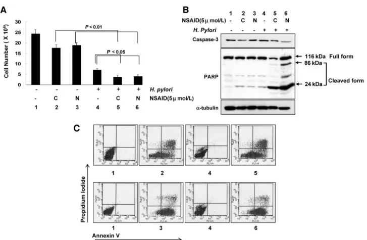
When AGS cells were treated with 5mol/L nimesulide (preferred COX-2 inhibitor) or 5mol/L celecoxib (selective COX-2 inhibitor) or H. pylori (2 ⫻ 106 CFU), a significant
decrease in cell survivals was observed with all three treatments, which was measured with cell counts (Fig. 6A). Significant attenuation in cell survivals was noted in cells treated with either celecoxib or nimesulide alone. These changes were more evi-dent in cells cotreated with a COX-2 inhibitor and H. pylori, suggesting that cell death processes were more activated than
with either COX-2 inhibitor or H. pylori alone. All of these findings that the combination of COX-2 inhibitor and H. pylori provoked and augmented apoptosis were more evidenced with Western blot of caspase-3 and PARP (Fig. 6B) and with flow cytometry analyses (Fig. 6C). Active caspase-3, cleavage of PARP, and increased fractions of positive annexin V— observed after COX-2 inhibitor treatment—were more apparently in-creased after the cotreatment of COX-2 inhibitor and H. pylori. These in vitro experiments explain the similar mechanistic basis of the cancer prevention of nimesulide treatment in H. pylori-associated gastric tumorigenesis as observed in in vivo animal experiment. Attenuated development of gastric tumor in previ-ous animal experiments could be achieved because both the COX-2 inhibitor and H. pylori acted synergistically to increase apoptosis, a possible chemopreventive mechanism of nimesu-lide.
DISCUSSION
The present study shows that H. pylori-associated gastric tumorigenesis and carcinogenesis was effectively suppressed by Fig. 4 Expression of COX-2, Bax, and Bcl-2. A, immunohistochemical staining for COX-2. In Aa, COX-2 expression was mainly confined to stromal
cells composed of macrophages, lymphocytes, and interstitium in gastric adenocarcinoma. Ab, gastric adenoma epithelial cells expressing COX-2. In
Ac and Ad, COX-2 expression was attenuated in nimesulide (NIM)-treated animals (Ad) compared with non-NIM-treated animals (Ac);⫻200. B,
COX-2 protein expression levels in the experimental groups. Western blot analysis of COX-2 protein expression in stomach tissues (Hp, H. pylori;
kDa, Mrin thousands). C, Bax and Bcl-2 expression levels in the experimental group. Bax and Bcl-2 expressions in stomach tissues were demonstrated according to group, and the ratio (Bax/Bcl2) of the expression was depicted. kDa, Mrin thousands.
the selective COX-2 inhibitor nimesulide. COX-2 protein was highly expressed in stromal cells of H. pylori-associated tumor tissues, which expression levels substantially decreased after nimesulide administration. In addition, significant inductions of apoptosis were observed. The results of in vitro experiments suggested that nimesulide and H. pylori synergistically operated to induce more apoptosis in AGS cells. These findings indicate that the inhibition of COX-2 activity might be a novel target for the treatment and prevention of H. pylori-associated gastric cancers, which are similar to colon cancers.
Cancer is the result of a multistep molecular and cellular process. Chemoprevention might be a better way to manage cancer rather than the current chemotherapy with cytotoxic agents in every aspect of efficacy, compliance, and feasibility. For this purpose, noncytotoxic nutrients or pharmacological compounds that might protect against the development and progression of mutant clones into malignant cells have been used in experimental and clinical studies (31). Nimesulide, a preferred selective inhibitor of COX-2 belonging to the sulfon-amide class, has been used clinically as an anti-inflammatory drug with less ulcerogenic effects in the gastrointestinal tract than classical NSAIDs. It is regarded as a good candidate agent in chemopreventive trials against colon and breast cancers (32). A large body of recent epidemiologic data suggests regular NSAID use can reduce both the incidence of, and mortality from, colorectal cancer and other solid tumors such as breast cancer (19, 23–25, 33).
It has been postulated that the protective effects of NSAIDs are mediated through the inhibition of cyclooxygenase enzymes, COX-1 and COX-2 (19, 22). COX-2 leads to increased synthe-sis of prostaglandins in both inflammatory and malignant tis-sues, and may support the development and progression of human malignancy. Recently, a more direct causal link between COX-2 expression and malignancy was shown in studies of COX-2-overexpressing transgenic mice (34), which displayed mammary gland hyperplasia and transformation in breast tissue overexpressing COX-2. Similarly, COX-2 “knockout” mice showed reduced intestinal polyp development (26), again sup-porting a causative role of COX-2 in tumorigenesis. Studies of human colorectal tumors revealed that COX-2 is overexpressed in more than 80% of carcinomas and in at least 50% of prema-lignant adenomas (22). Furthermore, COX-2 inhibitors effec-tively reduced colorectal tumors induced by azoxymethane treatment in rats, and spontaneous colorectal tumors in Apc⫺/⫺
Min mice (24, 35). On the basis of preclinical profiles that
COX-2 was highly expressed in polyp tissues obtained from patients with familial adenomatous polyposis (FAP), celecoxib entered a phase III randomized trial in 77 FAP patients. After 6 months of 400-mg intake twice daily, celecoxib caused a 28% reduction in polyp burden in the rectum (25).
Several studies have shown enhanced COX-2 expression in gastric cancer tissues (18, 27). Overexpression of COX-2 was a property shared by both intestinal and diffuse gastric carcino-mas. It seems that COX-2 might play an important role during the early stage of gastric carcinogenesis (16, 27). H. pylori infection is a major risk factor for gastric carcinoma. H. pylori up-regulated COX-2 mRNA expression and stimulated release of prostaglandin E2in a gastric cancer cell line (10), as well as
in the gastric mucosa of animal models and in humans (12, 14). Therefore, high levels of H. pylori infection might up-regulate COX-2 expression, which, in turn, could lead to the develop-ment of gastric carcinogenesis (27). Experidevelop-mentally, Xiao et al. (14) demonstrated that COX-2 was directly involved in hyper-plastic changes in mice infected with H. pylori. Consistent with the data from the above-mentioned studies, the present findings strongly support the contention that pharmacological inhibition of COX-2 overexpression may be useful against H. pylori-associated gastric cancer development and progression.
H. pylori and NSAIDs are known to be risk factors in the
pathogenesis of gastric ulcers through apparently different mechanisms, reciprocally or independently. H. pylori infection may induce strong mucosal inflammation, stimulate cytokine release, and provoke apoptosis. H. pylori had been known to be responsible for significant apoptosis, by which gastric ulcer-ations or gastric atrophy (significant apoptotic cell death of gastric stem cells or parietal cells) can be developed. For this, several cytotoxins of CagA or VacA, ammonia generated from urease actions, chemokines, or cytokines like interleukin (IL)-8, interferon-␥, IL-12, tumor necrosis factor ␣, oxidative stress, and HNP (helicobacter neutrophil-activating peptide), and so forth, had been known to be responsible for apoptosis after
H. pylori infection. In contrast, NSAIDs may inhibit mucosal
prostaglandin synthesis, leading to a weakening in the gastric mucosal barrier, and impaired resistance to acid injury. There-fore, one might expect increased incidence of gastric mucosa damage in the presence of these two risk factors (36). However, Fig. 5 TUNEL staining and apoptotic index. A, TUNEL staining
pos-itive cells were increased after H. pylori infection (Group 1), but the apoptotic activities were markedly decreased in gastric adenoma tissue (Group 3). However, nimesulide administration provoked increased apoptotic activities either in nontumor tissue of Group 4 or in tumor tissue of Group 5. B, mean index (M⫾SD) of apoptosis is shown, resulting in statistically increased apoptotic index in group treated with nimesulide irrespective of the presence of gastric tumor.
we found no evidence of any gastric mucosal damages in mice stomachs (group 4) assessed either by gross inspection or his-tologic evaluation, although the combination of COX-2 inhibi-tor and H. pylori did augment apoptosis in in vitro experiments (Fig. 6). The presence of H. pylori and coexisting gastritis has been shown to increase (37), have no effect (38), and even decrease (39), the risk of ulcer bleeding among patients ingesting aspirin or NSAIDs. In the present study, we found that despite long-term exposure to these two risk factors in groups 4 and 5, there was no increase in gastric inflammation or mucosal destruction compared with groups 1 and 2, which were exposed to a single risk factor. As for the explanations, we inferred that gastric defense mechanisms operated well in the groups exposed to both H. pylori and COX-2 inhibitor, and nimesulide administered to the H. pylori-infected mice must have blocked the effects of COX-2 on cellular pro-liferation, release of inflammatory mediators, and cell adhe-sion to matrix, which cause increased gastric inflammation (40). Moreover, H. pylori-alone provoked cellular apoptosis,
whereas NSAID-alone also contributed to apoptosis either by COX-2 inhibition or by direct activation of other cellular targets such as peroxisome proliferator-activated receptor␥ observed in our present experiment (data not shown; ref. 41).
In vitro experiments in this study showed the synergistic
effects of cotreatment with COX-2 inhibitor and H. pylori on apoptosis and inhibition of proliferation. Recently, Kern et
al. (42) suggested that hepatocarcinogenesis could be
pre-vented by COX-2 inhibitors, based on the induction of apo-ptosis and the inhibition of proliferation; and data from Reddy et al. (19) strengthened the argument that selective COX-2 inhibitors possess chemopreventive activity against colon carcinogenesis.
In conclusion, we found that gastric tumorigenesis was significantly attenuated by long-term administration of the COX-2 inhibitor, nimesulide, in an H. pylori-associated gastric cancer mouse model, and we propose that selective COX-2 inhibitors may be clinically useful in protecting against gastric cancer development.
Fig. 6 In vitro experiments with AGS cells. A, cell survivals after each treatment analyzed by cell counts. Statistically significant decreases in cell
survival were observed in AGS cells treated with both COX-2 inhibitor and H. pylori than were seen in cells treated with COX-2 inhibitor alone (P⬍ 0.01) or H. pylori alone (P⬍ 0.05). B, Western blot analysis of active form of caspase-3 and PARP. Significant decrease in the expression of active caspase-3 and cleavage of PARP were observed after both H. pylori and COX-2 inhibitor, signifying the occurrence of apoptotic cells death in cells treated with both COX-2 inhibitor and H. pylori infection. C stands for 5mol/L celecoxib, N stands for 5 mol/L nimesulide. C, flow cytometry. Fluorescence-activated cell sorter analysis for annexin V, and propidium iodide staining of AGS cells. The percentages give the proportion of cells in the respective quadrant. Apoptosis was greatest in cells treated with both COX-2 inhibitor and H. pylori. 1, control cells (AGS cells, gastric cancer cells); 2, cells treated with celecoxib (5mol/L); 3, cells treated with nimesulide (5 mol/L); 4, cells infected with H. pylori (2 ⫻ 106CFU); 5, cells treated with celecoxib (5mol/L) ⫹ H. pylori (2 ⫻ 106CFU); 6, cells treated with nimesulide (5mol/L) ⫹ H. pylori (2 ⫻ 106CFU). (kDa, M
r in thousands.)
REFERENCES
1. Blaser MJ, Chyou PH, Nomura A. Age at establishment of Helico-bacter pylori infection and gastric carcinoma, gastric ulcer, and duode-nal ulcer risk. Cancer Res 1995;55:562–5.
2. International Agency for Research on Cancer Working Group on the Evaluation of Carcinogenic Risks to Humans. Schistosomes, liver flukes and Helicobacter pylori: IARC Working Group on the Evaluation of Carcinogenic Risks to Humans. IARC monographs on the evaluation of carcinogenic risks to humans. Lyon, France: IARC; 1994. p. 218 –20. 3. Correa P. Human gastric carcinogenesis: a multistep and multifac-torial process—First American Cancer Society Award Lecture on Can-cer Epidemiology and Prevention. CanCan-cer Res 1992;52:6735– 40. 4. Kikuchi S, Crabtree JE, Forman D, Kurosawa M. Association be-tween infections with CagA-positive or -negative strains of Helicobacter pylori and risk for gastric cancer in young adults. Research Group on Prevention of Gastric Carcinoma among Young Adults. Am J Gastro-enterol 1999;94:3455–9.
5. Wang J, Chi DS, Kalin GB, et al. Helicobacter pylori infection and oncogene expressions in gastric carcinoma and its precursor lesions Dig Dis Sci 2002;47:107–13.
6. Shimizu N, Ikehara Y, Inada K, et al. Eradication diminishes en-hancing effects of Helicobacter pylori on glandular stomach carcino-genesis in Mongolian gerbils. Cancer Res 2000;60:1512– 4.
7. Hahm KB, Lee KJ, Choi SY, et al. Possibility of chemoprevention by the eradication of Helicobacter pylori: oxidative DNA damage and apoptosis in H. pylori infection. Am J Gastroenterol 1997;92:1853–7. 8. Bresalier RS. Helicobacter pylori and gastric cancer: a not so simple association Gastroenterology 1998;114:408 –9.
9. Webb PM, Yu MC, Forman D, et al. An apparent lack of association between Helicobacter pylori infection and risk of gastric cancer in China. Int J Cancer 1996;67:603–7.
10. Romano M, Ricci V, Memoli A, et al. Helicobacter pylori up-regulates cyclooxygenase-2 mRNA expression and prostaglandin E2 synthesis in MKN 28 gastric mucosal cells in vitro J Biol Chem 1998;273:28560 –3.
11. Tatsuguchi A, Sakamoto C, Wada K, et al. Localisation of cy-clooxygenase 1 and cycy-clooxygenase 2 in Helicobacter pylori related gastritis and gastric ulcer tissues in humans. Gut 2000;46:782–9. 12. Fu S, Ramanujam KS, Wong A, et al. Increased expression and cellular localization of inducible nitric oxide synthase and cyclooxygenase 2 in Helicobacter pylori gastritis. Gastroenterology 1999;116:1319 –29. 13. McCarthy CJ, Crofford LJ, Greenson J, Scheiman JM. Cyclooxy-genase-2 expression in gastric antral mucosa before and after eradication of Helicobacter pylori infection. Am J Gastroenterol 1999;94:1218 –23. 14. Xiao F, Furuta T, Takashima M, Shirai N, Hanai H. Involvement of cyclooxygenase-2 in hyperplastic gastritis induced by Helicobacter pylori infection in C57BL/6 mice. Aliment Pharmacol Ther 2001;15:875– 86. 15. Sung JJ, Leung WK, Go MY, et al. Cyclooxygenase-2 expression in Helicobacter pylori-associated premalignant and malignant gastric le-sions. Am J Pathol 2000;157:729 –35.
16. Saukkonen K, Nieminen O, van Rees B, et al. Expression of cyclooxygenase-2 in dysplasia of the stomach and in intestinal-type gastric adenocarcinoma. Clin Cancer Res 2001;7:1923–31.
17. Zimmermann KC, Sarbia M, Weber AA, Borchard F, Gabbert HE, Schror K. Cyclooxygenase-2 expression in human esophageal carci-noma. Cancer Res 1999;59:198 –204.
18. Ristimaki A, Honkanen N, Jankala H, Sipponen P, Harkonen M. Expression of cyclooxygenase-2 in human gastric carcinoma. Cancer Res 1997;57:1276 – 80.
19. Reddy BS, Rao CV, Seibert K. Evaluation of cyclooxygenase-2 inhibitor for potential chemopreventive properties in colon carcinogen-esis. Cancer Res 1996;56:4566 –9.
20. DuBois RN, Radhika A, Reddy BS, Entingh AJ. Increased cyclooxygenase-2 levels in carcinogen-induced rat colonic tumors. Gastroenterology 1996;110:1259 – 62.
21. Eberhart CE, Coffey RJ, Radhika A, et al. Up-regulation of cy-clooxygenase 2 gene expression in human colorectal adenomas and adenocarcinomas. Gastroenterology 1994;107:1183– 8.
22. Sano H, Kawahito Y, Wilder RL, et al. Expression of cyclooxyge-nase-1 and -2 in human colorectal cancer. Cancer Res 1995;55:3785–9. 23. Giardiello FM, Hamilton SR, Krush AJ, et al. Treatment of colonic and rectal adenomas with sulindac in familial adenomatous polyposis. N Engl J Med 1993;328:1313– 6.
24. Jacoby RF, Seibert K, Cole CE, Kelloff G, Lubet RA. The cyclooxygenase-2 inhibitor celecoxib is a potent preventive and thera-peutic agent in the min mouse model of adenomatous polyposis. Cancer Res 2000;60:5040 – 4.
25. Steinbach G, Lynch PM, Phillips RK, et al. The effect of celecoxib, a cyclooxygenase-2 inhibitor, in familial adenomatous polyposis. N Engl J Med 2000;342:1946 –52.
26. Oshima M, Dinchuk JE, Kargman SL, et al. Suppression of intes-tinal polyposis in Apc delta716 knockout mice by inhibition of cyclooxygenase-2 (COX-2). Cell 1996;87:803–9.
27. Lim HY, Joo HJ, Choi JH, et al. Increased expression of cyclooxygenase-2 protein in human gastric carcinoma. Clin Cancer Res 2000;6:519–25. 28. Han SU, Kim YB, Joo HJ, et al. Helicobacter pylori infection promotes gastric carcinogenesis in a mice model J Gastroenterol Hepa-tol 2002;17:253– 61.
29. Nam KT, Oh SY, Ahn B, et al. Decreased Helicobacter pylori-associated gastric carcinogenesis in mice lacking inducible nitric oxide synthase. Gut 2004;53:1250 –5.
30. Leininger JR, Jokinen MP. Tumours of the oral cavity, pharynx, oesophagus and stomach. In: Turusov VS, Mohr U, editors. Pathology of tumours in laboratory animals. Lyon: IARC; 1994. p. 167–93. 31. De Flora S, Bennicelli C, Bagnasco M. Rationale and mechanisms of cancer chemoprevention. Recent Results Cancer Res 1999;151:29 – 44. 32. Davis R, Brogden RN. Nimesulide. An update of its pharmacody-namic and pharmacokinetic properties, and therapeutic efficacy. Drugs 1994;48:431–54.
33. Harris RE, Alshafie GA, Abou-Issa H, Seibert K. Chemoprevention of breast cancer in rats by celecoxib, a cyclooxygenase 2 inhibitor. Cancer Res 2000;60:2101–3.
34. Liu CH, Chang SH, Narko K, et al. Overexpression of cyclooxy-genase-2 is sufficient to induce tumorigenesis in transgenic mice. J Biol Chem 2001;276:18563–9.
35. Rao CV, Rivenson A, Simi B, et al. Chemoprevention of colon carcinogenesis by sulindac, a nonsteroidal anti-inflammatory agent. Cancer Res 1995;55:1464 –72.
36. Leong RW, Chan FK, Sung JJ. Helicobacter pylori and nonsteroidal anti-inflammatory drugs. Approaching the end of the controversy in the new millennium, or room for more debate? J Gastroenterol 2001;36:731–9. 37. Aalykke C, Lauritsen JM, Hallas J, Reinholdt S, Krogfelt K, Lauritsen K. Helicobacter pylori and risk of ulcer bleeding among users of nonsteroidal anti-inflammatory drugs: a case-control study Gastroen-terology 1999;116:1305–9.
38. Cullen DJ, Hawkey GM, Greenwood DC, et al. Peptic ulcer bleed-ing in the elderly: relative roles of Helicobacter pylori and non-steroidal anti-inflammatory drugs. Gut 1997;41:459 – 62.
39. Santolaria S, Lanas A, Benito R, Perez-Aisa M, Montoro M, Sainz R. Helicobacter pylori infection is a protective factor for bleeding gastric ulcers but not for bleeding duodenal ulcers in NSAID users Aliment Pharmacol Ther 1999;13:1511– 8.
40. Katori M, Majima M. Cyclooxygenase-2: its rich diversity of roles and possible application of its selective inhibitors. Inflamm Res 2000;49:367–92. 41. Chen GG, Lee JF, Wang SH, Chan UP, Ip PC, Lau WY. Apoptosis induced by activation of peroxisome-proliferator activated receptor gamma is associated with Bcl-2 and NF-B in human colon cancer. Life Sci 2002;70:2631– 46.
42. Kern MA, Schubert D, Sahi D, et al. Proapoptotic and antiprolif-erative potential of selective cyclooxygenase-2 inhibitors in human liver tumor cells. Hepatology 2002;36:885–94.