Contents lists available atScienceDirect
Stem Cell Research
journal homepage:www.elsevier.com/locate/scr
Stem cell passage a
ffects directional migration of stem cells in electrotaxis
Seung Hee Hong
a,b, Mi Hee Lee
a, Min-Ah Koo
a,b, Gyeung Mi Seon
a,b, Ye Jin Park
a,c, Dohyun Kim
a,
Jong-Chul Park
a,b,c,⁎aCellbiocontrol Laboratory, Department of Medical Engineering, Yonsei University College of Medicine, Seoul, 03722, Republic of Korea bBrain Korea 21 PLUS Project for Medical Science, Yonsei University College of Medicine, Seoul, 03722, Republic of Korea cDepartment of Medical Device Industry, Yonsei University College of Medicine, Seoul, 03722, Republic of Korea
A R T I C L E I N F O Keywords: Stem cell Directional migration Electrotaxis Senescence A B S T R A C T
Stem cells can differentiate into various body tissues and organs and thus are considered as promising tools for cell therapy and tissue engineering. Early passage stem cells have high differentiation ability compared to late passage stem cells. Thus, it is important to use early passage stem cells in cell therapy. Here, we investigated whether cell migration could be used to compare young and senescent cells. We used‘electrotaxis’ where cells under electric treatment move towards the anode or cathode. Without an electric stimulus, stem cells moved randomly. However, under a direct electric current, the cells moved with directionality. Under stimulation with a direct electric current, early passage stem cells moved towards the anode; when the cells became senescent with increasing passages, the percentage of cells migrating to the anode decreased. These results suggest that the behavior of stem cells under the influence of a direct electric current is also related to their passage number. Therefore, electrotaxis migration analysis can be used to distinguish between young cell and senescent cells.
1. Introduction
Cell therapy has been widely examined and is used to treat various disease. Stem cells are promising sources for tissue engineering and cell therapy (Thomson et al., 1998). Particularly, mesenchymal stem cells (MSCs) have been explored as attractive therapeutic agents because of their capacity to proliferate and differentiate into multiple cell lineages such as osteocytes, adipocytes, and chondrocytes (Borougeni et al., 2012). MSCs are a class of multipotent stem cells that can be isolated from various tissues including adipose tissue, bone marrow, and per-ipheral blood. However, stem cells are associated with safety issue such as tumorigenicity (Ding et al., 2015). Previous studies showed that when undifferentiated stem cells are implanted, they can form a ter-atoma (Brivanlou et al., 2003;Prokhorova et al., 2009;Ben-David and Benvensity, 2011;Hentze et al., 2009;Barkholt et al., 2013;Nussbaum et al., 2007;Dazzi and Horwood, 2007). Therefore, to enhance the ef-fects of stem cells in cell therapy, it is important to identify appropriate cell sources. It is well-known that cell differentiation and function de-cline with passage number and thus early passage stem cells should be used in cell-based therapies.
The proliferation rates and other properties of cells gradually change with increasing passage numbers (Gu et al., 2016). In vitro, stem
cells undergo senescence as the passage number increases and also lose their innate characteristics (Kuilman et al., 2010). Senescent cells have altered morphology, produce senescence-associated heterochromatin foci, show decreased proliferation and differentiation abilities, exhibit senescence-associated ß-galactosidase activity, and have altered activ-ities of cell cycle inhibitors such as p16, p15, p21 and p53 (Kuilman et al., 2010;Chen et al., 2015;Simonsen et al., 2002). Stem cells also show a decreased ability to differentiate during senescence (Choudhery et al., 2014;Roobrouck et al., 2008;Fehrer and Lepperdinger, 2005). These changes may influence stem cell therapy.
Application of a direct electric current can induce the directional migration of many cell types, including mesenchymal stem cells, fi-broblasts and endothelial cells. This phenomenon is known as ‘elec-trotaxis’ (Iwasa et al., 2017). During electrotaxis, cells move towards the anode or cathode under a direct electric current. Exogenous electric stimulation can be a cue that controls cell behavior. Electrotaxis is a simple method for controlling cell migration. However, the migration direction and rate can be influenced by external stimulation and innate cellular features (Kim et al., 2017).
In this study, we investigated the effect of stem cell passage number on directional migration and evaluated passage-related reactions oc-curring in response to electric stimulus. Additionally, we examined
https://doi.org/10.1016/j.scr.2019.101475
Received 29 January 2019; Received in revised form 20 May 2019; Accepted 28 May 2019
⁎Corresponding author at: Department of Medical Engineering, Yonsei University College of Medicine, 50–1 Yonsei-ro, Seodaemun-gu, Seoul, 03722, Republic of Korea.
E-mail address:[email protected](J.-C. Park).
Available online 30 May 2019
1873-5061/ © 2019 The Authors. Published by Elsevier B.V. This is an open access article under the CC BY-NC-ND license (http://creativecommons.org/licenses/BY-NC-ND/4.0/).
senescent properties about the cells we used in this study. 2. Materials and methods
2.1. Cell cultures
Human mesenchymal stem cells (hMSCs, Lonza, Basel, Switzerland) were cultured in MSC growth medium (Lonza). Adipose-derived MSCs (ADSC, Lonza) were cultured in adipose-derived stem cell growth medium (Lonza). Tonsil-derived mesenchymal stem cells (TMSC, pro-vided by Dr. Jo in Ewha Woman's University (Seoul, Korea)) were cultured in Dulbecco's Modified Eagle's Medium (Welgene, Seoul, Korea) containing 10% fetal bovine serum and 1% antibiotics (Welgene). Cells were incubated at 37 °C in a 5% CO2atmosphere in a humidified incubator. Fresh complete medium replacement was con-ducted every 3–4 days. When the cells reached 90% confluence in the flask, adherent cells were detached using 0.25% trypsin EDTA (Welgene) and then passaged. In this study, we selected passages 5, 10, and passage 15 as representative of early, middle, and late passage cells.
2.2. Senescence associated ß -galactosidase staining & activity
Senescence-associated ß-galactosidase staining was performed using a Cellular Senescence Detection kit (Cell Biolabs, San Diego, CA, USA) according to the manufacturer's protocol. Briefly, passage 5, 10, and 15 TMSCs, hMSCs, and ADSCs were cultured at a density of 1 × 10⁵per well in 12-well plates and incubated overnight. The cells were washed with Dulbecco's phosphate-buffered saline (DPBS) and fixed with 4% paraformaldehyde for 10 min. After washing the cells with DPBS, they were stained with cell staining working solution for 24 h in an in-cubator at 37 °C in the dark. Senescent cells were observed under a bright-field microscope (Olympus CKX41, Tokyo, Japan).
Senescence associated ß-galactosidase activity was evaluated using the cellular senescence assay kit from Cell Biolabs according to the manufacturer's protocol. Passage 5, 10, and 15 TMSCs, hMSCs, ADSCs were seeded at a density of 5 × 10⁴per well in 48-well plates and in-cubated overnight. After washing the cells with cold DPBS, the cells were lysed with lysis buffer for 10 min at 4 °C. Whole cell lysates were centrifuged for 10 min at 4 °C in 14,000 rpm. The supernatants were transferred into black 96-well plates combined with an equal amount of reaction buffer, and incubated at 37 °C with protection from light for 3 h. The reaction mixtures were neutralized by adding stop solution and the activity of senescence-associated ß –galactosidase was measured with a fluorescence plate reader (Flexstation3, Molecular Devices, Sunnyvale, CA, USA) at excitation and emission wavelengths of 360 and 465 nm, respectively. The data were expressed as the mean relative fluorescence reading normalized to the total protein concentration. 2.3. MTT assay for cell proliferation
The cell proliferation of TMSCs, hMSCs, and ADSCs was assessed by 3-(4,5-dimethylthiazol-2-yl)-2,5-diphenyl-2-H-tetrazolium bromide (MTT) assay at passages 5, 10, and 15. Cells were seeded into 48-well plates at a density of 1000 cells per well. After 1, 2, 3, 4, 5, 6, and 7 days, the cells were washed with DPBS, MTT was added to each well, and the cells were incubated for 3 h at 37 °C in the dark. MTT solution was aspirated from the cells and a mixture of dimethyl sulfoxide and glycine buffer was added. After purple formazan crystals were fully dissolved, they were transferred to a 96-well plate. The absorbance values of the samples were read on a microplate reader at a wavelength of 570 nm (Molecular Devices).
2.4. Western blot analysis
Cells were lysed in RIPA buffer supplemented with 1 mM PMSF.
Proteins were separated by 15% sodium dodecyl sulfate-polyacrylamide gel electrophoresis and transferred onto polyvinylidene difluoride membranes. The primary antibodies recognized the following proteins: p16, p21, proliferating cell nuclear antigen (PCNA) andβ-actin (Cell Signaling Technology, Danvers, MA, USA). After incubation with horseradish peroxidase-linked anti-rabbit or anti-mouse IgG secondary antibody (Cell Signaling Technology), protein expression was evaluated with SignalFire ECL reagent (Cell Signaling Technology).
2.5. Application of electrotaxis stimulation to stem cells
To directly apply electric stimulation to the stem cells, a customized agar-salt electrotaxis chamber with an incubator system was used. This equipment consisted of an electrotaxis incubator connected to a mi-croscope to observe live cells and a chamber system for applying the direct electric current to the cells (Kim et al., 2017). Electrotaxis ana-lysis was performed in the incubator system (CCP ver 3.8, Live Cell Instrument, Seoul, Korea) equipped with an inverted microscope (Olympus). The stem cells were seeded at a density of 4 × 10³cells on a slide glass with a silicon O-ring with an inner diameter of 16 mm and incubated overnight in a CO2incubator. To place the cells in the elec-trotaxis incubator, the silicon O-ring was removed and the cell-seeded slide glass was placed in the bottom chamber and upper chamber. A silicon gasket was placed on the top of the slide glass. For electric current application, agar-salt bridges were used to connect the elec-trodes at each sides of the chamber. The DC pulse power supply (Daedo Powertronics Co. Ltd., Seoul, Korea) supplied the electric current to the chamber through the 2% agar salt bridge immersed in Steinberg's so-lution. During electric stimulation, the current was monitored with a digital power meter (Fluke, Everett, WA, USA) to ensure a constant electric current. Cell images were captured every 5 min using a charge-coupled device camera attached to an inverted microscope. An electric current of 1000μA was applied for 3 h (Kim et al., 2017).
2.6. Analysis of cell migration
Cells were visualized with a microscope using a 10× objective lens. The cell migration speed was calculated by manual tracking and che-motaxis tool plugin (v.1.01, distributed by ibidi GmbH, Munchen Germany) in the ImageJ program (ImageJ 1.37v, National Institutes of Health, Bethesda, MD, USA). Without an electric current, 40 stem cells were tracked at passages 5, 10, and 15. Under an electric current at 1000μA for 3 h, 80 stem cells were tracked at passages 5, 10 and 15. Cell migration was divided into the anode (direction of electric current, right side) and cathode (opposite direction of electric current, left side) reaction by using the chemotaxis tool plugin.
2.7. Statistical analysis
Statistical analyses were performed with SPSS 23.0 software (SPSS, Inc., Chicago, IL, USA). Comparison within the studied groups was performed by paired t-test, while different groups were evaluated by analysis of variance. A value of p < .05 is considered to indicate sta-tistical significance.
3. Results & discussion
3.1. Comparison of senescence properties in early, middle, and late passages Various methods can be used to monitor MSC senescence. One of the most convenient indicators is the number of cell passages. However, the passage number can be affected by various factors, such as the seeding density and the length of incubation. Therefore, senescence properties are need to be investigated. Staining of senescent cells based on accu-mulation of SA-β-gal is a widely used approach. Senescent cells show common biochemical markers such as SA-ß-gal activity. When stem
cells become senescent during serial passaging, the numbers of SA-ß-gal-positive cells increase (Wagner et al., 2008). We confirmed the different abilities of cell growth for stem cells passaged for different amounts of time. Innate factors in the cell change during long-term culture, resulting in difference in proliferation ability (Kippin et al., 2005;Amit et al., 2000) Additionally, evaluation of senescence-specific
gene expression markers can be used to identify senescent cells. In this study, TMSCs, hMSCs, and ADSCs were serially passaged 5 to 15 times, and these cells were used to confirm senescence by SA-β-gal, pro-liferation ability, and senescent gene expression. In many studies, stem cells passaged more than 10 times are defined as senescent (Izadpanah et al., 2008;Rilianawati et al., 2018;Osipova et al., 2011;Zhuang et al., Fig. 1. Senescence characteristic of stem cells during serial passaging.
(A) Senescence-associated ß-galactosidase staining for TMSCs, hMSCs, and ADSCs at passages 5, 10, and 15. Red arrow indicates senescence-associated ß-ga-lactosidase-positive stained cells. Scale bar = 100μm. (B) Senescence-associated ß-galactosidase activation measured by fluorometric analysis of TMSCs, hMSCs, and ADSCs at passages 5, 10, and 15. * p < .05. (C) Cell proliferation analysis of TMSCs, hMSCs, and ADSCs by MTT. TMSCs, hMSCs, and ADSCs -at passages 5, 10, and 15 were evaluated from 1 day after seeding to 7 days after seeding. Formazan crystal formation was measured at 570 nm. (D)Western blot analysis to detect senescence markers p16 and, p21 and proliferation marker PCNA. (For interpretation of the references to color in thisfigure legend, the reader is referred to the web version of this article.)
2015).
In SA-β-gal staining using fluorometric substrate, we detected strong positive staining ofβ-galactosidase with increasing passage of all stem cells (Fig. 1A). SA-ß-gal activity was normalized by the total protein concentration to obtain quantitative results for each passages. In TMSCs, middle passage cells showed similar activities compared to early passage cells, but late passage cells showed 200% higher activity. Similar tendencies were observed for ADSCs. Late passage ADSCs had 130% higher activity than early passage. In hMSCs, middle and late passage cells showed 130% higher activity than early passage cells (Fig. 1B). As a result, late passage cells showed high SA-ß-gal activity
compared to early passage cells in all cell types, indicating the higher ages of late compared to early passage cells.
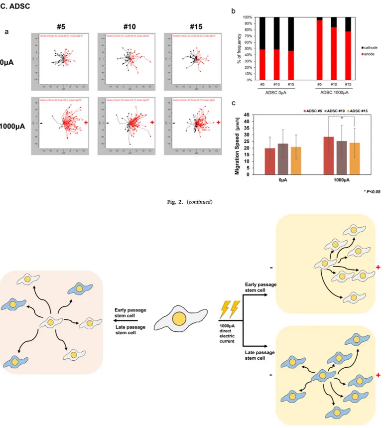
The proliferation rates of TMSCs, hMSCs and ADSCs were measured at passages 5, 10, and 15 by MTT assay (Fig. 1C). Early and late passage stem cells showed significant differences at 3 days after seeding. At 7 days after seeding, late passage TMSCs and hMSCs showed only 37% proliferation ability in the early passage. Late passage ADSCs showed 43% proliferation ability compared to early passage cells at 7 days after seeding. All stem cells showed passage-dependent proliferation rates. Early and late passage cells showed significant differences in their growth rates at the passages evaluated in this study. Therefore, the Fig. 2. Stem cell migration induced by electrotaxis.
(A) TMSC migration by electrotaxis. (a) TMSCs were manually tracked and cells migration was divided into the right (anode) and left (cathode). (b)Frequency of cell movement towards the anode and cathode. (c)Cell migration speed was measured at passages 5, 10, and 15 when direct current was applied to the cells. (B) hMSC migration by electrotaxis. (C) ADSC migration by electrotaxis. * p < .05.
passage interval selected is suitable for detecting differences in stem cell senescence.
To identify gene expression changes during increasing passage, we detected expression of the senescence markers p16 and p21 and pro-liferation marker PCNA by western blotting. β-Actin was used as a
loading control. Data for each protein quantified by the loading control and band intensity was normalized to the corresponding loading con-trol. We considered early passage expression as the control level and calculated the relative intensities in the middle and late passages. The expression of p16 was found to be related to the passage number Fig. 2. (continued)
Fig. 3. Schematic illustration of the study.
Early passage cells are presented in white color and late passage cells in blue color. Stem cells migrate randomly under normal conditions in both early and late passages. However, when stem cells are stimulated by a direct electric current, early passage stem cells migrated primarily towards the anode. Late passage stem cells showed a decreased tendency to migrate towards the anode. (For interpretation of the references to color in thisfigure legend, the reader is referred to the web version of this article.)
(Fig. 1D). p16 expression increased during serial passage. p21 was in-creased at the middle passage and dein-creased at the late passage in TMSCs and ADSCs, indicating that senescence was initiated in the middle passage. In contrast, in hMSCs, p21 was consistently increased during the late passage. In all cell types, PCNA decreased with in-creasing passage numbers. p16 and p21 signal transduction cascades commonly activate the senescence pathway. Particularly, p16 is ele-vated during senescence process and act critical in senescence cycle. However, p21 decreases from its maximum value at the initiation of senescence (Herranz and Gil, 2018;Stein et al., 1999). Therefore, p21 is important in the onset of senescence, whereas p16 maintains senes-cence. PCNA is an important factor in proliferation and is decreased during senescence.
Stem cells are considered as promising tools for tissue engineering and cell therapies. However, stem cells have a limited lifespan, limiting their applications in tissue engineering and cell therapies (Herranz and Gil, 2018). Increasing the passage number by long-term culture, re-ferred as late passage stem cells, alters their innate characteristics be-cause of senescence (Gu et al., 2016). Serially passaging in vitro culture results in a decreased cell differentiation potential, including lineage-dependent effects due to senescence. (Kretlow et al., 2008;Zhou et al., 2008). As the passage number of bone marrow-derived MSCs is in-creased, adipogenic differentiation shows a significant difference be-tween early and late passage cells. Late passage bone marrow-derived MSCs have a low ability to differentiate compared to early passage cells (Kretlow et al., 2008). Implanting undifferentiated stem cells can cause side effects such as tumorigenicity. Thus, using early passage cells is important for improving cell therapy outcomes.
3.2. Migration of stem cells in electrotaxis is passage-related
“Electrotaxis” referes to the direction of cell direction in response to electric stimulation. Using electrotaxis, cells are stimulated by an electric current, which is an easy method for controlling cellular posi-tioning, precisely and rapidly revealing changes compared to other methods. Cells preferentially moved towards one of the electrodes, as polarized effects are induced by ion transport or redistribution of charged cell-surface molecules (Cortese et al., 2014). Our system used an agar-salt bridge to apply a stable direct current between the power supply and customized electrotaxis chamber into which the cells were seeded.
To analyze migration upon direct electric current stimulation of TMSCs, hMSCs, and ADSCs, the cells were tracked in 5-min intervals for 3 h in the electrotaxis chamber. The migration speed was calculated as the accumulated migration distance divided by the total migration observation time. The percentage of cells migrating towards the anode and cathode was divided based on a center point. Cell movement to the right side of the center point, was considered as directional movement towards the anode while cell migration to left side of the center point was considered as migration towards the cathode.
TMSCs showed passage-dependent speed differences in the presence and absence of electric current stimulation. No significant differences in the migration speed at hMSCs and ADSCs were found in different pas-sages. Therefore, evaluating the migration speed does not explain passage-related differences in all cell types (Fig. 2Ac, 2Bc, 2Cc). TMSCs, hMSCs, and ADSCs moved randomly in the absence of a direct current at all passages (Fig. 2Aab, 2Bab, 2Cab). However, under stimulation with a direct current, the cells migrated in a directional manner. In TMSCs, hMSCs, and ADSCs, the cells moved towards anode when a direct current was applied. Interestingly, under a direct current, cell migration towards the anode tended to decrease with serial passaging. In TMSCs 95% of passage 5 cells moved towards the anode, while 78% of passage 15 cells moved towards the anode. In hMSCs 96% of passage 5 cells moved towards the anode, while 81% of passage 15 cells moved towards the anode. In ADSCs, 95% of passage 5 cells moved towards the anode, while 76% of passage 15 cells moved towards the anode.
The migration orientation did not change with increasing passage numbers in stem cells. Stem cells moved towards the electrode under stimulation with an electric current at 1000μA for 3 h, which did not affect cell viability (Kim et al., 2017). Additionally, weak electrotaxis does not cause cell differentiation (Zhao et al., 2011). Therefore, ap-plying a direct electric current for 3 h at a power of 1000μA does not alter the viability or osteogenic potency of stem cells.
Long-term serial passaging of MSCs causes the cells to become se-nescent with reduced osteogenic differentiation potential and decreased proliferation potential (Zaim et al., 2012;Siddappa et al., 2007). This may explain why cell lose their migratory directional potency, parti-cularly in electrotaxis. Cell electrotaxis is passage-dependent, and long-term culture cells showed a decreased directional frequency. Thus, different migration tendencies may be related to cell innate character-istics (Zhao et al., 2011).
Identifying cell sources in stem cell therapy is important and better results are obtained when early passage stem cells are used. Increasing the passage number by serial subculture in vitro causes cell senescence and alters cellular characteristics. Migration characteristics are also influenced by external and internal stimulation. In this study, we used electrotaxis to compare early and late passage stem cells. Without a direct electric current, TMSCs, hMSCs, and ADSCs moved, making it difficult to analyze passage-dependent movement. However, when stem cells were under a direct electric current, cells migrated towards the anode. As the stem cells passage number increased, the percent of mi-gration towards to anode decreased. In conclusion, our results de-monstrate that analyzing stem cell migration with electrotaxis is useful for comparing early passage and late passage stem cells (Fig. 3). Declarations of interest
None.
Acknowledgement
This research was supported by the Bio & Medical Technology Development Program of the National Research Foundation (NRF) funded by the Ministry of Science & ICT, Republic of Korea (No. 2017M3A9B3063638).
References
Amit, Michal, Carpenter, Melissa K., Inokuma, Margaret S., Chiu, Choy-Pik, Harris, Charles P., Waknitz, Michelle A., Itskovitz-Eldor, Joseph, Thomson, James A., 2000. Clonally derived human embryonic stem cell lines maintain pluripotency and pro-liferative potential for prolonged periods of culture. Dev. Biol. 227, 271–278.
Barkholt, Lisbeth, Flory, Egbert, Jekerle, Veronika, Lucas-Samuel, Sophie, Ahnert, Peter, Bisset, Lousie, Buscher, Dirk, Fibbe, Willem, Foussat, Arnaud, Kwa, Marcel, Lantz, Olivier, Maciulaitis, Romaldas, Palomaki, Tiina, Schneider, Christian K., Sensebe, Luc, Tachdjian, Gerard, Tarte, Karin, Tosca, Lucie, Salmikangas, Paula, 2013. Risk of tumorigenicity in mesenchymal stromal cell-based therapies-bridging scientific ob-servations and regulatory view points. Cytotherapy 15, 753–759.
Ben-David, Uri, Benvensity, Nissim, 2011. The tumorigenicity of human embryonic and induced pluripotent stem cells. Nat. Can. 11, 268–277.
Borougeni, M.E., Gowda, P., Johnson, J., Rao, J., Saremy, S., 2012. The proliferation and differentiation capacity of bone marrow derived-human mesenchymal stem cells in early and late doubling. Asian J. Biochem. 7 (1), 27–36.
Brivanlou, A.H., Gage, F.H., Jaenisch, R., Jessell, T., Melton, D., Rossant, J., 2003. Setting standards for human embryonic stem cells. Science 300, 913–916.
Chen, Aijun, Huang, Xin, Xue, Zhenan, Cao, Di, Huang, Kun, Chen, Jin, Pan, Yun, Gao, Yongliang, 2015. The role of p21 in apoptosis, proliferation, cell cycle arrest, and antioxidant activity in UVB-irradiated human HaCaT keratinocytes. Med. Sci. Monit. Basic Res. 21, 86–95.
Choudhery, Mahmood S., Badowski, Michael, Muise, Angela, Pierce, John, Harris, David T., 2014. Donor age negatively impacts adipose tissue-derived mesenchymal stem cell expansion and differentiation. J. Transl. Med. 12, 8.
Cortese, Barbara, Palama, Ilaria Elena, D'Amone, Stefania, Gigli, Giuseppe, 2014. Influence of electrotaxis on cell behavior. Intergrative Biol. 6 (9), 817–830.
Dazzi, Francesco, Horwood, Nicole J., 2007. Potential of mesenchymal stem cell therapy. Curr. Opin. Oncol. 19, 650–655.
Ding, Dah-Ching, Chang, Yu-Hsun, Shyu, Woei-Cherng, Lin, Shinn-Zong, 2015. Human umbilical cord mesenchymal stem cells: a new era for stem cell therapy. Cell Transplant. 24, 339–347.
Fehrer, Christine, Lepperdinger, Gunter, 2005. Mesenchymal stem cell aging. Exp. Gerontol. 4, 926–930.
Gu, Yajun, Li, Tao, Ding, Yanling, Sun, Lingxian, Tu, Tao, Zhu, Wei, Hu, Jiabo, Sun, Xiaochun, 2016. Changes in mesenchymal stem cells following long-term culture in vitro. Mol. Med. Rep. 13, 5207–5215.
Hentze, Hannes, Soong, Poh Loong, Wang, Siew Tein, Phillips, Blaine W., Putti, Thomas C., Dunn, N. Ray, 2009. Teratoma formation by human embryonic stem cells: eva-luation of essential parameters for future safety studies. Stem Cell Res. 2, 198–210.
Herranz, Nicolas, Gil, Jesus, 2018. Mechanisms and functions of cellular senescence. J. Clin. Invest. 128 (4), 1238–1246.
Iwasa, Stephanie N., Babona-Pilipos, Robart, Morshead, Cindi M., 2017. Environmental factors that influence stem cell migration: an “electric field”. Stem Cells Int. 2017.
Izadpanah, Reza, Kaushal, Deepak, Kriedt, Christopher, Tsien, Fern, Patel, Bindiya, Dufour, Jason, Bunnell, Bruce A., 2008. Long-term in invitro expansion alters the biology of adult mesenchymal stem cells. Cancer Res. 68 (11), 4229–4238.
Kim, Min Sung, Lee, Mi Hee, Kwon, Byeong-Ju, Kim, Dohyun, Koo, Min-Ah, Seon, Gyeung Mi, Park, Jong-Chul, 2017. Homogeneity evaluation of mesenchymal stem cells based on electrotaxis analysis. Sci. Rep. 7, 9582.
Kippin, Tod E., Martens, David J., van der Kooy, Derek, 2005. p21 loss compromises the relative quiescence of forebrain stem cell proliferation leading to exhaustion of their proliferation capacity. Genes Dev. 19, 756–767.
Kretlow, James D., Jin, Yu-Qing, Liu, Wei, Zhang, Wen Jie, Hong, Tan-Hui, Zhou, Guangdong, Scott Baggett, L., Mikos, Antonios G., Cao, Yilin, 2008. Donor age and cell passage affects differentiation potential of murine bone marrow-derived stem cells. BMC Cell Biol. 9, 60.
Kuilman, Thomas, Michaloglou, Chrysiis, Mooi, Wolter J., Peeper, Daniel S., 2010. The essence of senescence. Genes Dev. 24, 2463–2479.
Nussbaum, Jeannette, Minami, Elina, Laflamme, Michael A., Virag, Jitka A.I., Ware, Carol B., Masino, Amanda, Muskheli, Veronica, Pabon, Lil, Reinecke, Hans, Murry, Charles E., 2007. Transplantation of undifferentiated murine embryonic stem cells in the heart: teratoma formation and immune response. FASEB J. 21, 1345–1357.
Osipova, E.Y., shamanskaya, T.V., Kurakina, O.A., Nikitina, V.A., Purbueva, B.B., Ustugov, A.Y., Kachanov, D.Y., Skorobogatova, E.V., Dishlevaya, Z.M., Roumiantsev, S.A., 2011. Biological characteristics of mesenchymal stem cells during ex vivo ex-pansion. Br. J. Med. Med. Res. 1 (3), 85–95.
Prokhorova, T.A., Harkness, L.M., Frandsen, U., Ditzel, N., Schroder, H.D., Burns, J.S., Kassem, M., 2009. Teratoma formation by human embryonic stem cells is site de-pendent and enhanced by the presence of Matrigel. Stem Cells Dev. 18, 47–54.
Rilianawati, R., Bratakencana, Jennifer, Harlim, Ago, 2018. Differentiation potential of
adipose-derived mesenchymal stem cells to osteoblast cell in early, middle and late passage. J. Stem Cell Res. Ther. 8 (5), 1000426.
Roobrouck, Valerie D., Ulloa-Montoya, Fernando, Verfaillie, Catherine M., 2008. Self-renewal and differentiation capacity of your and aged stem cells. Exp. Cell Res. 314, 1937–1944.
Siddappa, Ramakrishnaiah, Licht, Ruud, van Blitterswijk, Clemens, de Boer, Jan, 2007. Donor variation and loss of multipotency during in vitro expansion of human me-senchymal stem cells for bone tissue engineering. J. Orthop. Res. 25, 1029–1041.
Simonsen, Janne L., Rosada, Cecilia, Serakinci, Nedime, Justesen, Jeannette, Stenderup, Karin, Rattan, Suresh I.S., Jensen, Thomas G., Kassem, Moustapha, 2002. Telomerase expression extends the proliferative life-span and maintains the osteogenic potential of human bone marrow stromal cells. Nat. Biotechnol. 20, 592–596.
Stein, Gretchen H., Drullinger, Linda F., Soulard, Alexandre, Dulic, Vjekoslav, 1999. Differential roles for cyclin-dependent kinase inhibitors p21 and p16 in the me-chanisms of senescence and differentiation in human fibroblast. Mol. Cell. Biol. 2109–2117.
Thomson, J.A., Itskovitz-Eldor, J., Shapiro, S.S., Waknitz, M.A., Swiergiel, J.J., Marshall, V.S., Jones, J.M., 1998. Embryonic stem cell lines derived from human blastocysts. Science. 282, 1145–1147.
Wagner, Wolfgang, Horn, Patrick, Castoldi, Mirco, Diehlmann, Anke, Bork, Simone, Saffrich, Rainer, Benes, Vladimir, Blake, Jonathon, Pfister, Stefan, Eckstein, Volker, Ho, Anthony D., 2008. Replicative senescence of mesenchymal stem cells: a con-tinuous and organized process. PLoS One 3 (5), e2213.
Zaim, Merve, Karaman, Serap, Cetin, Guven, Isik, Sevim, 2012. Donor age and long-term culture affect differentiation and proliferation of human bone marrow mesenchymal stem cells. Ann. Hematol. 91, 1175–1186.
Zhao, Zhiqiang, Watt, Carolyn, Karystinou, Alexandra, Roelofs, Anke J., McCaig, Colin D., Gibson, Iain R., De Bari, Cosimo, 2011. Directed migration of human bone marrow mesenchymal stem cells in a physiological direct current electricfield. Eur. Cells Mater. 22, 344–358.
Zhou, Shuanhu, Greenberger, Joel S., Epperly, Michael W., Goff, Julie P., Adler, Carolyn, LeBoff, Meryl S., Glowacki, Julie, 2008. Age-related intrinsic changes in human bone-marrow derived mesenchymal stem cells and their differentiation to osteoblasts. Aging Cell 7, 335–343.
Zhuang, Yong, Li, Dong, Fu, Jinqiu, Shi, Qing, Lu, Yuanyuan, Ju, Xiuli, 2015. Comparison of biological properties of umbilical cord-derived mesenchymal stem cells from early and late passages: immunomodulatory ability is enhanced in aged cells. Mol. Med. Rep. 11, 166–174.