Verification of Current Risk Scores for Kawasaki Disease in
Korean Children
The purpose of this study was to evaluate and assess the compatibility of current risk scoring systems from Japan that were developed to predict intravenous immunoglobulin (IVIG) resistance in patients with Kawasaki disease (KD). The authors previously investigated another prediction model for patients with refractory KD in Korea. A retrospective study involving 350 patients with KD who were admitted between January 2014 and December 2015 was performed. Patients younger than 2 years were excluded for the propensity score matching in this study. Patients were classified into IVIG responders and IVIG resistance groups. The well-known Harada, Kobayashi, and Egami risk scores were calculated for each patient, and the proportion of high-risk patients was compared between the two groups for each risk score. Logistic regression analysis revealed that platelets, C-reactive protein (CRP) levels, and aspartate aminotransferase levels were independent predictors of IVIG resistance. Multivariate analysis suggested that platelets and CRP were risk factors. Risk-scoring systems from Japan have good specificity but low sensitivity. Among the three risk scoring systems, the Kobayashi risk score demonstrated significant differences between the IVIG resistance and IVIG responder groups in Korean patients with KD. It is very important to identify IVIG-resistant patients to protect them from ongoing coronary arterial lesion(s); therefore, early prediction and timely optimal additional treatment is of significant benefit. It would be helpful to construct a highly sensitive, exclusive scoring system for Korean patients with KD.
Keywords: Kawasaki Disease; Intravenous Immunoglobulin; Resistance; Prediction; Risk Score
Jaeeun Shin, Heeyoung Lee, and Lucy Eun
Division of Pediatric Cardiology, Department of Pediatrics, Yonsei University College of Medicine, Gangnam Severance Hospital, Seoul, Korea Received: 14 April 2017
Accepted: 15 September 2017 Address for Correspondence: Lucy Eun, MD, PhD
Division of Pediatric Cardiology, Department of Pediatrics, Yonsei University College of Medicine, Gangnam Severance Hospital, 211 Eonju-ro, Gangnam-gu, Seoul 06273, Korea
E-mail: [email protected]
https://doi.org/10.3346/jkms.2017.32.12.1991 • J Korean Med Sci 2017; 32: 1991-1996
INTRODUCTION
Kawasaki disease (KD) is an acute systemic vasculitis of unknown etiology that involves small- and medium-size arteries (1). The classic diagnostic criteria for KD include 5 days of fever, and at least 4 of 5 clinical features, including non-exudative bilateral conjunctival injection; erythema of the lips and oral cavity; atyp-ical rash; edema or erythema of hands, and feet; and cervatyp-ical lymphadenopathy (2). In the recently revised guidelines from the Joint Working Group in 2014, at least 5 of 6 items should be satisfied for diagnosis of KD. However, patients with 4 items of the principal symptoms can be diagnosed with KD when coro-nary aneurysm or dilation is recognized using two-dimensional echocardiography or coronary angiography (3). Patients with four or fewer principal symptoms indicative of KD are diagnosed with incomplete KD (4).
Prompt treatment with high-dose (2 g/kg) intravenous immu-noglobulin (IVIG) and oral acetylsalicylic acid has been shown to resolve manifestations of KD, and to significantly decrease the prevalence of coronary artery abnormalities. However, most studies have indicated that 10% to 15% of patients with KD
ex-perience persistent or recurrent fever after completion of initial IVIG administration, indicating treatment resistance (5). Chil-dren with IVIG resistance are at higher risk for development of coronary artery aneurysms. Recent research has focused on identification of predictors of IVIG resistance to implement ad-ditional therapies early in the course of illness and prevent cor-onary lesions.
Several different risk scores are used to predict IVIG resistance in Japanese children with KD. We chose the Harada, Kobayashi, and Egami risk scores (6-8) (Table 1), which are most common-ly used in clinical practices in Japan, and assessed their perfor-mance in Korean patients with KD. In the previous studies, there have been several trials to compare usefulness of Japanese scor-ing system for Korean children with KD (9,10). The purpose of this study was to investigate clinical risk factors to identify re-fractory KD, which is appropriate for Korean children.
MATERIALS AND METHODS
Patients and data collection
The patients with KD at Gangnam Severance Hospital (Seoul,
ORIGINAL ARTICLE
Pediatrics 2017-03-16
Korea), between January 2014 and December 2015 were enrolled. Retrospective review of clinical records was performed with lab-oratory data regarding admission, age, sex, duration of fever (in days) at diagnosis, and the results of echocardiography were obtained.
During the study period, 6,633 patients were admitted to our pediatric department, and 350 of them (5.28%) were diagnosed with KD. KD patients were divided into IVIG responders (n = 245) and IVIG resistance (n = 105) groups. IVIG resistant patients re-quired a second dosage of IVIG or steroid therapy because of a persistent or reappearance of fever within 36 hours after the ini-tial IVIG treatment.
Single high-dose IVIG (2 g/kg) infused over approximately 12–24 hours to all the patients. If aspartate aminotransferase (AST) and alanine aminotransferase (ALT) were both within the reference ranges or only mildly elevated, the medium-dose aspirin (30 mg/kg/day) was administered. When body temper-ature presented below 37.8°C with symptomatic improvement, the low-dose aspirin therapy (5 mg/kg/day) started. IVIG-resis-tant patients received additional IVIG (2 g/kg) or methylpred-nisolone (30 mg/kg/day) as the second-line therapy.
Echocardiography
Two-dimensional echocardiography was performed at the time of diagnosis and repeated approximately 6–8 weeks after diag-nosis by a pediatric cardiologist. Dimensions of the proximal left main coronary artery, proximal left anterior descending cor-onary artery, and proximal right corcor-onary artery were adjusted for body surface area and presented as Z-scores based on a for-mula reported by Haycock et al. (11). Coronary dilatation was defined as a Z-score > 2.5 (12).
Statistical analysis
SAS version 9.4 (SAS Inc., Cary, NC, USA) and R version 3.3.0 (The R Foundation for Statistical Computing, Vienna, Austria) were used for statistical calculations. Data were expressed as the mean ± standard deviation for continuous variables, and
median with range or count with percentage, as appropriate, for categorical variables. Propensity score matching was per-formed for the variable of age at diagnosis because it has a sta-tistically significant difference between IVIG resistant and IVIG-responsive children. Student’s t-test and Mann-Whitney U tests were used to compare groups, and the χ2 test was applied to categorical data; P < 0.05 was considered to be statistically sig-nificant. To identify independent predictors of IVIG resistance, multivariate logistic regression models were constructed using variables selected by univariate analysis. Continuous variables were converted to dichotomous variables according to the up-per or lower quartile for each independent predictor identified by the multivariate logistic regression. Area under the receiver operating characteristic (ROC) curves was calculated to evalu-ate the capacity of the model. To identify personal risk probabil-ity that would be used in the nomogram, similar univariate and multivariate logistic analyses were performed using variables to derive a final variable formula.
Ethics statement
The present study protocol was reviewed and approved by the Institutional Review Board of Yonsei University College of Med-icine (No. 2017-0099-001). Informed consent was submitted by all subjects when they were enrolled.
RESULTS
Study subjects include a total of 350 KD patients, consisting of 200 typical KD patients (57.1%) and 150 incomplete KD pati-ents (42.9%). The number of patipati-ents with resistance was 105 (30%) and the remaining 245 were responders. Of the 105 resis-tant patients, 46 (43.8%) patients were incomplete KD, whereas of the 245 responder patients, 104 (42.4%) patients were incom-plete KD. There was statistically significant difference in vari-able of age. Finally, we performed propensity score matching of 102 patients in the present study (Fig. 1).
The baseline characteristics of each group are summarized in Table 1. Age ranged from 23 to 53 months (median 38.5 months) in the IVIG-responder group, and from 23 to 51 months (medi-an 39 months) in the IVIG-resist(medi-ance group, which showed no statistical difference (P = 0.909). The total duration of fever was 2 to 17 days (mean 5.3 days) for the responder group, and 1 to 10 days (mean 5.1 days) for the resistant group. The one-day fe-ver patient had BCG site injection, strawberry tongue, and cor-onary ectasia, so we diagnosed atypical Kawasaki and treated IVIG. There were no significant differences in duration of fever, sex, and incidence of recurrent KD.
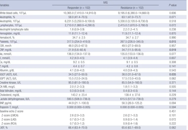
Regarding laboratory parameters, IVIG-resistant individuals were found to have lower platelet levels and higher levels of C-reactive protein (CRP) and AST; these differences were statisti-cally significant (Table 2). There were no other statististatisti-cally sig-Table 1. Baseline characteristics
Variables IVIG P value
Responder (n = 102) Resistance (n = 102) Age, mon 38.5 (23.0–53.0) 39.0 (23.0–51.0) 0.909 Fever*, day 5.0 (3.0–7.0) 5.0 (3.0–5.0) 0.085 Sex 0.782 Male 60 (58.8) 58 (56.9) Female 42 (41.2) 44 (43.1) Recurrent 0.847 No 86 (84.3) 87 (85.3) Yes 16 (15.7) 15 (14.7)
Values are presented as median (range) or number (%). IVIG = intravenous immunoglobulin.
Table 2. Comparison of laboratory variables
Variables IVIG P value
Responder (n = 102) Resistance (n = 102)
White blood cells, 103/μL 10,365.0 (7,410.0–14,910.0) 9,195.0 (6,380.0–14,660.0) 0.606
Neutrophils, % 58.0 (41.4–70.5) 63.1 (47.0–73.7) 0.071
Neutrophils, 103/μL 6,231.5 (3,250.0–9,100.0) 5,330.0 (3,120.0–9,730.0) 0.518 Lymphocytes, 103/μL 2,710.0 (1,860.0–4,290.0) 2,415.0 (1,670.0–3,780.0) 0.072
Neutrophil lymphocyte ratio 1.8 (0.9–3.9) 2.2 (1.2–4.1) 0.238
Hemoglobin, mg/dL 11.8 (11.1–12.4) 11.9 (11.1–12.4) 0.870 Hematocrit, % 34.7 ± 2.5 34.7 ± 2.7 0.990 Platelets, 103/μL 317.5 (254.0–419.0) 267.5 (226.0–345.0) 0.015 ESR, mm/h 48.0 (25.0–67.0) 49.5 (27.0–69.0) 0.957 CRP, mg/dL 21.9 (5.6–60.4) 34.7 (11.5–95.6) 0.019 Na, mmol/L 136.0 (134.0–137.0) 135.0 (133.0–136.0) 0.077 K, mmol/L 4.2 (4.0–4.5) 4.1 (3.9–4.4) 0.607 Ca, mg/dL 9.2 ± 0.5 9.1 ± 0.5 0.308 P, mg/dL 4.4 ± 0.7 4.2 ± 0.8 0.141 Albumin, g/dL 4.1 (3.8–4.2) 4.0 (3.8–4.3) 0.543
SGOT (AST), IU/L 34.5 (27.0–50.0) 38.0 (31.0–67.0) 0.035
SGPT (ALT), IU/L 15.5 (12.0–24.0) 17.5 (13.0–43.0) 0.062
Creatine kinase, U/L 95.0 (61.0–180.0) 85.5 (54.0–160.0) 0.371
CK-MB, mcg/L 2.0 (1.2–3.3) 1.8 (1.1–3.2) 0.505
Total bilirubin, mg/dL 0.3 (0.3–0.4) 0.4 (0.3–0.5) 0.144
Cholesterol, mg/dL 143.2 ± 23.4 138.4 ± 27.6 0.229
Lactate dehydrogenase, IU/L 590.5 (508.0–736.0) 615.5 (517.0–738.0) 0.279
BNP, pg/mL 44.9 (21.1–100.0) 58.3 (26.0–125.2) 0.094
Troponin-T, mcg/L 0.000 (0.000–0.005) 0.000 (0.000–0.005) 0.594
Baseline echo Z-score 0.451
Z-score (LMCA) 2.6 (2.0–3.2) 2.6 (2.1–3.2) 0.101
Z-score (LAD) 0.7 (0.3–1.2) 0.9 (0.5–1.4) 0.072
Z-score (RCA) 0.7 (0.3–1.2) 0.9 (0.4–1.6) 0.222
LVEF, % 66.4 (63.4–70.0) 65.6 (63.1–69.0) 0.062
Data presented as median (range) or mean ± standard deviation, unless otherwise indicated. Bolded values indicate statistically significant differences.
IVIG = intravenous immunoglobulin, ESR = erythrocyte sedimentation rate, CRP = C-reactive protein, SGOT = serum glutamic oxaloacetic transaminase, AST = aspartate ami-notransferase, SGPT = serum glutamic pyruvic transaminase, ALT = alanine amiami-notransferase, CK-MB = creatine kinase myocardial b fraction, BNP = brain natriuretic peptide, LMCA = left main coronary artery, LAD = left anterior descending coronary artery, RCA = right coronary artery, LVEF = left ventricular ejection fraction.
Fig. 1. Jitter plots: comparison for age matching.
Age (month) Group 0 1 350 300 250 200 150 100 50 0 Before matching Age (month) Group 0 1 120 100 80 60 40 20 0 After matching
nificant differences in indicators between the two groups. The Harada, Kobayashi, and Egami scoring systems were ap-plied to the patients in this study, and the proportion of high-risk patients identified with each of the three high-risk scores were compared (Table 3). Among the three risk scores, Kobayashi
showed significant differences between the IVIG-responder and IVIG-resistant groups (P = 0.014).
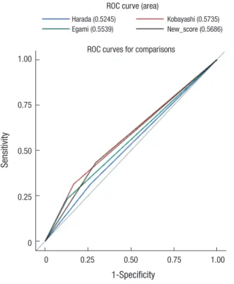
The sensitivity, specificity, positive predictive value (PPV), and negative predictive value (NPV) for each risk score system is shown in Table 4. The sensitivity of each scoring system was low (31.4%, 31.4%, and 23.5%, respectively) and the specificity was high (73.5%, 83.3%, and 87.3%, respectively). Risk scoring systems from Japan typically have good specificity but low sen-sitivity for predicting IVIG resistance in Korean KD patients. In multivariate analysis, platelet counts and CRP levels were selected as independent predictors of IVIG-resistant KD (Table 5). The cut-off values, which were identified by the ROC curve, were applied in a stepwise procedure (Fig. 2).
From the result, a nomogram was derived for personal risk probability of IVIG resistance in KD patients (Fig. 3). The under-lying logistic model is given by the equation:
Probability = 0.5765 + (0.0606) × sex + (−0.0062) × age + (−1.0653) × platelets + (1.5756) × CRP
From this, probabilities for age, sex, platelets, and CRP vari-Table 3. Comparison of scoring systems
Scoring systems IVIG P value Responder (n = 102) Resistance (n = 102) Harada score 0.466 Low risk 75 (73.5) 70 (68.6) High risk 27 (26.5) 32 (31.4) Kobayashi score 0.014 Low risk 85 (83.3) 70 (68.6) High risk 17 (16.7) 32 (31.4) Egami score 0.063 Low risk 89 (87.3) 78 (76.5) High risk 13 (12.8) 24 (23.5)
Data presented as number (%). Bolded value indicates statistically significant differ-ence.
IVIG = intravenous immunoglobulin.
Table 4. Diagnostic performance
Risk scores Sensitivity Specificity PPV NPV Accuracy
Harada 31.4 (23.2–40.9) 73.5 (64.2–81.2) 54.2 (41.7–66.3) 51.7 (43.7–59.7) 52.5 (45.6–59.3) Kobayashi 31.4 (23.2–40.9) 83.3 (74.8–89.4) 65.3 (51.3–77.1) 54.8 (47.0–62.5) 57.4 (50.6–64.1) Egami 23.5 (16.3–32.7) 87.3 (79.3–92.5) 64.9 (48.7–78.2) 53.3 (45.7–60.7) 55.4 (48.6–62.2) Data presented as percentage (range).
PPV = positive predict value, NPV = negative predict value.
Table 5. Multiple conditional logistic regression analysis
Risk factors OR 95% CI P value
Platelets 278 × 103/μL 0.997 0.994–0.999 0.012 CRP 111.4 mg/dL 1.008 1.001–1.014 0.016 OR = odds ratio, CI = confidence interval, CRP = C-reactive protein.
Fig. 2. ROC curve and area under the curve comparison. ROC = receiver operating characteristic.
Sensitivity 1-Specificity 0 0.25 0.50 0.75 1.00 1.00 0.75 0.50 0.25 0
ROC curves for comparisons ROC curve (area) Harada (0.5245)
Egami (0.5539)
Kobayashi (0.5735) New_score (0.5686)
Fig. 3. Nomograms predicting the probability of IVIG resistance in patients with KD. IVIG = intravenous immunoglobulin, KD = Kawasaki disease, Plt = platelets, CRP = C-reactive protein, mo = months, k = 103/μL.
Points Sex Age_mo Plt_k CRP Total points Probability 0 10 20 30 40 50 60 70 80 90 100 130 100 70 50 30 10 0 20 40 60 80 100 140 180 220 0.2 0.3 0.4 0.5 0.6 0.7 0.8 0.9 1 1 0 0 2 1
ables can be estimated.
For example, if a 100-month-old female KD patient with a platelet count of 250,000/μL and CRP level of 120 mg/dL, female is linked to sex 2 and converted to 5 points and 100 months would be altered to 12.5 points. Moreover, platelets would be convert-ed into 67.5 points, and CRP was given 100 points. The total point axis in Fig. 3 can reach a maximum of 185 points, with a predict-ed probability range of approximately 0.84.
DISCUSSION
In Japan, Harada (6), Kobayashi et al. (7), and Egami et al. (8) proposed scoring systems for predicting IVIG non-responders using various parameters, which is well matched in Japanese patients with complete KD. However, in a previous study, Sleep-er et al. (13) concluded that these Japanese scoring systems wSleep-ere not suitable for the North American population. A random co-hort study in a United States population found a sensitivity of 42% and a specificity of 85% for the Egami score, and a sensitiv-ity of 42% and specificsensitiv-ity of 87% for the Kobayashi score. Davies et al. (14) used the Kobayashi scoring system for children with KD from the United Kingdom, and found a sensitivity of 58% and a specificity of 35%. These results indicate that the Kobayas-hi scoring system is not useful in the British population. Fu et al. (15) predicted IVIG resistance in Chinese children with KD, and found a sensitivity of 21% and specificity of 87% for Egami scores, and a sensitivity of 49% and sensitivity of 72% for the Ko-bayashi score. Based on these findings, Japanese KD scoring systems were considered to have limited predictive value for IVIG response in other countries. Korea is home to the second largest KD population in the world; therefore, we have evaluat-ed whether these scoring systems would be suitable there. According to a recent nationwide survey in Korea in 2011, the 11.6% of patients did not respond to initial IVIG treatment and coronary artery dilation was noted in 16.4% of patients, based on the Japanese Ministry of Health and Welfare criteria (16). De-spite the timely administration of IVIG, 10% to 20% of patients do not respond to treatment. These refractory patients are at in-creased risk of developing coronary artery complications. It is very important to identify high-risk patients for protecting against ongoing coronary arteriopathy. The significance of coronary outcomes emphasizes the importance of early prediction and timely optimal additional treatment of IVIG-resistant patients. Previous studies have reported risk factors for refractory KD, which include high neutrophil count; low hemoglobin; throm-bocytopenia; low albumin; high CRP; high lactate dehydroge-nase; hyponatremia; elevated AST; abnormal findings in initial echocardiography; and recurrent KD (17-19). Most risk factors are related to severe inflammatory reactions and are not sup-pressed by IVIG alone. In IVIG-resistance, fever represents on-going inflammatory reactions in vessels and may reflect defects
in the control of immunological stimuli (20).
Previous reports to predict IVIG resistance KD have mostly excluded incomplete KD. However, our study included patients with high rates of incomplete KD. This may have been a factor in determining whether previous studies were appropriate for our patients.
Our study tested the utility of current risk scores in Korean patients with KD. They have good specificity but low sensitivity for predicting IVIG resistance. The scoring system suggested by Kobayashi et al. (7) can help predict IVIG-resistant KD and lead to timely management and better clinical outcomes. In addi-tion, from the results of our study, we identified a few indepen-dent predictive parameters for refractory KD — platelets and CRP level — that suggest a more focused importance of vascu-lar inflammatory reactions in the disease process.
This study had some limitations, the first of which was its ret-rospective, single-center design; consequently, we could not obtain sufficient data regarding clinical characteristics and lab-oratory findings. Second, many patients were excluded because of a lack of adequate data or results in age matching. The inci-dence of coronary complications is highly reported in extreme age group, but in our study, there is a limit due to age matching. Third, we could not exclude patients with comorbidities from other febrile illnesses.
In conclusion, Korean KD patients may need an exclusive scoring system. The result of our study may be helpful in select-ing patients who are at high-risk for IVIG resistance or coronary artery complications. Further multicenter studies are needed to develop a better and more comprehensive evaluation system to provide individualized treatment planning.
DISCLOSURE
The authors have no potential conflicts of interest to disclose.
AUTHOR CONTRIBUTION
Conceptualization: Shin J, Eun L. Data curation: Shin J, Lee H, Eun L. Writing - original draft: Shin J, Eun L. Writing - review & editing: Eun L.
ORCID
Jaeeun Shin https://orcid.org/0000-0003-3089-9347 Heeyoung Lee https://orcid.org/0000-0002-8294-0914 Lucy Eun https://orcid.org/0000-0002-4577-3168
REFERENCES
1. Burns JC, Glodé MP. Kawasaki syndrome. Lancet 2004; 364: 533-44.
M, Harada K; Kawasaki Disease Research Committee. Revision of diag-nostic guidelines for Kawasaki disease (the 5th revised edition). Pediatr Int 2005; 47: 232-4.
3. JCS Joint Working Group. Guidelines for diagnosis and management of cardiovascular sequelae in Kawasaki disease (JCS 2013). Digest version.
Circ J 2014; 78: 2521-62.
4. Cimaz R, Sundel R. Atypical and incomplete Kawasaki disease. Best Pract Res Clin Rheumatol 2009; 23: 689-97.
5. Kibata T, Suzuki Y, Hasegawa S, Matsushige T, Kusuda T, Hoshide M, Taka-hashi K, Okada S, Wakiguchi H, Moriwake T, et al. Coronary artery lesions and the increasing incidence of Kawasaki disease resistant to initial im-munoglobulin. Int J Cardiol 2016; 214: 209-15.
6. Harada K. Intravenous gamma-globulin treatment in Kawasaki disease.
Acta Paediatr Jpn 1991; 33: 805-10.
7. Kobayashi T, Inoue Y, Takeuchi K, Okada Y, Tamura K, Tomomasa T, Ko-bayashi T, Morikawa A. Prediction of intravenous immunoglobulin unre-sponsiveness in patients with Kawasaki disease. Circulation 2006; 113:
2606-12.
8. Egami K, Muta H, Ishii M, Suda K, Sugahara Y, Iemura M, Matsuishi T. Prediction of resistance to intravenous immunoglobulin treatment in pa-tients with Kawasaki disease. J Pediatr 2006; 149: 237-40.
9. Kim BY, Kim D, Kim YH, Ryoo E, Sun YH, Jeon IS, Jung MJ, Cho HK, Tchah H, Choi DY, et al. Non-responders to intravenous immunoglobulin and coronary artery dilatation in Kawasaki disease: predictive parameters in Korean children. Korean Circ J 2016; 46: 542-9.
10. Park HM, Lee DW, Hyun MC, Lee SB. Predictors of nonresponse to intra-venous immunoglobulin therapy in Kawasaki disease. Korean J Pediatr
2013; 56: 75-9.
11. Haycock GB, Schwartz GJ, Wisotsky DH. Geometric method for measur-ing body surface area: a height-weight formula validated in infants, chil-dren, and adults. J Pediatr 1978; 93: 62-6.
12. Manlhiot C, Millar K, Golding F, McCrindle BW. Improved classification of coronary artery abnormalities based only on coronary artery z-scores after Kawasaki disease. Pediatr Cardiol 2010; 31: 242-9.
13. Sleeper LA, Minich LL, McCrindle BM, Li JS, Mason W, Colan SD, Atz AM, Printz BF, Baker A, Vetter VL, et al. Evaluation of Kawasaki disease risk-scoring systems for intravenous immunoglobulin resistance. J Pediatr
2011; 158: 831-835.e3.
14. Davies S, Sutton N, Blackstock S, Gormley S, Hoggart CJ, Levin M, Herberg JA. Predicting IVIG resistance in UK Kawasaki disease. Arch Dis Child
2015; 100: 366-8.
15. Fu PP, Du ZD, Pan YS. Novel predictors of intravenous immunoglobulin resistance in Chinese children with Kawasaki disease. Pediatr Infect Dis J
2013; 32: e319-23.
16. Kim GB, Han JW, Park YW, Song MS, Hong YM, Cha SH, Kim DS, Park S. Epidemiologic features of Kawasaki disease in South Korea: data from nationwide survey, 2009–2011. Pediatr Infect Dis J 2014; 33: 24-7.
17. Sato S, Kawashima H, Kashiwagi Y, Hoshika A. Inflammatory cytokines as predictors of resistance to intravenous immunoglobulin therapy in Ka-wasaki disease patients. Int J Rheum Dis 2013; 16: 168-72.
18. Sano T, Kurotobi S, Matsuzaki K, Yamamoto T, Maki I, Miki K, Kogaki S, Hara J. Prediction of non-responsiveness to standard high-dose gamma-globulin therapy in patients with acute Kawasaki disease before starting initial treatment. Eur J Pediatr 2007; 166: 131-7.
19. Tremoulet AH, Best BM, Song S, Wang S, Corinaldesi E, Eichenfield JR, Martin DD, Newburger JW, Burns JC. Resistance to intravenous immu-noglobulin in children with Kawasaki disease. J Pediatr 2008; 153:
117-21.
20. Matsubara T, Ichiyama T, Furukawa S. Immunological profile of periph-eral blood lymphocytes and monocytes/macrophages in Kawasaki dis-ease. Clin Exp Immunol 2005; 141: 381-7.