doi:10.3748/wjg.v20.i14.3938 © 2014 Baishideng Publishing Group Co., Limited. All rights reserved.
Endoscopic submucosal dissection for undifferentiated-type
early gastric cancer: Do we have enough data to support
this?
Choong Nam Shim, Sang Kil Lee
Choong Nam Shim, Sang Kil Lee, Department of Internal Medicine, Institute of Gastroenterology, Yonsei University Col-lege of Medicine, Seoul 120-752, South Korea
Author contributions: Shim CN contributed to study concept and design, data collection, analysis, and drafting of the manu-script; Lee SK contributed to study concept and design, critical revision of the manuscript, and study supervision.
Correspondence to: Sang Kil Lee, MD, PhD, Department of Internal Medicine, Institute of Gastroenterology, Yonsei Univer-sity College of Medicine, 134 Shinchon-dong, Seodaemun-gu, Seoul 120-752, South Korea. [email protected]
Telephone: +82-2-2228996 Fax: +82-2-3936884 Received: October 27, 2013 Revised: January 18, 2014 Accepted: February 17, 2014
Published online: April 14, 2014
Abstract
Although endoscopic submucosal dissection (ESD) is
now accepted for treatment of early gastric cancers
(EGC) with negligible risk of lymph node (LN)
metas-tasis, ESD for intramucosal undifferentiated type EGC
without ulceration and with diameter ≤ 2 cm is
re-garded as an investigational treatment according to the
Japanese gastric cancer treatment guidelines. This
con-sideration was largely based on the analysis of
surgi-cally resected EGCs that contained undifferentiated type
EGCs; however, results from several institutes showed
some discrepancies in sample size and incidence of LN
metastasis. Recently, some reports about the safety and
efficacy of ESD for undifferentiated type EGC meeting
the expanded criteria have been published.
Nonethe-less, only limited data are available regarding long-term
outcomes of ESD for EGC with undifferentiated
histolo-gy so far. At the same time, endoscopists cannot ignore
the patients’ desire to guarantee quality of life after
the relatively non-invasive endoscopic treatment when
compared to conventional surgery. To satisfy the needs
WJG 20
thAnniversary Special Issues (8): Gastric cancer
TOPIC HIGHLIGHT
of patients and provide solid evidence to support ESD
for undifferentiated EGC, we need more delicate tools
to predict undetected LN metastasis and more data that
can reveal predictive factors for LN metastasis.
© 2014 Baishideng Publishing Group Co., Limited. All rights reserved.
Key words: Early gastric cancer; Endoscopic
submuco-sal dissection; Undifferentiated histology; Indications
Core tip: Endoscopic submucosal dissection (ESD) for
intramucosal undifferentiated (UD) type early gastric
cancer (EGC) without ulceration and with diameter
≤ 2 cm is regarded as an investigational treatment
according to the Japanese gastric cancer treatment
guidelines. In contrast, the controversial results about
the safety of ESD for UD-EGC fulfilling the criteria have
been reported and a little is known about the long-term
outcomes. Therefore, in this review, we focused on the
safety and therapeutic efficacy of ESD for UD-EGC with
reference to risks for lymph node metastasis within the
proposed criteria as well as the short-term and
long-term outcomes of ESD for UD-EGC.
Shim CN, Lee SK. Endoscopic submucosal dissection for undif-ferentiated-type early gastric cancer: Do we have enough data to support this? World J Gastroenterol 2014; 20(14): 3938-3949 Available from: URL: http://www.wjgnet.com/1007-9327/full/ v20/i14/3938.htm DOI: http://dx.doi.org/10.3748/wjg.v20. i14.3938
INTRODUCTION
Early gastric cancer (EGC) is defined as gastric cancer
that is confined to the mucosa or submucosa, irrespective
of the presence of regional lymph node (LN)
metasta-ses
[1]. In the Eastern hemisphere, up to 70% of all gastric
cancers are diagnosed as EGCs (due to mass population
screening)
[2-4], whereas in the Western hemisphere, the
rate of gastric cancers identified as EGCs accounts for
only about 15%
[5,6]. EGC reveals a favorable prognosis
compared with advanced gastric cancer, with 5-year
sur-vival rates being in excess of 90% to 95%, based on
Ko-rea, Japan, and European data
[7-13].
In Eastern countries, endoscopic resection (ER),
including endoscopic mucosal resection (EMR) and
en-doscopic submucosal dissection (ESD), has been widely
accepted as a minimally invasive treatment for EGC with
a negligible risk of LN metastasis
[14-18]. Recently,
consid-erable data have also been reported from the Western
world as ER is gaining wide acceptance
[19-21]. Tumors
in-dicated for ER as a standard treatment are
differentiated-type adenocarcinomas without ulceration, of which the
depth of invasion is clinically diagnosed as mucosal layer
and the diameter is
≤
20 mm
[22]. Gotoda
et al
[23]studied
surgically resected specimens of EGC and suggested the
following four expanded indication criteria for
endoscop-ic treatment of EGC without LN metastasis: (1)
differen-tiated intramucosal cancer without ulceration, regardless
of size; (2) differentiated intramucosal cancer with
ulcer-ation and diameter
≤
30 mm; (3) differentiated minute
submucosal penetrative cancer in diameter
≤
30 mm;
and (4) undifferentiated (UD) type intramucosal cancer
without ulceration and diameter
≤
20 mm. In particular,
surgery was still considered in the UD-EGC meeting the
expanded criteria because endoscopic
en-bloc removal was
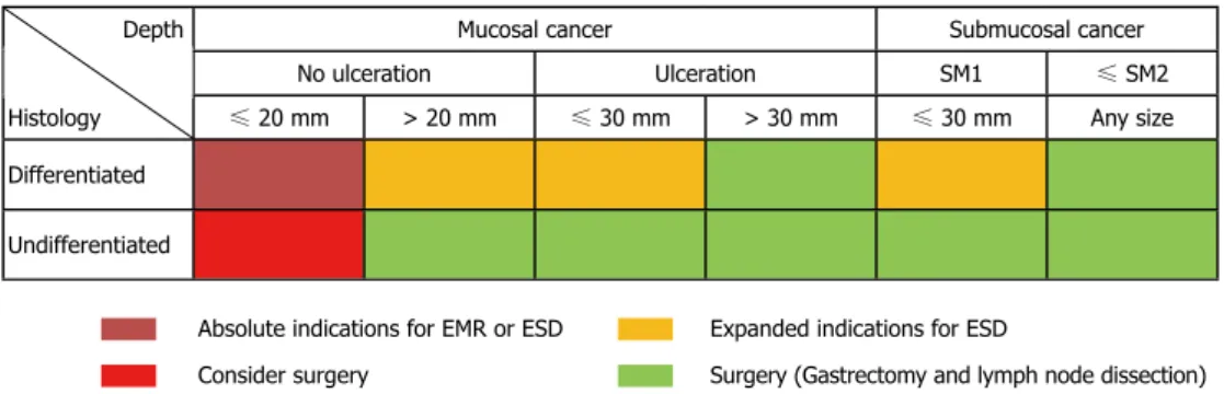
sometimes difficult in this type of tumors (Figure 1)
[24,25].
However, Hirasawa
et al
[26]added to the body of evidence
that there is no LN metastasis in patients with
UD-EGC within the expanded criteria. This study revealed
the 95%CI of the calculated risk of metastasis to nodes
was 0%-0.96%, while the earlier study by Gotoda
et al
[23]showed that of risk was 0%-2.6% due to small sample
size (
n = 141), which may potentially be inferior to the
outcomes of surgical resection.
Along these lines, the Japanese gastric cancer
treat-ment guidelines (2010, ver. 3) state that ER for these
UD-EGCs is regarded as an investigational treatment, and
that ESD, not EMR, should be employed. In contrast,
clinical practice guidelines, according to both National
Comprehensive Cancer Network
[27]and European
Soci-ety for Medical Oncology
[28], do not yet recognize ER for
EGCs meeting the expanded criteria as safe. Moreover,
the controversial results about the safety of ESD for
UD-EGC fulfilling the criteria have been reported and a
little is known about the long-term outcomes. Therefore,
in this review, we focused on the safety and therapeutic
efficacy of ESD for UD-EGC with reference to risks for
LN metastasis within the proposed criteria as well as the
short-term and long-term outcomes of it.
PREOPERATIVE ASSESSMENT OF LN
METASTASIS
The most important factor concerning endoscopic
treat-ment with curative intent is the prediction of regional LN
metastasis before treatment
[22,27,28]. Reported rates of LN
metastasis in EGC range from 5.7% to 20% based on
the analysis of surgically resected specimen of EGC
[29-34].
UD-EGC demonstrates 4.2% to 4.9% and 19.0% to
23.8%of LN metastasis in the mucosal and submucosal
invasive tumors, respectively
[23,26]. To date, no imaging
modality has been proven to be consistently accurate in
assessing LN metastasis in EGC
[35,36]. Endoscopic
ultra-sound (EUS) is one of most studied procedures for the
locoregional staging of gastric cancer. Reported
sensitivi-ties and specificisensitivi-ties of EUS to detect LN metastases
in gastric cancers varied widely, between 16.7% and
95.3%, and between 48.4% and 100%, respectively
[35].
EUS demonstrated a moderate accuracy that seems to
describe advanced T stage (T3 and T4) better than N or
less advanced T stage
[37,38]. Although a clinically relevant
benefit of EUS to distinguish intramucosal lesions from
submucosal lesions should be further improved
[39], EUS
is an important imaging modality for preoperative
as-sessment to exclude LN metastasis as well as to confirm
deeper wall invasion including the proper muscle layer.
Nevertheless, we should consider that UD histology
would cause under-diagnosis and affect the accuracy of
EUS compared to the differentiated histology
[40].
In addition to the diagnostic role of magnifying
en-doscopy with narrow-band imaging (ME-NBI) for
de-termining tumor margin in EGC
[41,42], ME-NBI has been
suggested as a supporting tool for the assessment of
Depth Mucosal cancer Submucosal cancer
No ulceration Ulceration SM1 ≤ SM2
Histology ≤ 20 mm > 20 mm ≤ 30 mm > 30 mm ≤ 30 mm Any size
Differentiated Undifferentiated
Absolute indications for EMR or ESD Expanded indications for ESD
Consider surgery Surgery (Gastrectomy and lymph node dissection)
Figure 1 Absolute and expanded indication for endoscopic mucosal resection and endoscopic submucosal dissection for early gastric cancer. SM1:
Tu-mor invasion into the upper third of the submucosa (≤ 500 μm); SM2: Tumor invasion into the mid-third of the submucosa (> 500 μm). EMR: Endoscopic mucosal resection; ESD: Endoscopic submucosal dissection.
invasion depth in EGC
[43-46]. In contrast to the usefulness
of ME-NBI for evaluating invasive depth in esophageal
or colon cancer
[47,48], the utility of ME-NBI for
determin-ing invasion depth in EGC is not conclusive, because the
invasive tumor is often not exposed at the surface and the
mucosal structure remains, even when cancer invades the
submucosa. Therefore, it is difficult to estimate reliably
the depth of invasion by surface appearance
[49]. ME-NBI
should also distinguish findings suggestive of
submuco-sal invasion from those indicative of the UD histologic
type
[44,45]. The findings of a nonstructural pattern in the
neoplastic lesion of the stomach on ME
[45]or no surface
pattern and sparse microvessels (markedly distorted,
isolated, heterogeneous) or with avascular areas on
ME-NBI
[44]are indicative of undifferentiated type
adenocarci-noma or differentiated cancer with deep submucosal
inva-sion. In contrast, ME-NBI images of UD-EGC were very
closely related to the histopathological findings in other
study
[50], and therefore, this imaging tool can be useful
in the pretreatment assessment of the histopathological
patterns of cancer development and the lateral extent of
UD-EGC. Thus, the role of ME-NBI in differentiation
of histologic types in addition to invasive depth should be
validated through further prospective studies.
Other imaging modalities including abdominal
ul-trasound (AUS), computed tomography (CT), magnetic
resonance imaging (MRI), and positron emission
tomog-raphy (PET) achieved limited success to stage
preopera-tive LN status
[35,36]. A meta-analysis by Seevaratnam
et al
[36]showed that imaging modalities range in overall accuracy
from 53.4% (MRI) to 68.1% (AUS), in sensitivity from
40.3% (PET) to 85.3% (MRI), and in specificity from
75.0% (MRI) to 97.7% (PET), with no significant
differ-ences between modalities. To date, there are no clinically
relevant imaging tools to detect the submucosal invasion
and the LN metastasis in EGC that are critical conditions
for determining proper candidates for ER.
RISK FACTORS FOR LN METASTASIS
AND PROPOSED CRITERIA FOR ESD
Because currently available imaging modalities fail to
curately evaluate nodal status, endoscopic resectability
ac-cording to nodal status in EGC and subsequent curability
are still determined by means of the presence or absence
of certain tumor characteristics which were obtained
from the analysis of surgically resected EGC. According
to the Japanese gastric cancer treatment guidelines
[22], the
main risk factors predictive of LN metastasis in EGC
are histologic type, depth of invasion, ulceration, size,
and lymphovascular invasion largely based on two
large-scale datasets
[23,26]. These factors consist of absolute and
expanded indications as well as curability of ER with
en
bloc resection and negative lateral/vertical margin (Table
1)
[22,51]. A meta-analysis by Kwee
et al
[52]identified the
characteristics related to LN metastasis in EGC, including
age, gender, location, size, macroscopic type, ulceration,
histologic type in accordance with Japanese and Lauren
classification, lymphovascular invasion, submucosal
vas-cularity, a proliferating cell nuclear antigen labeling index,
a matrix metalloproteinase-9-positivity, a gastric mucin
phenotype, and a vascular endothelial growth
factor-C-positivity. These factors revealed partially different
cor-relations with LN metastasis in intramucosal and
submu-cosal EGCs, respectively.
With regard to LN metastasis particularly in
UD-EGC, many recent studies investigated the risk factors
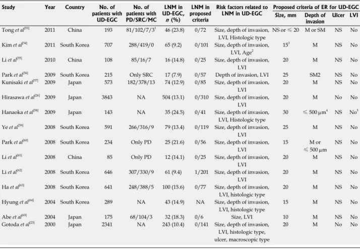
and suggested their criteria for ER of UD-EGC (Table
2)
[23,26,53-65]. The overall rates of LN metastasis in
UD-EGC varied from 7.9% to 24.5%; however, the
hetero-geneous composition in subtypes of UD histology in
lesions from 15 studies should be taken into account.
Size of lesion
Although the intramucosal lesions without ulceration and
diameter
≤
20 mm have been considered as rational
cri-teria for ESD in UD-EGC by Japanese researchers
[23,26],
different ER criteria have also been suggested with
vari-ous standards in size, depth of invasion, and presence of
ulcerative finding
[53-65]. Concerning lesion size, a majority
of recent studies (11/15, 73.3%) suggested that a
diam-eter of 20 mm to 30 mm would be the upper limit of the
size criterion for UD-EGC to be amenable to treatment
with ESD; however, the remaining four studies proposed
a diameter of 10 or 15 mm as the upper limit of the
cri-terion, based on their results suggesting the possibility of
LN metastasis even in smaller UD-EGC
[54,60,64,65]. Debates
over the size criterion were highlighted by several reports
of LN metastasis of UD-EGCs within the expanded
criteria, including a diameter
≤
20 mm
[31,66-70]. Moreover,
the size discrepancy between pathologic size and
en-doscopic size should be resolved, because we can only
determine the indications of ER based on the
endoscopi-cally estimated size. While a previous study revealed that
endoscopic visual estimation method was found to show
Table 1 Curability for endoscopic resection of early gastriccancer Curability criteria
Curative resection
En bloc resection, no lateral and vertical margin positivity, no lymphovascular invasion
Intramucosal cancer, differentiated histology, size ≤ 20 mm, No ulcerative finding
Curative resection for expanded indications
En bloc resection, no lateral and vertical margin positivity, no lymphovascular invasion
Intramucosal cancer, differentiated histology, size > 20 mm, no ulcerative finding
Intramucosal cancer, differentiated histology, size ≤ 30 mm, presence of ulcerative finding
SM 1 depth of invasion, differentiated histology, size ≤ 30 mm, no ulcerative finding
Intramucosal cancer, undifferentiated histology, size ≤ 20 mm, no ulcerative finding
Non-curative resection
Any resection that does not satisfy one of the above criteria
for ER, were relatively small compared with other
stud-ies. More importantly, the majority of recent studies
reported the LN metastasis in a depth of submucosal
invasion
[23,26,54,55,57,59,61-65].
Ulceration
Ulceration within the lesion is the representative finding
with heterogeneity. More than moderate heterogeneity
was identified at previous meta-analysis with possible
ex-planation for this heterogeneity due to the interobserver
variability between studies for the assessment of tumor
ulcerations
[52]. Furthermore, this may be due to the
differ-ent definitions in addition to the interobserver variability
for the assessment of ulcerations
[52,67,75,80]. Though most
of the recent studies (13/15, 86.7%) did not consider the
ulcer finding in their proposed criteria, patients with
tu-mor ulcerations had a significantly higher risk of LN
me-tastasis in intramucosal EGC irrespective of histological
type at meta-analysis
[52]. And ulcerous change decreases
the accuracy of EUS diagnosis for the invasive depth
of EGC
[81]. Therefore, we do not consider ER for
UD-EGCs with ulceration as safe.
reliable agreement with pathologic measurements in
EGC treated with ER
[71], other earlier ESD series showed
the mean size discrepancies ranged from 5.8 mm to 6.8
mm, which are not negligible in ER for EGC
[72,73]. In
UD-EGC, the margins of the lesion tend to be obscured
compared to the differentiated histology, which was
found to cause frequent margin failure of ESD in our
previous report
[74]. Thus, a standard reliable measurement
method is required through further prospective studies
[75].
Submucosal invasion
Some studies suggest that a shallow submucosal
inva-sion is an acceptable depth of invainva-sion in ESD for
UD-EGC
[53,56,58,60]. However, this suggestion should be
reserved until EUS is more reliable for determination of
invasive depth, because there is a high chance of
endo-sonographically underestimated depth of invasion and
subsequently higher vertical margin positivity in
poorly-differentiated EGC
[40,74], in addition to the difficult
assess-ment of depth of invasion in UD-EGC
[76-79].
Addition-ally, the numbers of enrolled UD-EGCs in these studies,
suggesting a minute submucosal invasion as a criterion
Table 2 Proposed criteria for endoscopic resection of undifferentiated type early gastric cancer Study Year Country No. of
patients with UD-EGC No. of patients with PD/SRC/MC LNM in UD-EGC, n (%) LNM in proposed criteria
Risk factors related to
LNM in UD-EGC Proposed criteria of ER for UD-EGCSize, mm Depth of invasion Ulcer LVI
Tong et al[53] 2011 China 193 81/102/7/31 46 (23.8) 0/72 Size, depth of invasion,
LVI, Histologic type
NS or ≤ 20 M or SM NS No
Kim et al[54] 2011 South Korea 707 288/419/0 65 (9.2) 0/101 Size, depth of invasion,
LVI, Age2
153 M NS No
Li et al[55] 2010 China 108 85/16/7 16 (14.8) 0/25 Size, depth of invasion,
LVI
20 M NS No
Park et al[56] 2009 South Korea 215 Only SRC 17 (7.9) 0/57 Depth of invasion, LVI 25 SM2 NS No
Kunisaki et al[57] 2009 Japan 573 182/378/13 74 (12.9) 0/85 Size, depth of invasion,
LVI
20 M NS No
Hirasawa et al[26] 2009 Japan 3843 NA 504 (13.1) 0/310 Size, depth of invasion,
LVI
20 M No No
Hanaoka et al[58] 2009 Japan 143 NA 35 (24.5) 0/41 Size, depth of invasion,
LVI, Histologic type
30 ≤ 500 μm4 NS No5
Ye et al[59] 2008 South Korea 591 266/316/9 79 (13.4) 0/119 Size, depth of invasion,
LVI
25 M NS No
Park et al[60] 2008 South Korea 234 Only PD 25 (21.6) 0/56 Size, depth of invasion,
LVI
15 M or
≤ 500 μm
NS No
Li et al[61] 2008 China 85 Only PD 12 (14.1) 0/25 Size, depth of invasion,
LVI
20 M NS No
Li et al[62] 2008 South Korea 646 307/330/9 61 (9.4) 1/201 Size, depth of invasion,
LVI
20 M NS No
Ha et al[63] 2008 South Korea 641 248/388/5 100 (15.6) 0/77 Size, depth of invasion,
LVI, histologic type
20 M NS No
Hyung et al[64] 2004 South Korea 289 NA 43 (14.9) NA Size, depth of invasion,
LVI, histologic type
15 M NS No
Abe et al[65] 2004 Japan 175 68/104/3 32 (18.3) 0/6 Size, LVI 10 M NS No
Gotoda et al[23] 2000 Japan 2341 NA 243 (10.4) 0/141 Size, depth of invasion,
LVI, histologic type, ulcer, macroscopic type
20 M No No
1Three patients had EGCs with histology of undifferentiated adenocarcinoma; 2Young age less than 45 years was related to the lymph node metastasis of
only poorly-differentiated carcinoma; 3Size criteria were ≤ 25 mm in poorly-differentiated adenocarcinomas and ≤ 15 mm in signet-ring cell carcinomas,
respectively; 4The depth of invasion in proposed criteria was ≤ 500 μm or no more from the lower margin of the muscularis mucosae; 5Hanaoka et al also
suggested the proportion of undifferentiated components < 50% as one of criteria. UD-EGC: Undifferentiated type early gastric cancer; PD: Poorly-differen-tiated adenocarcinoma; SRC: Signet-ring cell carcinoma; MC: Mucinous carcinoma; LNM: Lymph node metastasis; ER: Endoscopic resection; LVI: Lympho-vascular invasion; M: Mucosa; SM: Submucosa; NS: Not significant; NA: Not available.
Lymphovascular invasion
Only the absence of lymphovascular invasion was the
cri-terion included by all studies, which was consistent with
the results of a meta-analysis revealing that lymphatic
tumor invasion is the strongest predictor for LN
metas-tasis in both mucosal and submucosal gastric cancer
[52].
For this reason, EGCs with lymphovascular invasion in
endoscopically resected specimen should be treated by
further surgery
[22]. However, the Japanese gastric
can-cer treatment guidelines are not based on the status of
lymphovascular invasion. The lymphovascular invasion
is involved in the decision of curability of ER, since its
evaluation can only be available in specimens obtained
by ER. Moreover, the determination of lymphovascular
invasion sometimes lacks objectivity possibly because of
the inability to distinguish lymphatics from blood vessels
on conventional hematoxylin-eosin staining
[82]. Several
studies suggested an endoscopic elevated macroscopic
type
[83]and a stromal cell-derived factor-1α as risk factors
of lymphovascular invasion
[84]with reports of usefulness
of immunohistochemical staining for detection
[82,85,86].
Considering the importance of lymphovascular invasion
for prediction of LN metastasis, prospective studies of
preoperative prediction for lymphovascular invasion are
warranted.
CLINICAL CHARACTERISTICS
Clinical characteristics of recent representative studies on
ER for UD-EGC are summarized in Table 3
[73,74,76,80,87-90].
All eight studies were analyzed retrospectively. The
num-bers of lesions ranged from 46 to 103 lesions and were
not large enough to elicit conclusive results. Six studies
performed solely ESD
[73,76,80,87,89,90]and the rest carried
out both EMR and ESD
[74,88]. Inclusion criteria of these
studies were based on the expanded criteria except those
of two studies by Kim
et al
[80]and Kang
et al
[73]. The study
by Kim
et al
[80]included patients who refused surgery and
were treated by ESD as an experimental treatment. The
study by Kang
et al
[73]included patients with UD-EGC
with ulceration. Submucosal invasion and ulcers were
noted in 9.7%-19.6% and 1.0%-9.3% of lesions satisfying
the expanded criteria, respectively. The two studies that
included patients who refused surgery and lesions with
ulcerations in endoscopic finding showed relatively high
submucosal invasion and ulceration rates. The inaccurate
endoscopic size estimation in UD-EGCs is well noted in
the studies, because the lesions with size > 20 mm were
noted in up to 45.5% of lesions
[88]. Particularly, the study
including intramucosal UD-EGC with size
≤
20 mm
regardless of ulcerations revealed notably higher SM
in-vasion (28.3%), ulcer finding (28.3%), and size > 20 mm
(51.7%) rates
[73]. The overall inaccuracies of assessment
of depth of invasion, ulcerative findings, and size of
UD-EGC tumors fulfilling the expanded criteria are not
negligible, and thus ESD criteria based on endoscopic
and histologic findings in UD-EGC should have more
re-strictions compared to differentiated EGC. To overcome
this limitation, new methods beyond the current level of
technology are strongly needed.
SHORT-TERM OUTCOMES
In addition to a very low possibility of LN metastasis, the
safety of ESD for UD-EGC can be established based
on the feasibility of curative resection with acceptable
complication rates and consequently favorable long-term
outcomes.
Short-term outcomes, including
en bloc resection,
com-plete resection, curative resection, and complication rates,
of ER for UD-EGC are listed in Table 4
[73,74,76,80,87-90].
Whereas homogeneous definitions of
en bloc resection
applied for the studies, the definitions of complete
resec-tion category were heterogeneous depending on the
in-volvement of
en bloc resection or lymphovascular invasion
or submucosal invasion
[73,74,80,87,90]. Additionally, the
defini-tions of curative resection in some studies did not clarify
the involvement of
en bloc resection
[80,89,90]. The overall
rates of
en bloc resection, complete resection, and curative
resection of ER for UD-EGCs varied from 83.1% to
100%, from 55.0% to 90.7%, and from 31.1% to 82.5%,
respectively, while those of ESD for UD-EGCs
meet-ing the expanded criteria ranged from 91.3% to 99.0%,
from 89.7% to 90.7%, and from 63.9% to 82.5%,
respec-Table 3 Clinical characteristics of representative studies on endoscopic resection for undifferentiated type early gastric cancer Study Year Country No. of patientswith UD-EGC No. of patients with PD/SRC Age (yr)
1 Sex (male) SM invasion Ulcer Size (mm)1 Size > 20 mm Kim et al[80] 2013 South Korea 74 55/19 61.8 ± 12.0 40 (54.1) 16 (21.6) 11 (14.9) 19.9 ± 12.5 36 (48.6)
Abe et al[87] 2013 Japan 97 18/77/22 62.0 (35.0-88.0)3 55 (56.7) 19 (19.6) 9 (9.3) 12.03 14 (14.4)
Park et al[88] 2012 South Korea 77 47/154 60.9 (33.0-82.0) 49 (63.6) 12 (15.6) 4 (5.2) 23.3 ± 14.0 35 (45.5)
Okada et al[89] 2012 Japan 1035 12/91 59.0 (34.0-91.0) 48 (46.6) 10 (9.7) 1 (1.0) 8.0 (1.0-33.0)3 NA
Kamada et al[76] 2012 Japan 46 NA 65.5 (29.0-90.0) 24 (52.2) 7 (15.2) 1 (2.2) NA 8 (17.4)
Yamamoto et al[90] 2010 Japan 58 48/10 64.0 (33.0-81.0) 31 (53.4) 7 (12.1) 2 (3.4) 11.0 (2.0-28.0) 5 (8.6)
Kang et al[73] 2010 South Korea 60 30/30 56.7 ± 10.4 31 (51.7) 17 (28.3) 17 (28.3) 26.3 ± 12.9 31 (51.7)
Kim et al[74] 2009 South Korea 58 17/41 55.0 (26.0-81.0) 26 (44.8) NA 0 (0) 13.3 ± 6.5 4 (6.9)
Data are expressed as absolute numbers (percentage) or mean ± SD. 1Data are expressed as mean with standard deviation or range; 2Two patients had EGCs
with histology of moderately to poorly differentiated adenocarcinoma; 3Data are expressed as median with or without range; 4Fifteen patients had EGCs
with mixed type histology; 5A total of 103 EGCs in 101 patients were enrolled. UD-EGC: Undifferentiated typr early gastric cancer; PD:
tively
[76,87,89,90]. The results of ESD for cases within the
expanded criteria were comparable with the outcomes
of ESD for differentiated EGCs fulfilling the criteria
of 93.0% to 95.7% and 81.0% to 91.1% for
en bloc and
complete resection rates, respectively
[91-93]. In contrast, the
curative resection rate seems to be lower than that of
dif-ferentiated EGCs, which is 91.1%
[93]. This may arise from
less accurate endoscopic size estimation in UD-EGC due
to an ill-defined margin of tumor infiltration
[41,94,95]and
several distinct features of UD-EGC, including a larger
size and submucosal infiltration that can lead to higher
rates of lymphovascular invasion
[73,82,90,96-98], compared
with EGCs with differentiated histology. Therefore, the
achievement of reasonable curative resection rate in ESD
for UD-EGC is critical by means of more precisely
de-fining of curable lesions.
Further surgical treatments were performed in 26.2%
to 60.0% of patients with incomplete or non-curative
ER. The presence of residual tumor and LN metastasis
in surgical specimens after incomplete or non-curative
ER were detected in 4.8% to 44.4% and 0% to 13.3%
of cases. The overall rates of bleeding and perforation
varied from 1.4% to 13.8% and from 1.0% to 4.3%,
respectively, whereas those of ESD for UD-EGCs
meet-ing the expanded criteria ranged from 4.1% to 8.7% and
from 1.0% to 4.3%, respectively. The results from lesions
within the criteria were comparable with the bleeding
and perforation rates of ESD for differentiated EGCs
fulfilling the criteria, which were 2.1% to 4.9% and 2.4%
to 6.6%, respectively
[91-93]. In terms of procedure-related
complications, ESD for UD-EGC appears not to be
infe-rior to ESD for EGC with differentiated histology.
LONG-TERM OUTCOMES
Only limited data are available regarding long-term
out-comes of ESD for UD-EGC
[51,80,87,89], although the
recur-rences after ER have been shown in 0% to 6.9% with
fol-low-up durations ranging from 16 to 45.6 mo
[73,74,76,88,90].
Okada
et al
[89]reported the first study regarding long-term
outcomes of ESD for UD-EGC with limited median
follow-up periods. The 5-year cause-specific survival rate
among 78 patients with curative resection of UD-EGC
was 100%, which was as high as the reported data for
gastrectomy
[99,100]; however, the median follow-up period
was only 36 mo. The cumulative 3- and 5-year
disease-free survival rates are 96.7% (95%CI: 92.0%-100%) and
96.7% (95%CI: 92.0%-100%), respectively. During the
follow-up period, all patients survived, and no cases of
local recurrence and/or distant metastasis were observed.
There were only second ESDs for one synchronous
le-sion of one patient 6 mo after the primary ESD (1/78,
1.3%) and two metachronous lesions of another patient
after 23 mo (1/78, 1.3%).
Abe
et al
[87]analyzed the overall 5-year survival of
79 UD-EGC patients that underwent ESD, while they
enrolled 97 patients for short-term outcomes analyses.
Of the 46/79 patients in the long-term outcome group
who had curative resection, none had local recurrence or
LN or distant metastasis, and none died of gastric cancer
during a median follow-up of 76.4 mo. The 5-year overall
survival rate after curative resection was 93.0%, and no
patient died of gastric cancer. These favorable results are
comparable to long-term outcomes of those who
un-derwent ESD for differentiated EGC and surgery for
in-tramucosal gastric cancer, which have the overall survival
rates of 92.4% to 97.1%
[101-103]and 93.5%
[104], respectively.
The 5-year cumulative incidence of metachronous gastric
cancer was 11.4% in the patients with curative resection
and they were treated with ESD.
Kim
et al
[80]reported consistent results showing a local
recurrence rate of 5.5% and a 5-year overall survival rate
of 93.7% among 74 enrolled patients with median
fol-low-up period of 34 mo (range 7-81 mo). All 4 recurred
lesions did not meet the expanded indications and all
underwent noncurative resection. There was no mortality
related to ESD for treatment of EGC during follow-up,
whereas a total of five patients died after ESD due to
un-derlying diseases (four patients) and lung metastasis (one
patient).
The questionnaire study on long-term outcomes of
curative ESD for EGC at six Japanese institutions with
follow-up rates of at least 90% over a minimum 5-year
period was reported by Oda
et al
[51]. Of a total of 1289
patients with curative resections for the expanded
in-dications, the long-term outcomes of 58 patients with
Table 4 Short-term outcomes of endoscopic resection for undifferentiated early gastric cancer n (%)Study LMP VMP LVI En bloc
resection Complete resection resectionCurative OP after ER
1 Residual tumor2 LNM 2 Bleeding Perforation Kim et al[80] NA NA 10 (12.5) 67 (90.5) 54 (73.0) 23 (31.1) 19/51 (37.3) NA NA 1 (1.4) 3 (4.1) Abe et al[87] 5 (5.2) 4 (4.1) 3 (3.1) 96 (99.0) 88 (90.7) 62 (63.9) 21/35 (60.0) 1/21 (4.8) 2/21 (9.5) 4 (4.1) 4 (4.1) Park et al[88] 12 (15.6)3 5 (6.5) 64 (83.1) NA 35 (45.5) 11/42 (26.2) NA 0/11 (0.0) NA NA Okada et al[89] 5 (4.9)3 2 (2.0) 102 (99.0) NA 85 (82.5) 10/18 (55.6) 2/10 (20.0) 0/10 (0.0) 9 (8.7) 1 (1.0) Kamada et al[76] 5 (10.9) 4 (8.7) 4 (8.7) 42 (91.3) NA NA 5 1/5 (20.0) NA 2/46 (4.3) 2 (4.3) Yamamoto et al[90] 1 (1.7) 0 (0.0) 2 (3.4) 57 (98.3) 52 (89.7) 46 (79.3) 8/12 (66.7) 2/8 (25.0) 0/8 (0.0) 5 (8.6) 2 (3.4) Kang et al[73] 14 (23.3) 11 (18.3) 15 (25.0) 60 (100) 33 (55.0) NA 15/27 (55.6) 6/15 (40.0) 2/15 (13.3) 1 (1.7) 1 (1.7) Kim et al[74] 10 (52.6) 9 (47.4) NA 49 (84.5) 39 (67.2) NA 9/19 (47.4) 4/9 (44.4) 1/9 (11.1) 8 (13.8) 1 (1.7) 1Proportions are ratio of additional operation to incomplete or non-curative endoscopic resection; 2Data are the incidence of residual tumor or lymph node
metastasis in specimens obtained by additional operation; 3Data are cases with lateral and/or vertical margin positivity. LMP: Lateral margin positivity;
intramucosal UD-EGC
≤
20 mm in size without
ulcer-ations were analyzed, and 96.6% of them (56/58) were
followed up for at least 5 years. The overall mortality rate
was 10.7% (6/56), and there was no local recurrence, or
distant metastasis, or gastric cancer-related death during
their long-term follow-up periods.
In addition to the 5-year survival outcomes, the
long-term data on metachronous EGCs after ESD for
UD-EGC are also lacking. The cumulative incidences of
metachronous lesions varied from 1.3% to 11.4%
dur-ing median follow-up periods with a range of 36-76.4
mo
[87-89]. This finding is comparable to the annual
inci-dences of metachronous lesions after ESD for
differ-entiated EGC, which ranged from 1.9% to 3.9%
[105,106]as well as reports of remnant gastric cancers occurring
in 1.8% to 5% of patients who have had surgical
treat-ment for gastric cancer
[107,108]. Therefore, careful periodic
endoscopic surveillance should be performed, because
UD histology is a possible risk factor associated with the
occurrence of metachronous lesions after ER
[109].
Al-though the clinical importance of scheduled endoscopic
surveillance after curative resection are recently
evalu-ated through large-volume multicenter study
[110], further
studies on surveillance follow-up after curative ESD for
UD-EGC, compared with curative cases in differentiated
EGC, are warranted.
PROSPECTS FOR THE FUTURE
A combination of laparoscopic sentinel node biopsy
and ESD for UE-EGC is an attractive option as a novel,
whole stomach-preserved, minimally invasive approach
with histological confirmation of LN metastasis.
How-ever, a number of technical controversies should be
resolved to accept the laparoscopic sentinel node
map-ping and consequent intraoperative ESD as an acceptable
treatment. These include the accuracy of intraoperative
pathological diagnosis, the necessity of full-thickness
re-section, and the possibility of cancer cells being present
in afferent lymphatic vessels leading to sentinel nodes
[111].
In particular, a well-designed, multicenter feasibility study
of laparoscopic sentinel node mapping and biopsy for
UD-EGC should be conducted, though the accuracy of
determining LN status by laparoscopic sentinel node
bi-opsy is generally acceptable in cases with EGC
[112-114].
Natural orifice transluminal endoscopic surgery
(NOTES) is another promising area to supplement ESD
by providing for the means for performing secure gastric
closure at the time of the accidental perforation without
recourse to surgical operation, or as a complement for
endoscopic sentinel node biopsy
[115-117]. The potential
indications of NOTES have been suggested with a wide
spectrum of upper gastrointestinal diseases, including
submucosal malignancy and morbid obesity in female
pa-tients
[118-120]. Furthermore, the first prospective study of
14 patients with EGC who had a risk for LN metastasis
and who were treated by hybrid NOTES was reported
and suggested that hybrid NOTES may be useful as a
bridge between ER and laparoscopic surgery
[121].
Never-theless, given the relatively technical complexity and
lim-its, NOTES has not been proven to remarkably superior
to laparoscopic means so far.
CONCLUSION
Based on the results of studies on short- and long-term
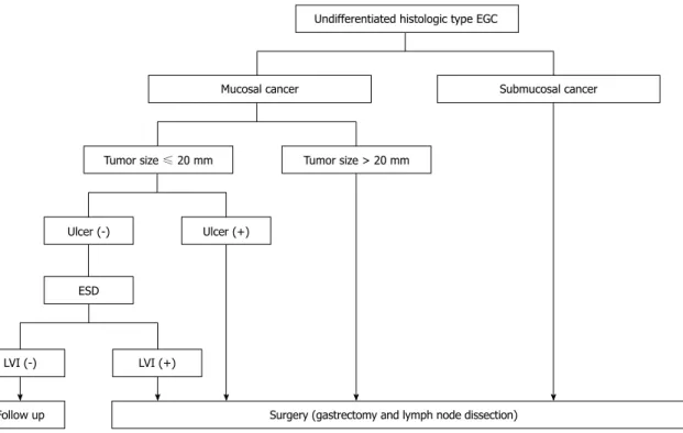
Undifferentiated histologic type EGC
Mucosal cancer Submucosal cancer
Tumor size ≤ 20 mm Tumor size > 20 mm
Ulcer (-) Ulcer (+)
ESD
LVI (-) LVI (+)
Follow up Surgery (gastrectomy and lymph node dissection)
Figure 2 Treatment algorithm for undifferentiated type early gastric cancer according to depth of invasion, tumor size, ulceration, and lymphovascular invasion. EGC: Early gastric cancer; ESD: Endoscopic submucosal dissection; LVI: Lymphovascular invasion.
outcomes, the expanded criteria for ESD of UD-EGC
are feasible with reference to therapeutic efficacy and
safety in the long-term period if curative resection is
accomplished, although more long-term outcomes are
needed. We now suggest the treatment algorithm for
UD-EGC according to depth of invasion, tumor size,
ulceration, and lymphovascular invasion (Figure 2). This
is consistent with the conditions of curative resection
according to the Japanese gastric cancer treatment
guide-lines
[23,26]. However, we should recognize the limitation
of current diagnostic and histological tools to predict LN
metastasis. The innovative improvement of preoperative
imaging modalities and well-defined criteria predictive
of LN metastasis from multicenter, prospective studies
would reduce the limitation.
REFERENCES
1 Japanese Gastric Cancer Association. Japanese classification
of gastric carcinoma: 3rd English edition. Gastric Cancer 2011;
14: 101-112 [PMID: 21573743 DOI: 10.1007/s10120-011-0041-5]
2 Ahn YO, Park BJ, Yoo KY, Kim NK, Heo DS, Lee JK, Ahn
HS, Kang DH, Kim H, Lee MS. Incidence estimation of stomach cancer among Koreans. J Korean Med Sci 1991; 6: 7-14 [PMID: 1888453]
3 Nakamura K, Ueyama T, Yao T, Xuan ZX, Ambe K, Adachi
Y, Yakeishi Y, Matsukuma A, Enjoji M. Pathology and prog-nosis of gastric carcinoma. Findings in 10,000 patients who underwent primary gastrectomy. Cancer 1992; 70: 1030-1037 [PMID: 1515980]
4 Shimizu S, Tada M, Kawai K. Early gastric cancer: its
surveil-lance and natural course. Endoscopy 1995; 27: 27-31 [PMID: 7601031 DOI: 10.1055/s-2007-1005628]
5 Siewert JR. Gastric cancer: the dispute between East and
West. Gastric Cancer 2005; 8: 59-61 [PMID: 15864709 DOI: 10.1007/s10120-005-0323-x]
6 Everett SM, Axon AT. Early gastric cancer in Europe. Gut
1997; 41: 142-150 [PMID: 9301490]
7 Okamura T, Tsujitani S, Korenaga D, Haraguchi M, Baba H,
Hiramoto Y, Sugimachi K. Lymphadenectomy for cure in patients with early gastric cancer and lymph node metasta-sis. Am J Surg 1988; 155: 476-480 [PMID: 3344913]
8 Noguchi Y, Imada T, Matsumoto A, Coit DG, Brennan MF.
Radical surgery for gastric cancer. A review of the Japanese experience. Cancer 1989; 64: 2053-2062 [PMID: 2680049] 9 Sue-Ling HM, Martin I, Griffith J, Ward DC, Quirke P,
Dixon MF, Axon AT, McMahon MJ, Johnston D. Early gas-tric cancer: 46 cases treated in one surgical department. Gut 1992; 33: 1318-1322 [PMID: 1446852]
10 Sue-Ling HM, Johnston D, Martin IG, Dixon MF, Lansdown MR, McMahon MJ, Axon AT. Gastric cancer: a curable dis-ease in Britain. BMJ 1993; 307: 591-596 [PMID: 8401015] 11 Sano T, Sasako M, Kinoshita T, Maruyama K. Recurrence of
early gastric cancer. Follow-up of 1475 patients and review of the Japanese literature. Cancer 1993; 72: 3174-3178 [PMID: 8242540]
12 Park CH, Song KY, Kim SN. Treatment results for gastric cancer surgery: 12 years’ experience at a single institute in Korea. Eur J Surg Oncol 2008; 34: 36-41 [PMID: 17442532 DOI: 10.1016/j.ejso.2007.03.004]
13 Kojima T, Parra-Blanco A, Takahashi H, Fujita R. Outcome of endoscopic mucosal resection for early gastric cancer: re-view of the Japanese literature. Gastrointest Endosc 1998; 48: 550-54; discussion 550-54; [PMID: 9831855]
14 Tada M, Murakami A, Karita M, Yanai H, Okita K. Endo-scopic resection of early gastric cancer. Endoscopy 1993; 25:
445-450 [PMID: 8261986 DOI: 10.1055/s-2007-1010365] 15 Hirao M, Masuda K, Asanuma T, Naka H, Noda K,
Mat-suura K, Yamaguchi O, Ueda N. Endoscopic resection of early gastric cancer and other tumors with local injection of hypertonic saline-epinephrine. Gastrointest Endosc 1988; 34: 264-269 [PMID: 3391382]
16 Gotoda T, Kondo H, Ono H, Saito Y, Yamaguchi H, Saito D, Yokota T. A new endoscopic mucosal resection procedure using an insulation-tipped electrosurgical knife for rectal flat lesions: report of two cases. Gastrointest Endosc 1999; 50: 560-563 [PMID: 10502182]
17 Ohkuwa M, Hosokawa K, Boku N, Ohtu A, Tajiri H, Yoshi-da S. New endoscopic treatment for intramucosal gastric tu-mors using an insulated-tip diathermic knife. Endoscopy 2001;
33: 221-226 [PMID: 11293753 DOI: 10.1055/s-2001-12805]
18 Lee JH, Kim JJ. Endoscopic mucosal resection of early gas-tric cancer: Experiences in Korea. World J Gastroenterol 2007;
13: 3657-3661 [PMID: 17659722]
19 Probst A, Pommer B, Golger D, Anthuber M, Arnholdt H, Messmann H. Endoscopic submucosal dissection in gastric neoplasia - experience from a European center. En-doscopy 2010; 42: 1037-1044 [PMID: 20972955 DOI: 10.1055/ s-0030-1255668]
20 Ribeiro-Mourão F, Pimentel-Nunes P, Dinis-Ribeiro M. En-doscopic submucosal dissection for gastric lesions: results of an European inquiry. Endoscopy 2010; 42: 814-819 [PMID: 20886399 DOI: 10.1055/s-0030-1255778]
21 Farhat S, Chaussade S, Ponchon T, Coumaros D, Chara-chon A, Barrioz T, Koch S, Houcke P, Cellier C, Heresbach D, Lepilliez V, Napoleon B, Bauret P, Coron E, Le Rhun M, Bichard P, Vaillant E, Calazel A, Bensoussan E, Bellon S, Mangialavori L, Robin F, Prat F. Endoscopic submucosal dissection in a European setting. A multi-institutional report of a technique in development. Endoscopy 2011; 43: 664-670 [PMID: 21623560 DOI: 10.1055/s-0030-1256413]
22 Japanese Gastric Cancer Association. Japanese gastric can-cer treatment guidelines 2010 (ver. 3). Gastric Cancan-cer 2011; 14: 113-123 [PMID: 21573742 DOI: 10.1007/s10120-011-0042-4] 23 Gotoda T, Yanagisawa A, Sasako M, Ono H, Nakanishi Y,
Shimoda T, Kato Y. Incidence of lymph node metastasis from early gastric cancer: estimation with a large number of cases at two large centers. Gastric Cancer 2000; 3: 219-225 [PMID: 11984739]
24 Gotoda T. Endoscopic resection of early gastric cancer. Gastric Cancer 2007; 10: 1-11 [PMID: 17334711 DOI: 10.1007/ s10120-006-0408-1]
25 Soetikno R, Kaltenbach T, Yeh R, Gotoda T. Endoscopic mucosal resection for early cancers of the upper gastrointes-tinal tract. J Clin Oncol 2005; 23: 4490-4498 [PMID: 16002839 DOI: 10.1200/jco.2005.19.935]
26 Hirasawa T, Gotoda T, Miyata S, Kato Y, Shimoda T, Tani-guchi H, Fujisaki J, Sano T, YamaTani-guchi T. Incidence of lymph node metastasis and the feasibility of endoscopic resection for undifferentiated-type early gastric cancer. Gas-tric Cancer 2009; 12: 148-152 [PMID: 19890694 DOI: 10.1007/ s10120-009-0515-x]
27 Ajani JA, Bentrem DJ, Besh S, D’Amico TA, Das P, Den-linger C, Fakih MG, Fuchs CS, Gerdes H, Glasgow RE, Hay-man JA, Hofstetter WL, Ilson DH, Keswani RN, Kleinberg LR, Korn WM, Lockhart AC, Meredith K, Mulcahy MF, Orringer MB, Posey JA, Sasson AR, Scott WJ, Strong VE, Varghese TK, Warren G, Washington MK, Willett C, Wright CD, McMillian NR, Sundar H. Gastric cancer, version 2.2013: featured updates to the NCCN Guidelines. J Natl Compr Canc Netw 2013; 11: 531-546 [PMID: 23667204]
28 Okines A, Verheij M, Allum W, Cunningham D, Cervantes A. Gastric cancer: ESMO Clinical Practice Guidelines for di-agnosis, treatment and follow-up. Ann Oncol 2010; 21 Suppl 5: v50-v54 [PMID: 20555102 DOI: 10.1093/annonc/mdq164] 29 Guadagni S, Reed PI, Johnston BJ, De Bernardinis G,
Cat-P- Reviewers Bener A S- Editor Wen LL L- Editor Cant MR E- Editor Ma S P- Reviewers Bener A S- Editor Song XX L- Editor Stewart GJ E- Editor Ma S
arci M, Valenti M, di Orio F, Carboni M. Early gastric can-cer: follow-up after gastrectomy in 159 patients. Br J Surg 1993; 80: 325-328 [PMID: 8472141]
30 Choi HJ, Kim YK, Kim YH, Kim SS, Hong SH. Occurrence and prognostic implications of micrometastases in lymph nodes from patients with submucosal gastric carcinoma. Ann Surg Oncol 2002; 9: 13-19 [PMID: 11829425]
31 Seto Y, Shimoyama S, Kitayama J, Mafune K, Kaminishi M, Aikou T, Arai K, Ohta K, Nashimoto A, Honda I, Yamagishi H, Yamamura Y. Lymph node metastasis and preopera-tive diagnosis of depth of invasion in early gastric cancer. Gastric Cancer 2001; 4: 34-38 [PMID: 11706625 DOI: 10.1007/ s101200100014]
32 Boku T, Nakane Y, Okusa T, Hirozane N, Imabayashi N, Hioki K, Yamamoto M. Strategy for lymphadenectomy of gastric cancer. Surgery 1989; 105: 585-592 [PMID: 2705096] 33 Lee E, Chae Y, Kim I, Choi J, Yeom B, Leong AS.
Prognos-tic relevance of immunohistochemically detected lymph node micrometastasis in patients with gastric carcinoma. Cancer 2002; 94: 2867-2873 [PMID: 12115374 DOI: 10.1002/ cncr.10562]
34 Ren G, Cai R, Zhang WJ, Ou JM, Jin YN, Li WH. Prediction of risk factors for lymph node metastasis in early gastric cancer. World J Gastroenterol 2013; 19: 3096-3107 [PMID: 23716990 DOI: 10.3748/wjg.v19.i20.3096]
35 Kwee RM, Kwee TC. Imaging in assessing lymph node status in gastric cancer. Gastric Cancer 2009; 12: 6-22 [PMID: 19390927 DOI: 10.1007/s10120-008-0492-5]
36 Seevaratnam R, Cardoso R, McGregor C, Lourenco L, Ma-har A, SutradMa-har R, Law C, Paszat L, Coburn N. How useful is preoperative imaging for tumor, node, metastasis (TNM) staging of gastric cancer? A meta-analysis. Gastric Cancer 2012; 15 Suppl 1: S3-18 [PMID: 21837458 DOI: 10.1007/ s10120-011-0069-6]
37 Puli SR, Batapati Krishna Reddy J, Bechtold ML, Antillon MR, Ibdah JA. How good is endoscopic ultrasound for TNM staging of gastric cancers? A meta-analysis and systematic review. World J Gastroenterol 2008; 14: 4011-4019 [PMID: 18609685]
38 Cardoso R, Coburn N, Seevaratnam R, Sutradhar R, Louren-co LG, Mahar A, Law C, Yong E, Tinmouth J. A systematic review and meta-analysis of the utility of EUS for preopera-tive staging for gastric cancer. Gastric Cancer 2012; 15 Suppl 1: S19-S26 [PMID: 22237654 DOI: 10.1007/s10120-011-0115-4] 39 Mocellin S, Marchet A, Nitti D. EUS for the staging of
gastric cancer: a meta-analysis. Gastrointest Endosc 2011; 73: 1122-1134 [PMID: 21444080 DOI: 10.1016/j.gie.2011.01.030] 40 Kim JH, Song KS, Youn YH, Lee YC, Cheon JH, Song SY,
Chung JB. Clinicopathologic factors influence accurate en-dosonographic assessment for early gastric cancer. Gastroin-test Endosc 2007; 66: 901-908 [PMID: 17963876 DOI: 10.1016/ j.gie.2007.06.012]
41 Nagahama T, Yao K, Maki S, Yasaka M, Takaki Y, Matsui T, Tanabe H, Iwashita A, Ota A. Usefulness of magnifying endoscopy with narrow-band imaging for determining the horizontal extent of early gastric cancer when there is an un-clear margin by chromoendoscopy (with video). Gastrointest Endosc 2011; 74: 1259-1267 [PMID: 22136775 DOI: 10.1016/ j.gie.2011.09.005]
42 Kiyotoki S, Nishikawa J, Satake M, Fukagawa Y, Shirai Y, Hamabe K, Saito M, Okamoto T, Sakaida I. Usefulness of magnifying endoscopy with narrow-band imaging for determining gastric tumor margin. J Gastroenterol Hepa-tol 2010; 25: 1636-1641 [PMID: 20880172 DOI: 10.1111/ j.1440-1746.2010.06379.x]
43 Kikuchi D, Iizuka T, Hoteya S, Yamada A, Furuhata T, Yamashita S, Domon K, Nakamura M, Matsui A, Mitani T, Ogawa O, Watanabe S, Kaise M. Usefulness of magnify-ing endoscopy with narrow-band imagmagnify-ing for determin-ing tumor invasion depth in early gastric cancer.
Gastro-enterol Res Pract 2013; 2013: 217695 [PMID: 23401676 DOI: 10.1155/2013/217695]
44 Li HY, Dai J, Xue HB, Zhao YJ, Chen XY, Gao YJ, Song Y, Ge ZZ, Li XB. Application of magnifying endoscopy with narrow-band imaging in diagnosing gastric lesions: a pro-spective study. Gastrointest Endosc 2012; 76: 1124-1132 [PMID: 23025977 DOI: 10.1016/j.gie.2012.08.015]
45 Yoshida T, Kawachi H, Sasajima K, Shiokawa A, Kudo SE. The clinical meaning of a nonstructural pattern in early gastric cancer on magnifying endoscopy. Gastrointest Endosc 2005; 62: 48-54 [PMID: 15990819]
46 Kobara H, Mori H, Fujihara S, Kobayashi M, Nishiyama N, Nomura T, Kato K, Ishihara S, Morito T, Mizobuchi K, Iwa-ma H, Masaki T. Prediction of invasion depth for submuco-sal differentiated gastric cancer by magnifying endoscopy with narrow-band imaging. Oncol Rep 2012; 28: 841-847 [PMID: 22752002 DOI: 10.3892/or.2012.1889]
47 Yoshida T, Inoue H, Usui S, Satodate H, Fukami N, Kudo SE. Narrow-band imaging system with magnifying endos-copy for superficial esophageal lesions. Gastrointest Endosc 2004; 59: 288-295 [PMID: 14745410]
48 Kanao H, Tanaka S, Oka S, Hirata M, Yoshida S, Chayama K. Narrow-band imaging magnification predicts the histol-ogy and invasion depth of colorectal tumors. Gastrointest Endosc 2009; 69: 631-636 [PMID: 19251003 DOI: 10.1016/ j.gie.2008.08.028]
49 Uedo N, Fujishiro M, Goda K, Hirasawa D, Kawahara Y, Lee JH, Miyahara R, Morita Y, Singh R, Takeuchi M, Wang S, Yao T. Role of narrow band imaging for diagnosis of early-stage esophagogastric cancer: current consensus of experienced endoscopists in Asia-Pacific region. Dig Endosc 2011; 23 Suppl 1: 58-71 [PMID: 21535204 DOI: 10.1111/ j.1443-1661.2011.01119.x]
50 Okada K, Fujisaki J, Kasuga A, Omae M, Hirasawa T, Ishiyama A, Inamori M, Chino A, Yamamoto Y, Tsuchida T, Nakajima A, Hoshino E, Igarashi M. Diagnosis of undifferentiated type early gastric cancers by magnification endoscopy with narrow-band imaging. J Gastroenterol Hepatol 2011; 26: 1262-1269 [PMID: 21443667 DOI: 10.1111/j.1440-1746.2011.06730.x]
51 Oda I, Oyama T, Abe S, Ohnita K, Kosaka T, Hirasawa K, Ishido K, Nakagawa M, Takahashi S. Preliminary results of multicenter questionnaire study on long-term outcomes of curative endoscopic submucosal dissection for early gastric cancer. Dig Endosc 2014; 26: 214-219 [PMID: 23826719 DOI: 10.1111/den.12141]
52 Kwee RM, Kwee TC. Predicting lymph node status in early gastric cancer. Gastric Cancer 2008; 11: 134-148 [PMID: 18825308 DOI: 10.1007/s10120-008-0476-5]
53 Tong JH, Sun Z, Wang ZN, Zhao YH, Huang BJ, Li K, Xu Y, Xu HM. Early gastric cancer with signet-ring cell histologic type: risk factors of lymph node metastasis and indications of endoscopic surgery. Surgery 2011; 149: 356-363 [PMID: 20727560 DOI: 10.1016/j.surg.2010.07.006]
54 Kim HM, Pak KH, Chung MJ, Cho JH, Hyung WJ, Noh SH, Kim CB, Lee YC, Song SY, Lee SK. Early gastric cancer of signet ring cell carcinoma is more amenable to endoscopic treatment than is early gastric cancer of poorly differentiat-ed tubular adenocarcinoma in select tumor conditions. Surg Endosc 2011; 25: 3087-3093 [PMID: 21487870 DOI: 10.1007/ s00464-011-1674-5]
55 Li H, Lu P, Lu Y, Liu C, Xu H, Wang S, Chen J. Predictive factors of lymph node metastasis in undifferentiated early gastric cancers and application of endoscopic mucosal re-section. Surg Oncol 2010; 19: 221-226 [PMID: 20471826 DOI: 10.1016/j.suronc.2009.05.006]
56 Park JM, Kim SW, Nam KW, Cho YK, Lee IS, Choi MG, Chung IS, Song KY, Park CH, Jung CK. Is it reasonable to treat early gastric cancer with signet ring cell histology by en-doscopic resection? Analysis of factors related to lymph-node metastasis. Eur J Gastroenterol Hepatol 2009; 21: 1132-1135
[PMID: 19369881 DOI: 10.1097/MEG.0b013e32832a21d8] 57 Kunisaki C, Takahashi M, Nagahori Y, Fukushima T,
Makino H, Takagawa R, Kosaka T, Ono HA, Akiyama H, Moriwaki Y, Nakano A. Risk factors for lymph node metas-tasis in histologically poorly differentiated type early gastric cancer. Endoscopy 2009; 41: 498-503 [PMID: 19533552 DOI: 10.1055/s-0029-1214758]
58 Hanaoka N, Tanabe S, Mikami T, Okayasu I, Saigenji K. Mixed-histologic-type submucosal invasive gastric cancer as a risk factor for lymph node metastasis: feasibility of en-doscopic submucosal dissection. Endoscopy 2009; 41: 427-432 [PMID: 19418397 DOI: 10.1055/s-0029-1214495]
59 Ye BD, Kim SG, Lee JY, Kim JS, Yang HK, Kim WH, Jung HC, Lee KU, Song IS. Predictive factors for lymph node metastasis and endoscopic treatment strategies for undiffer-entiated early gastric cancer. J Gastroenterol Hepatol 2008; 23: 46-50 [PMID: 18171341 DOI: 10.1111/j.1440-1746.2006.04791. x]
60 Park YD, Chung YJ, Chung HY, Yu W, Bae HI, Jeon SW, Cho CM, Tak WY, Kweon YO. Factors related to lymph node metastasis and the feasibility of endoscopic mucosal resection for treating poorly differentiated adenocarcinoma of the stomach. Endoscopy 2008; 40: 7-10 [PMID: 18210339 DOI: 10.1055/s-2007-966750]
61 Li H, Lu P, Lu Y, Liu CG, Xu HM, Wang SB, Chen JQ. Pre-dictive factors for lymph node metastasis in poorly differen-tiated early gastric cancer and their impact on the surgical strategy. World J Gastroenterol 2008; 14: 4222-4226 [PMID: 18636670]
62 Li C, Kim S, Lai JF, Oh SJ, Hyung WJ, Choi WH, Choi SH, Zhu ZG, Noh SH. Risk factors for lymph node metastasis in undifferentiated early gastric cancer. Ann Surg Oncol 2008;
15: 764-769 [PMID: 18043971 DOI:
10.1245/s10434-007-9707-y]
63 Ha TK, An JY, Youn HK, Noh JH, Sohn TS, Kim S. Indica-tion for endoscopic mucosal resecIndica-tion in early signet ring cell gastric cancer. Ann Surg Oncol 2008; 15: 508-513 [PMID: 18071825 DOI: 10.1245/s10434-007-9660-9]
64 Hyung WJ, Cheong JH, Kim J, Chen J, Choi SH, Noh SH. Application of minimally invasive treatment for early gas-tric cancer. J Surg Oncol 2004; 85: 181-185; discussion 186 [PMID: 14991872 DOI: 10.1002/jso.20018]
65 Abe N, Watanabe T, Sugiyama M, Yanagida O, Masaki T, Mori T, Atomi Y. Endoscopic treatment or surgery for undif-ferentiated early gastric cancer? Am J Surg 2004; 188: 181-184 [PMID: 15249247 DOI: 10.1016/j.amjsurg.2003.12.060] 66 Lee JH, Choi MG, Min BH, Noh JH, Sohn TS, Bae JM, Kim
S. Predictive factors for lymph node metastasis in patients with poorly differentiated early gastric cancer. Br J Surg 2012; 99: 1688-1692 [PMID: 23023388 DOI: 10.1002/bjs.8934] 67 Chung JW, Jung HY, Choi KD, Song HJ, Lee GH, Jang SJ,
Park YS, Yook JH, Oh ST, Kim BS, Kim JH. Extended in-dication of endoscopic resection for mucosal early gastric cancer: analysis of a single center experience. J Gastroenterol Hepatol 2011; 26: 884-887 [PMID: 21198830 DOI: 10.1111/ j.1440-1746.2010.06611.x]
68 Hirasawa T, Fujisaki J, Fukunaga T, Yamamoto Y, Yamagu-chi T, Katori M, Yamamoto N. Lymph node metastasis from undifferentiated-type mucosal gastric cancer satisfying the expanded criteria for endoscopic resection based on routine histological examination. Gastric Cancer 2010; 13: 267-270 [PMID: 21128064 DOI: 10.1007/s10120-010-0577-9]
69 Haruta H, Hosoya Y, Sakuma K, Shibusawa H, Satoh K, Yamamoto H, Tanaka A, Niki T, Sugano K, Yasuda Y. Clini-copathological study of lymph-node metastasis in 1,389 patients with early gastric cancer: assessment of indications for endoscopic resection. J Dig Dis 2008; 9: 213-218 [PMID: 18959593 DOI: 10.1111/j.1751-2980.2008.00349.x]
70 Song SY, Park S, Kim S, Son HJ, Rhee JC. Characteristics of intramucosal gastric carcinoma with lymph node metastatic
disease. Histopathology 2004; 44: 437-444 [PMID: 15139991 DOI: 10.1111/j.1365-2559.2004.01870.x]
71 Choi J, Kim SG, Im JP, Kim JS, Jung HC. Endoscopic estima-tion of tumor size in early gastric cancer. Dig Dis Sci 2013; 58: 2329-2336 [PMID: 23589139 DOI: 10.1007/s10620-013-2644-7] 72 Kang KJ, Kim KM, Kim JJ, Rhee PL, Lee JH, Min BH,
Rhee JC, Kushima R, Lauwers GY. Gastric extremely well-differentiated intestinal-type adenocarcinoma: a chal-lenging lesion to achieve complete endoscopic resection. Endoscopy 2012; 44: 949-952 [PMID: 22987215 DOI: 10.1055/ s-0032-1310161]
73 Kang HY, Kim SG, Kim JS, Jung HC, Song IS. Clinical out-comes of endoscopic submucosal dissection for undiffer-entiated early gastric cancer. Surg Endosc 2010; 24: 509-516 [PMID: 19585066 DOI: 10.1007/s00464-009-0614-0]
74 Kim JH, Lee YC, Kim H, Song KH, Lee SK, Cheon JH, Kim H, Hyung WJ, Noh SH, Kim CB, Chung JB. Endoscopic resection for undifferentiated early gastric cancer. Gastroin-test Endosc 2009; 69: e1-e9 [PMID: 19327466 DOI: 10.1016/ j.gie.2008.10.040]
75 Lee HL, Choi CH, Cheung DY. Do we have enough evi-dence for expanding the indications of ESD for EGC? World J Gastroenterol 2011; 17: 2597-2601 [PMID: 21677826 DOI: 10.3748/wjg.v17.i21.2597]
76 Kamada K, Tomatsuri N, Yoshida N. Endoscopic submu-cosal dissection for undifferentiated early gastric cancer as the expanded indication lesion. Digestion 2012; 85: 111-115 [PMID: 22269290 DOI: 10.1159/000334681]
77 Okada K, Fujisaki J, Kasuga A, Omae M, Yoshimoto K, Hi-rasawa T, Ishiyama A, Yamamoto Y, Tsuchida T, Hoshino E, Igarashi M, Takahashi H. Endoscopic ultrasonography is valuable for identifying early gastric cancers meeting expanded-indication criteria for endoscopic submucosal dissection. Surg Endosc 2011; 25: 841-848 [PMID: 20734082 DOI: 10.1007/s00464-010-1279-4]
78 Akahoshi K, Chijiwa Y, Hamada S, Sasaki I, Nawata H, Kabemura T, Yasuda D, Okabe H. Pretreatment staging of endoscopically early gastric cancer with a 15 MHz ultra-sound catheter probe. Gastrointest Endosc 1998; 48: 470-476 [PMID: 9831834]
79 Hizawa K, Iwai K, Esaki M, Matsumoto T, Suekane H, Iida M. Is endoscopic ultrasonography indispensable in assess-ing the appropriateness of endoscopic resection for gastric cancer? Endoscopy 2002; 34: 973-978 [PMID: 12471541 DOI: 10.1055/s-2002-35851]
80 Kim YY, Jeon SW, Kim J, Park JC, Cho KB, Park KS, Kim E, Chung YJ, Kwon JG, Jung JT, Kim EY, Kim KO, Jang B, Lee SH, Yang CH. Endoscopic submucosal dissection for early gastric cancer with undifferentiated histology: could we extend the criteria beyond? Surg Endosc 2013; 27: 4656-4662 [PMID: 23943115 DOI: 10.1007/s00464-013-3099-9]
81 Akashi K, Yanai H, Nishikawa J, Satake M, Fukagawa Y, Okamoto T, Sakaida I. Ulcerous change decreases the ac-curacy of endoscopic ultrasonography diagnosis for the invasive depth of early gastric cancer. Int J Gastrointest Cancer 2006; 37: 133-138 [PMID: 18080789 DOI: 10.1007/ s12029-007-9004-9]
82 Sako A, Kitayama J, Ishikawa M, Yamashita H, Nagawa H. Impact of immunohistochemically identified lymphatic invasion on nodal metastasis in early gastric cancer. Gastric Cancer 2006; 9: 295-302 [PMID: 17235632 DOI: 10.1007/ s10120-006-0396-1]
83 Jung da H, Park YM, Kim JH, Lee YC, Youn YH, Park H, Lee SI, Kim JW, Choi SH, Hyung WJ, Noh SH. Clinical im-plication of endoscopic gross appearance in early gastric cancer: revisited. Surg Endosc 2013; 27: 3690-3695 [PMID: 23588711 DOI: 10.1007/s00464-013-2947-y]
84 Song IC, Liang ZL, Lee JC, Huang SM, Kim HY, Oh YS, Yun HJ, Sul JY, Jo DY, Kim S, Kim JM, Lee HJ. Expression of stromal cell-derived factor-1α is an independent risk