ORIGINAL ARTICLE
DOI: 10.4174/jkss.2011.81.2.104 Journal of the Korean Surgical Society
JKSS
pISSN 2233-7903ㆍeISSN 2093-0488
Received February 24, 2011, Accepted April 27, 2011 Correspondence to: Sung Joon Kwon
Department of Surgery, Hanyang University College of Medicine, 17 Haengdang-dong, Seongdong-gu, Seoul 133-792, Korea Tel: +82-2-2290-8453, Fax: +82-2-2281-0224, E-mail: [email protected]
cc Journal of the Korean Surgical Society is an Open Access Journal. All articles are distributed under the terms of the Creative Commons Attribution Non-Commercial License (http://creativecommons.org/licenses/by-nc/3.0/) which permits unrestricted non-commercial use, distribution, and reproduction in any medium, provided the original work is properly cited.
F18-fluorodeoxyglucose-positron emission
tomography and computed tomography is not
accurate in preoperative staging of gastric cancer
Tae Kyung Ha, Yun Young Choi
1, Soon Young Song
2, Sung Joon Kwon
Departments of Surgery, 1Nuclear Medicine, 2Diagnostic Radiology, Hanyang University College of Medicine, Seoul, Korea
Purpose: To investigate the clinical benefits of F18-fluorodeoxyglucose-positron emission tomography and computed to-mography (18F-FDG-PET/CT) over multi-detector row CT (MDCT) in preoperative staging of gastric cancer. Methods:
FDG-PET/CT and MDCT were performed on 78 patients with gastric cancer pathologically diagnosed by endoscopy. The ac-curacy of radiologic staging retrospectively was compared to pathologic result after curative resection. Results: Primary tu-mors were detected in 51 (65.4%) patients with 18F-FDG-PET/CT, and 47 (60.3%) patients with MDCT. Regarding detection of lymph node metastasis, the sensitivity of FDG-PET/CT was 51.5% with an accuracy of 71.8%, whereas those of MDCT were 69.7% and 69.2%, respectively. The sensitivity of 18F-FDG-PET/CT for a primary tumor with signet ring cell carcinoma was lower than that of 18F-FDG-PET/CT for a primary tumor with non-signet ring cell carcinoma (35.3% vs. 73.8%, P < 0.01).
Conclusion: Due to its low sensitivity, 18F-FDG-PET/CT alone shows no definite clinical benefit for prediction of lymph node metastasis in preoperative staging of gastric cancer.
Key Words: Gastric cancer, 18F-FDG-PET/CT, MDCT, Preoperative staging
INTRODUCTION
Gastric cancer remains the second most common cancer diagnosed in the world. It accounts for 9.9% of all new cas-es of cancer diagnosed and is rcas-esponsible for 12.1% of all cancer deaths [1]. Although the number of overall cancer deaths has declined, gastric cancer is still the leading cause of cancer death in Korea [2]. An essential step in managing gastric cancer is accurately assessing the preoperative stage and deciding on the adequate surgery including en-doscopic treatment, minimal invasive surgery, and
pallia-tive operation.
However, managing gastric cancer is challenging be-cause problems in determining treatment strategy remain as the prognosis has a wide range according to TNM clas-sification [3]. There are limitations in the treatment of ad-vanced gastric cancer (AGC) with surgery, whereas early gastric cancer (EGC) can be treated using minimal in-vasive surgery including endoscopy and laparoscopy with the limitation of lymphadenectomy [4,5]. Traditio-nally, computed tomography (CT) has been used for pre-operative staging of gastric cancer. CT provides useful
in-formation on tumors based on anatomical structure, but there is a limitation in the accuracy of detection of EGC [6]. It was reported that the diagnostic accuracy in T staging can be increased using multi-detector row CT (MDCT), but N staging which is one of the most important prog-nostic factor as deciding treatment strategy for EGC re-main unsatisfactory [7,8].
Recently, Positron emission tomography (PET) using 18
F-fluorodeoxyglucose (18F-FDG) is being widely used to determine status of many different cancers, considered a new perspective on staging approach in malignancy. This advanced technology more accurately displays functional image of cancer with altered glucose metabolism, but lacks precision in localizing the tumor. A combined image of 18F-FDG-PET and CT (18F-FDG-PET/CT) can provide additional information by using the characteristics of both modalities. Improved staging accuracy has been demon-strated with the use of 18F-FDG-PET/CT in patients with lung and colon cancers [9-11].
18
F-FDG-PET/CT has being widely used in Korea after the National Health Insurance Program decided to re-imburse 18F-FDG-PET/CT cost in 2006 [12]. There is a pauc-ity of data on the role of 18F-FDG-PET/CT in the pre-operative diagnosis of gastric cancer. The purpose of this study is to compare the effectiveness of 18F-FDG-PET/CT to MDCT in terms of preoperative T and N staging of gas-tric cancer.
METHODS
Patients
A retrospective analysis of 78 preoperative 18F-FDG- PET/CT and MDCT in patients with gastric cancer who had undergone curative gastrectomy between February 2007 and October 2008 was performed. Informed consent for 18F-FDG-PET/CT and MDCT for the purpose of pre-operative staging of gastric cancer was obtained from all patients. The patients comprised 53 men and 25 women with a median age of 61 years (range, 32 to 85 years). All of these patients underwent a preoperative staging proce-dure, including past history, physical examination, blood chemistry, abdominal MDCT, and
esophagogastroduo-denoscopy. 18F-FDG-PET/CT was performed within 4 we-eks before gastrectomy.
MDCT technique
With MDCT unit, abdominal MDCT images were obtained. The MDCT scanner used was a 64-detector row scanner (Brilliance CT 64, Philips Medical System, Cleve-land, OH, USA). All patients were given 4 g of effervescent granules (Top, Taejoon Pharmaceuticals, Seoul, Korea) to distend the stomach wall. The patients were placed in a su-pine position on the CT table. The acquisition volume in-cluded the whole abdomen from the dome of the dia-phragm to lower margin of the symphysis pubis. An 18-gauge intravenous cannula was inserted into a vein in the antecubital fossa, forearm or wrist. Scanning of the ab-domen was performed after intravenous injection with au-tomatic power injector of 2 mL/kg of contrast medium (Ultravist 300, Schering, Berlin, Germany) at a flow rate of 2 to 3.5 mL/sec, total 120 mL. The scan delay time was de-termined by automatic bolus tracking method. The region of interest (ROI) was positioned at the descending aorta, at the level of the diaphragm. The hepatic arterial phase im-age was started 7 seconds after the attenuation reached 200 HU. Additional CT scan for portal phase images was start-ed 60 seconds after the start of the contrast injection. The re-spective scanning parameters used for 16- and 64-MDCT scanners were 16 × 1.5 mm and 64 × 0.625 mm collimation, table feed of 24 and 46 mm per gantry rotation. X-ray tube voltage was 120 or 140 kVp, and amperage was determined by automatic dose reduction protocol. Volume data of MDCT scan were reconstructed as 5 mm thickness at 5 mm intervals. Coronal reformative images were reconstructed on a workstation, with a thickness of 3 mm at 3 mm intervals. All reconstructed images were sent to picture ar-chiving and communication system (PACS) for evaluation.
18
F-FDG-PET/CT technique
Patients were advised to fast for at least 6 hours prior to the intravenous injection of 5 to 6 MBq/kg of 18F-FDG. The 18
F-FDG-PET/CT scan was obtained approximately 1 hour after administration of the FDG, using Biograph 6 system (Siemens, Knoxville, TN, USA). Seven or eight bed posi-tions were imaged from the skull base to the mid thigh
Table 1. Characteristics of 78 patients with gastric cancer according to PET/CT results for primary tumor and LNM Characteristics PET/CT (−) (n = 27) PET/CT (+) (n = 51) P-value PET/CT LN (−) (n = 55) PET/CT LN (+) (n = 23) P-value Age (yr) 0.477 0.624 ≤60 15 (39.5) 23 (60.5) 28 (73.7) 10 (26.3) >60 12 (30.0) 28 (70.0) 27 (67.5) 13 (32.5) Gender 0.308 1.000 Male 16 (30.2) 37 (69.8) 37 (69.8) 16 (30.2) Female 11 (44.0) 14 (56.0) 18 (72.0) 7 (28.0) Tumor location 0.080 0.067 Upper 2 (25.0) 6 (75.0) 3 (37.5) 5 (62.5) Middle 15 (50.0) 15 (50.0) 21 (70.0) 9 (30.0) Lower 10 (25.0) 30 (75.0) 31 (77.5) 9 (22.5) Tumor size 0.002 0.330 ≤3.5 21 (51.2) 20 (48.8) 31 (75.6) 10 (24.4) >3.5 6 (16.2) 31 (83.8) 24 (64.9) 13 (35.1) Depth of tumor 0.005 <0.001 T1 20 (54.1) 17 (45.9) 34 (91.9) 3 (8.1) T2 6 (22.2) 21 (77.8) 14 (51.9) 13 (48.1) T3 1 (9.1) 10 (90.9) 6 (54.5) 5 (45.5) T4 0 (0.0) 3 (100.0) 1 (33.3) 2 (66.7) LNM 0.003 0.001 N0 23 (51.1) 22 (48.9) 39 (86.7) 6 (13.3) N1 3 (18.8) 13 (81.3) 9 (56.3) 7 (43.8) N2 0 (0.0) 4 (100.0) 2 (50.0) 2 (50.0) N3 1 (7.7) 12 (92.3) 5 (38.5) 8 (61.5) Histology 0.005 0.016 Non-SRCa) 16 (26.2) 45 (73.8) 39 (63.9) 22 (36.1) SRC 11 (64.7) 6 (35.3) 16 (94.1) 1 (5.9)
Values are presented as number (%).
PET/CT, positron emission tomography and computed tomography; LN, lymph node; LNM, lymph node metastasis; SRC, signet ring carcinoma.
a)
Papillary adenocarcinoma, well differentiated adenocarcinoma, moderately differentiated adenocarcinoma, poorly differentiated adenocarcinoma, and mucinous adenocarcinoma.
level. Patients were advised not to speak, chew, or move during scanning.
CT acquisition used the following parameters: 100 mA, 130 kVp tube voltage, helical thickness of 5 mm, and a heli-cal pitch of 1.5:1. Using these parameters, the CT scan from the skull base to the mid thighs was obtained in 20 seconds. Immediately following the CT scan, a PET emis-sion scan was acquired from the mid thigh to the base of skull with an acquisition time of 2.3 minutes per bed posi-tion based on body weight.
18
F-FDG-PET/CT and MDCT image interpretation
An experienced gastrointestinal radiologist interpreted the MDCT images. Transverse and coronal MDCT images were evaluated with PACS monitors in random order. A
le-sion was determined to be cancerous when the gastric wall showed a focal thickening of at least 6 mm or greater or when focal enhancement was seen in the gastric wall. Lymph nodes were considered metastatic if they were large than 8 mm in the short-axis diameter and oval [13-15].
FDG uptake lesions of gastric wall and of lymph node bearing areas, regardless of size detected on PET/CT were regarded as malignancy, and the maximum standardized uptake value (SUVmax) was obtained using volume ROI.
Operation
All patients underwent total gastrectomy (n = 19, 24.3%) or subtotal gastrectomy (n = 59, 75.7%) and standard lym-phadenectomy (at least D2 lymlym-phadenectomy). The ad-vanced gastric cancer in 41 patients and EGC in 37 patients
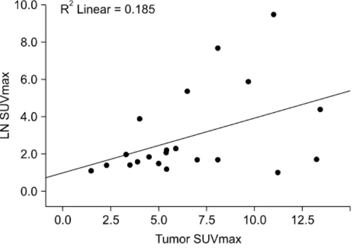
Fig. 1. Correlation analysis curve between primary tumor maximum standardized uptake value (SUVmax) and lymph node
(LN) SUVmax.
Table 2.Detection rate of primary tumor and lymph node metas-tasis in 18F-FDG-PET/CT and MDCT
Variable 18F-FDG-PET/CT MDCT P-valuea) Primary tumor in EGC 17/37 (45.9) 10/37 (27.0) 0.039 Primary tumor in AGC 34/41 (82.9) 37/41 (90.2) 0.508 LNM in EGC 0/37 (0.0) 1/37 (2.7) NA LNM in AGC 17/41 (41.5) 22/41 (53.7) 0.125 Values are presented as number/total number (%).
18
F-FDG-PET/CT, F18-fluorodeoxyglucose-positron emission tomo-graphy and computed tomotomo-graphy; MDCT, multi-detector row CT; EGC, early gastric cancer; AGC, advanced gastric cancer; LNM, lymph node metastasis; NA, not available.
a)
Chi-square McNemar test.
were revealed by pathologic diagnosis. According to the World Health Organization classification with Japanese modification [16], histopathological type of primary tu-mor was categorized as papillary adenocarcinoma (n = 1, 1.3%), well-differentiated adenocarcinoma (n = 4, 5.1%), moderately differentiated adenocarcinoma (n = 31, 39.7%), poorly differentiated adenocarcinoma (n = 22, 28.3%), mu-cinous adenocarcinoma (n = 3, 3.8%), and signet ring cell carcinoma (SRC) (n = 17, 21.8%).
Statistical analysis
We conducted Fisher’s exact test for comparing the dif-ference of respective categorical variables. The relation be-tween SUVmax and pTNM stage was determined using Kruskall-Wallis test. The sensitivity and specificity of 18
F-FDG-PET/CT and MDCT were calculated and com-pared using McNemar’s chi-square test. The correlation analysis was performed to determine the relationship be-tween tumor SUVmax and lymph node SUVmax, and also be-tween tumor SUVmax and tumor size. P-values less than 0.05 were considered to be significant. The statistical anal-ysis was carried out by the SPSS ver. 18.0 (SPSS Inc., Chicago, IL, USA).
RESULTS
Fifty one primary tumors were detected by
pre-operative 18F-FDG-PET/CT, and 27 patients had no notice-able results in 18F-FDG-PET/CT. Tumor size, depth of tu-mor, and lymph node metastasis (LNM) significantly af-fected the detection of primary tumor by 18F-FDG- PET/CT. As tumor size, depth of tumor, and LNM in-creased, the primary tumor was revealed more frequently by 18F-FDG-PET/CT. The detection rate of 18F-FDG- PET/CT for SRC was relatively lower than that for non-SRC (35.3% vs. 73.8%, P = 0.005). 18F-FDG-PET/CT de-tected LNM in 23 patients, and 55 patients showed no sig-nificant SUVmax of lymph node which was considered as metastasis. The results of 18F-FDG-PET/CT in detecting LNM were similar to the results of 18F-FDG-PET/CT in de-tecting primary tumor; however, tumor size had no associ-ation with detection of LNM on 18F-FDG-PET/CT (Table 1). The correlation analysis showed a positive correlation exist between SUVmax of primary tumor and that of meta-static lymph node (P = 0.05, R = 0.43) (Fig. 1). However, there was no significant correlation between SUVmax of primary tumor and tumor size.
When assessed using the Kruskall-Wallis test, the mean SUVmax of primary tumor showed no significant difference between pT1, pT2, pT3, and pT4 (6.0 ± 1.4, 6.3 ± 0.6, 6.2 ± 0.8, and 5.4 ± 1.7). In addition, there was no statistical difference of the mean SUVmax of metastatic lymph node between pN0, pN1, pN2, and pN3 (1.8 ± 0.5, 3.1 ± 0.9, 5.6 ± 3.8, and 2.4 ± 0.5).
The detection rate of primary tumor by 18F-FDG- PET/CT and MDCT showed no significant difference. Among the 37 patients with EGC, 18F-FDG-PET/CT preoperatively de-tected 17 patients who were suspected to have gastric
Table 3. The sensitivity, specificity, PPV, NPV, and accuracy of PET/CT and CT for LNM in gastric cancer
Sensitivity Specificity PPV NPV Accuracy
CT PET/CT CT PET/CT CT PET/CT CT PET/CT CT PET/CT
EGC 20.0 0.0 78.1 85.3 12.5 0.0 86.2 85.3 70.2 78.4 (1/5) (0/5) (25/32) (29/32) (1/8) (0/3) (25/29) (29/34) (26/37) (29/37) AGC 78.6 60.7 46.2 76.9 75.9 85.0 50.0 47.6 70.2 65.9 (22/28) (17/28) (6/13) (10/13) (22/29) (17/20) (6/12) (10/21) (26/37) (27/41) Total 69.7 51.5 75.6 86.7 62.2 73.9 75.6 70.9 69.2 71.8 (23/33) (17/33) (31/45) (39/45) (23/37) (17/23) (31/41) (39/55) (54/78) (56/78) Values are presented as % (number/total number).
PPV, positive predictive value; NPV, negative predictive value; PET/CT, positron emission tomography and computed tomography; LNM, lymph node metastasis; EGC, early gastric cancer; AGC, advanced gastric cancer.
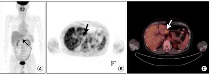
Fig. 2. Early gastric cancer in a 47-year-old man. Miaximum intensity projection image of (A) positron emission tomography (PET) and (B) axial PET scan demonstrate focal hypermetabolic lesion in gastric antrum (black arrow) with maximum standardized uptake value (SUVmax)
of 4.3. (C) PET/CT scan shows focal hypermtabolic intraluminal mass in posterior wall of gastric antrum (white arrow).
cancers. However, MDCT detected only 10 patients out of 37. The other 10 gastric cancer patients had no measurable le-sions on both 18F-FDG-PET/CT and MDCT. A statistical dif-ference between the detection rate of 18F-FDG-PET/CT and that of MDCT in patients with EGC was found (45.9% vs. 27.0%, P = 0.039). However, there was no significant differ-ence between the detection rate of 18F-FDG-PET/CT (34/41 patients) and that of MDCT (37/41 patients) in AGC (Table 2). The SUVmax of primary tumor between EGC and AGC dis-played similar result (6.0 ± 6.1, range 1.5 to 27.0 vs. 6.2 ± 2.9, range 2.2 to 13.2). However, the result of 18F-FDG-PET/CT (17 of 78 patients) for LNM confirmed pathologically was not significantly different from that of MDCT (23 of 78 patients) with respect to the detection of LNM (21.8% vs. 29.5%, P = 0.07). In 41 patients with AGC, MDCT revealed 22 LNM
com-pared to 17 LNM on FDG-PET/CT (53.7% vs. 39.0%, P = 0.125). In EGC, detection rate of 18F-FDG-PET/CT (0 of 37 pa-tients) for LNM did not show significant difference from that of MDCT (1 of 37 patients).
Overall, the accuracy of 18F-FDG-PET/CT was similar to that of MDCT in the diagnosis of LNM in gastric cancer (71.8% vs. 69.2%). However, the sensitivity of MDCT was superior to that of 18F-FDG-PET/CT (69.7% vs. 51.5%, P = 0.035). 18F-FDG-PET/CT was also superior to MDCT in terms of the specificity (86.7% vs. 75.6%, P = 0.029) (Table 3).
DISCUSSION
di-agnosis of gastric cancer, providing high-resolution ana-tomic details about staging, but is limited by the use of size criteria [17]. FDG-PET lacks accurate anatomical ori-entation when displaying metabolic information of malig-nancy [18]. The combination of PET and CT has emerged as the most accurate technology and provides good ana-tomical localization of functional data [19].
18
F-FDG-PET/CT is increasingly used in the pre-operative diagnosis of various cancers to determine stag-ing, as well as in the detection of recurrence after curative surgery [9]. And recently, for the purpose of the detection of recurrence for gastric cancer after curative gastrectomy, 18
F-FDG-PET/CT has been introduced and shown com-parable results to CT [20]. As shown in this study, the per-formance of 18F-FDG-PET/CT was comparable to that of MDCT with a similar 71.8% accuracy. The interesting re-sult of 18F-FDG-PET/CT over MDCT in this study was an improvement in the detection of EGC (45.9% vs. 27.0%), while there was no significant difference of SUVmax be-tween EGC and AGC (6.0 ± 6.1 vs. 6.2 ± 2.9). The metabolic alteration of tumor allowed 18F-FDG-PET/CT to detect le-sion regardless of tumor advancement (Fig. 2).
In the present study, the detection rate of primary tumor and LNM in 18F-FDG-PET/CT was significantly higher as the T and N stage increased, while the tumor size was not significantly associated with the detection of LNM in 18
F-FDG-PET/CT. The histological subtype had a strong in-fluence on the detection of primary tumor and LNM. The detection rate of SRC in 18F-FDG-PET/CT was lower than that of non-SRC in terms of detection of primary tumor and LNM. The FDG uptake in primary tumor and LNM in cases of signet ring cell carcinoma was less pronounced than that in non-SRC [21,22]. The results suggest that 18
F-FDG-PET/CT appears to play a less useful diagnostic role in such cases.
The evaluation of 18F-FDG-PET/CT compared to MD-CT in the diagnostic accuracy of EGC has not achieved consistent levels of accuracy. Previous reports have shown low detection rates for EGC in CT, with a range of 26 to 53% [23,24]. In one study, the detection rate of mucosal cancer was very low (16.7%) in comparison to that for sub-mucosal cancer (68.8%) in MDCT [25]. In the present study, the detection rate of mucosal cancer and
sub-mucosal cancer in 18F-FDG-PET/CT was 35.0% (7/20) and 58.8% (10/17), respectively, which is considerably higher detection rate for cases of mucosal cancer. From this result, 18
F-FDG-PET/CT displays greater superiority with respect to the detection of mucosal gastric cancer compared to MDCT, but has a limited role compared to other modal-ities [26].
We found a borderline statistical significant positive correlation in SUVmax between the primary tumor and lymph node. The high SUVmax of primary tumor is asso-ciated with better detection rate for LNM. High SUVmax in the primary tumor had a positive impact on the accuracy of 18F-FDG-PET/CT assessment of LNM. Gastric cancer with SRC tended to be associated with a lower detection rate in both the primary tumor and LNM in 18F-FDG- PET/CT, and 18F-FDG-PET/CT may not be sufficient for preoperative staging in gastric cancer with SRC.
FDG-PET has a low to moderate sensitivity of LNM due to its limited resolution; FDG-PET have a 4- to 5-mm reso-lution [27], but 14.5% of metastatic lymph node in gastric cancer has the largest diameter of less than 3 mm [28]. Another reason for the low sensitivity of FDG-PET is the masking of perigastric lymph node by the FDG uptake of the adjacent primary tumor. 18F-FDG-PET/CT provides both anatomical and functional information and displays more accurate localization of both the primary tumor and lymph node with increased SUV than FDG-PET alone. We found that the sensitivity and specificity of 18F-FDG- PET/CT in this study was comparable to a previous report in assessing lymph node status in gastric cancer (51.5% and 86.7% vs. 54.7% and 92.2%) [29].
The interesting finding from the present study was that both techniques had important informative role in pre-operative staging of gastric cancer. 18F-FDG-PET/CT was superior to MDCT in terms of the detection of the primary tumor in EGC, while MDCT provided more accuracy of LNM than 18F-FDG-PET/CT in AGC.
In conclusion, 18F-FDG-PET/CT has a better diagnostic performance than MDCT in detecting a primary tumor in EGC and comparable accuracy of detecting LNM. Howev-er, 18F-FDG-PET/CT shows no definite improvement over MDCT in detecting primary tumors and LNM in patients with gastric cancer. Moreover, 18F-FDG-PET/CT has clear
limitations in preoperative staging in case of patients with SRC. This limitation must be taken into consideration when assessing gastric cancer. Therefore, randomized controlled trial on the application of 18F-FDG-PET/CT in the diagnosis of EGC and AGC, respectively, is advocated.
CONFLICTS OF INTEREST
No potential conflict of interest relevant to this article was reported.
REFERENCES
1. Parkin DM, Pisani P, Ferlay J. Global cancer statistics. CA Cancer J Clin 1999;49:33-64, 1.
2. Bae JM, Jung KW, Won YJ. Estimation of cancer deaths in Korea for the upcoming years. J Korean Med Sci 2002; 17:611-5.
3. Fujii K, Isozaki H, Okajima K, Nomura E, Niki M, Sako S, et al. Clinical evaluation of lymph node metastasis in gas-tric cancer defined by the fifth edition of the TNM classi-fication in comparison with the Japanese system. Br J Surg 1999;86:685-9.
4. Cervantes A, Roselló S, Roda D, Rodríguez-Braun E. The treatment of advanced gastric cancer: current strategies and future perspectives. Ann Oncol 2008;19 Suppl 5:v103-7. 5. Nomura S, Kaminishi M. Surgical treatment of early
gas-tric cancer. Dig Surg 2007;24:96-100.
6. Woo SK, Kim S, Kim TU, Lee JW, Kim GH, Choi KU, et al. Investigation of the association between CT detection of early gastric cancer and ultimate histology. Clin Radiol 2008;63:1236-44.
7. Kumano S, Murakami T, Kim T, Hori M, Iannaccone R, Nakata S, et al. T staging of gastric cancer: role of multi-de-tector row CT. Radiology 2005;237:961-6.
8. Kim HJ, Kim AY, Oh ST, Kim JS, Kim KW, Kim PN, et al. Gastric cancer staging at multi-detector row CT gastrog-raphy: comparison of transverse and volumetric CT scanning. Radiology 2005;236:879-85.
9. Bar-Shalom R, Yefremov N, Guralnik L, Gaitini D, Frenkel A, Kuten A, et al. Clinical performance of PET/CT in evalu-ation of cancer: additional value for diagnostic imaging and patient management. J Nucl Med 2003;44:1200-9. 10. Cohade C, Osman M, Leal J, Wahl RL. Direct comparison
of (18)F-FDG PET and PET/CT in patients with colorectal carcinoma. J Nucl Med 2003;44:1797-803.
11. Lardinois D, Weder W, Hany TF, Kamel EM, Korom S, Seifert B, et al. Staging of non-small-cell lung cancer with integrated positron-emission tomography and computed
tomography. N Engl J Med 2003;348:2500-7.
12. Lee MC, Oh SW, Chung JK, Lee DS. The current status and future perspectives of nuclear medicine in Korea. Nucl Med Mol Imaging 2010;44:95-101.
13. Kim AY, Han JK, Seong CK, Kim TK, Choi BI. MRI in stag-ing advanced gastric cancer: is it useful compared with spi-ral CT? J Comput Assist Tomogr 2000;24:389-94.
14. Dorfman RE, Alpern MB, Gross BH, Sandler MA. Upper abdominal lymph nodes: criteria for normal size de-termined with CT. Radiology 1991;180:319-22.
15. Sohn KM, Lee JM, Lee SY, Ahn BY, Park SM, Kim KM. Comparing MR imaging and CT in the staging of gastric carcinoma. AJR Am J Roentgenol 2000;174:1551-7.
16. Sugano H, Nakamura K, Kato Y. Pathological studies of human gastric cancer. Acta Pathol Jpn 1982;32 Suppl 2:329-47.
17. Hopper KD, Singapuri K, Finkel A. Body CT and oncologic imaging. Radiology 2000;215:27-40.
18. Israel O, Mor M, Gaitini D, Keidar Z, Guralnik L, Engel A, et al. Combined functional and structural evaluation of cancer patients with a hybrid camera-based PET/CT sys-tem using (18)F-FDG. J Nucl Med 2002;43:1129-36.
19. Weber DA, Ivanovic M. Correlative image registration. Semin Nucl Med 1994;24:311-23.
20. Sim SH, Kim YJ, Oh DY, Lee SH, Kim DW, Kang WJ, et al. The role of PET/CT in detection of gastric cancer recur-rence. BMC Cancer 2009;9:73.
21. Kim SK, Kang KW, Lee JS, Kim HK, Chang HJ, Choi JY, et al. Assessment of lymph node metastases using 18F-FDG PET in patients with advanced gastric cancer. Eur J Nucl Med Mol Imaging 2006;33:148-55.
22. Chen J, Cheong JH, Yun MJ, Kim J, Lim JS, Hyung WJ, et al. Improvement in preoperative staging of gastric ad-enocarcinoma with positron emission tomography. Cancer 2005;103:2383-90.
23. Minami M, Kawauchi N, Itai Y, Niki T, Sasaki Y. Gastric tu-mors: radiologic-pathologic correlation and accuracy of T staging with dynamic CT. Radiology 1992;185:173-8. 24. Fukuya T, Honda H, Hayashi T, Kaneko K, Tateshi Y, Ro T,
et al. Lymph-node metastases: efficacy for detection with helical CT in patients with gastric cancer. Radiology 1995;197:705-11.
25. Shimizu K, Ito K, Matsunaga N, Shimizu A, Kawakami Y. Diagnosis of gastric cancer with MDCT using the wa-ter-filling method and multiplanar reconstruction: CT-his-tologic correlation. AJR Am J Roentgenol 2005;185:1152-8. 26. Gore RM. Upper gastrointestinal tract tumours: diagnosis
and staging strategies. Cancer Imaging 2005;5:95-8. 27. Rohren EM, Turkington TG, Coleman RE. Clinical
applica-tions of PET in oncology. Radiology 2004;231:305-32. 28. Mönig SP, Zirbes TK, Schröder W, Baldus SE, Lindemann
DG, Dienes HP, et al. Staging of gastric cancer: correlation of lymph node size and metastatic infiltration. AJR Am J Roentgenol 1999;173:365-7.
29. Kwee RM, Kwee TC. Imaging in assessing lymph node sta-tus in gastric cancer. Gastric Cancer 2009;12:6-22.