Letter to the Editor
398 Ann Dermatol
Received April 7, 2015, Revised May 26, 2015, Accepted for publication June 15, 2015
Corresponding author: Do Young Kim, Department of Dermatology, Severance Hospital, Cutaneous Biology Research Institute, Yonsei Uni-versity College of Medicine, 50-1 Yonsei-ro, Seodaemun-gu, Seoul 03722, Korea. Tel: 82-2-2228-2080, Fax: 82-2-393-9157, E-mail: [email protected] This is an Open Access article distributed under the terms of the Creative Commons Attribution Non-Commercial License (http://creativecommons. org/licenses/by-nc/4.0) which permits unrestricted non-commercial use, distribution, and reproduction in any medium, provided the original work is properly cited.
Copyright © The Korean Dermatological Association and The Korean Society for Investigative Dermatology
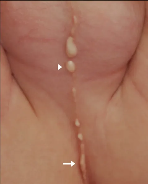
Fig. 1. Linearly arranged, multiple, whitish cysts along the perineal raphe. The arrow indicates the canalicular appearance of the cysts. The arrowhead indicates the biopsy site.
http://dx.doi.org/10.5021/ad.2016.28.3.398
A Case of Multiple Canaliform Median Raphe Cysts
Showing a Mixed Type Lining of Epithelium: A Case
Report and Review of the Literature
Sungsik Shin, Seung Kyung Hann
1, Do Young Kim
Department of Dermatology, Severance Hospital, Cutaneous Biology Research Institute, Yonsei University College of Medicine, 1Drs. Woo
and Hann’s Skin Center, Seoul, Korea
Dear Editor:
A 7-month-old boy presented with linearly arranged, mul-tiple pinhead-sized to rice-sized whitish cysts along the median raphe from the perineum to the penile shaft (Fig. 1), which were present from birth and progressively in-creased in size and number. The lesions spread along the perineal raphe and had a cordlike appearance. The infant showed no signs of pain, tenderness, pruritus, or other symptoms. He had no history of medical disease or con-genital anomaly, and the rest of the skin was normal. A skin biopsy was performed at the upper cystic portion of the lesion (Fig. 1), and the biopsy specimen showed the cysts located in the dermis with variable lining. Some parts were lined by stratified squamous epithelium with well-formed granular layer and keratin flakes. Other parts consisted of cuboidal urothelium-like epithelium with mu-cinous cells (Fig. 2). Thus, considering the site and the his-tologic features, a diagnosis of mixed-type median raphe cysts was made.
Median raphe cysts are rare, benign congenital lesions that can develop anywhere along the midline of the ven-tral side of the male genital area. They can form from the urethral meatus to the anus and the perineum along the perineal raphe1. Histologically, they are classified into four
types. The urethral type, the most common, consists of an urothelium-like epithelium, with a layer of columnar cells overlaid with several stratified layers of uniform small cells. The epidermoid type consists of stratified squamous cell epithelium. The glandular type consists of a well-formed intraepithelial glandular structure in the lining of the ure-thral epithelium. The mixed type, the second most com-mon and the type of this case, consists of more than one type of epithelium, including the urethral epithelium with squamous metaplasia, urethral epithelium with mucinous cells, or a combination of these three1.
Histomorphological features of the median raphe cyst are considered to be related to its embryonic origin and pathogenesis2. The pathogenesis is unclear, but several hy-potheses have been proposed. First, the ‘tissue trapping’
Letter to the Editor
Vol. 28, No. 3, 2016 399 Fig. 2. (A) The biopsy revealed cysts located in the dermis that showed variable lining (H&E, ×100). (B) Focal part of the cyst wall lined by stratified squamous epithelium with well-formed granular layer and keratin flakes (H&E, ×400). (C) Focal part of the cyst wall lined by cuboidal urothelium-like epithelium with mucinous cells (H&E, ×400). The arrows indicate the mucinous cells. theory states that median raphe cysts are caused by either
a defective fusion of the urethral folds or an anomalous outgrowth of the epithelium during the development of the urethra2. Second, median raphe cysts may result from the anomalous developmental rest of the periurethral glands of Littre. This hypothesis may help explain glan-dular type median raphe cysts and the involvement of mu-cinous cells in some cases3. A third hypothesis suggests that blockage of the paraurethral ducts may underlie the development of median raphe cysts4.
The canaliform median raphe cysts demonstrated in the present case are an uncommon presentation of this rare condition, and reports of these cysts are rare. Most pa-tients present with a single isolated from or a few cysts. The patient described here presented with multiple, con-tinuous cysts. This unique morphology makes diagnosis confusing due to its rarity and particular clinical features. Thus, histologic confirmation is often required5.
Complete local excision is recommended for the sympto-matic lesions to prevent possible complications. Furthermore,
whether associated congenital anomalies are present should also be assessed.
REFERENCES
1. Shao IH, Chen TD, Shao HT, Chen HW. Male median raphe cysts: serial retrospective analysis and histopathological classification. Diagn Pathol 2012;7:121.
2. Nagore E, Sánchez-Motilla JM, Febrer MI, Aliaga A. Median raphe cysts of the penis: a report of five cases. Pediatr Dermatol 1998;15:191-193.
3. Dini M, Baroni G, Colafranceschi M. Median raphe cyst of the penis: a report of two cases with immunohistochemical investigation. Am J Dermatopathol 2001;23:320-324. 4. Shiraki IW. Parametal cysts of the glans penis: a report of 9
cases. J Urol 1975;114:544-548.
5. Verma SB. Canal-like median raphe cysts: an unusual presentation of an unusual condition. Clin Exp Dermatol 2009;34:e857-e858.