Research Article
Automated Segmentation of Cerebellum Using Brain
Mask and Partial Volume Estimation Map
Dong-Kyun Lee,
1Uicheul Yoon,
2Kichang Kwak,
1and Jong-Min Lee
11Department of Biomedical Engineering, Hanyang University, Hangdang-dong, Sungdong-gu, Seoul 133-791, Republic of Korea 2Department of Biomedical Engineering, Catholic University of Daegu, Daegu, Gyeongsan-si 712-702, Republic of Korea
Correspondence should be addressed to Uicheul Yoon; [email protected] Received 22 December 2014; Revised 2 April 2015; Accepted 2 April 2015 Academic Editor: Michele Migliore
Copyright © 2015 Dong-Kyun Lee et al. This is an open access article distributed under the Creative Commons Attribution License, which permits unrestricted use, distribution, and reproduction in any medium, provided the original work is properly cited. While segmentation of the cerebellum is an indispensable step in many studies, its contrast is not clear because of the adjacent cerebrospinal fluid, meninges, and cerebra peduncle. Thus, various cerebellar segmentation methods, such as a deformable model or a template-based algorithm might exhibit incorrect segmentation of the venous sinuses and the cerebellar peduncle. In this study, we propose a fully automated procedure combining cerebellar tissue classification, a template-based approach, and morphological operations sequentially. The cerebellar region was defined approximately by removing the cerebral region from the brain mask. Then, the noncerebellar region was trimmed using a morphological operator and the brain-stem atlas was aligned to the individual brain to define the brain-stem area. The proposed method was validated with the well-known FreeSurfer and ITK-SNAP packages using the dice similarity index and recall and precision scores. As a result, the proposed method was significantly better than the other methods for the dice similarity index (0.93, FreeSurfer: 0.92, SNAP: 0.87) and precision (0.95, FreeSurfer: 0.90, ITK-SNAP: 0.93). Therefore, it could be said that the proposed method yielded a robust and accurate segmentation result. Moreover, additional postprocessing with the brain-stem atlas could improve its result.
1. Introduction
It is well known that the human cerebellum is responsible for controlling the timing of various functional activities
such as motor, balance, language, or distance measures [1–
3]. It also supports cognitive processes such as emotion and
attention [4,5]. Therefore, cerebellar volume quantification
using magnetic resonance imaging (MRI) has been widely used to investigate the cause of certain diseases, such as bipolar or motor disorders, to analyze cerebellar atrophy on traumatic brain injury, and to aid in the understanding of
brain development with age [6, 7]. The delineation of the
cerebellum is also important because it can be used as the reference region for intensity normalization of PIB or FDG
PET analysis [8,9].
Several methods for segmentation of the cerebellum, ranging from manual to automated, have been suggested
[10, 11]. While manual delineation has been adopted and
accepted as a gold standard, it suffers from inter- and intrarater variability, since it is tedious and time consuming
[12–14]. Two distinct automated methods have been proposed
to solve these problems: representative-deformable mod-els and template-based approaches. Various
representative-deformable models, such as active contour [10, 15],
gra-dient vector flow [16], and level set, have been suggested
since they are robust and unaffected by noise. Because the particular energy function, including intensity difference and sharpness, generally determines the resultant boundary, ambiguous boundaries and complex textures might trap the function. On the other hand, template-based approaches
are based on a nonlinear registration algorithm [17–19] that
computes the transformation from the reference volume to the target volume. Template-based approaches are the most widely used methods for brain segmentation, especially for lobar parcellation, since they have less constrained topology and can be applied to multiple segmentations simultaneously. However, these approaches might present the risk of system-atic error in anatomical labeling due to the relatively high variability of individual cerebellar structures.
Recently, it has been suggested that combined conven-tional segmentation algorithms, such as deformable models
Volume 2015, Article ID 167489, 10 pages http://dx.doi.org/10.1155/2015/167489
or template-based methods and tissue classification algo-rithms, might overcome several limitations of the methods described in the previous paragraph. S´egonne et al. (2004)
[20] presented a skull-stripping procedure combining the
deformable model approach and a watershed algorithm.
Kim et al. (2012) [21] constructed a deformable parametric
model for the hippocampus from seed features obtained from
multiple templates. Firbank et al. (2008) [22] integrated the
template-based approach with tissue classification for the
segmentation of the hippocampus. Shan et al. (2005) [11]
proposed combining the advantages of both template- and deformable-model-based approaches, where the cerebellar template was chosen as a seed for the active contour. These approaches showed better segmentation results than any sin-gle method, especially when applied to complex structures.
Although all these approaches exhibit relatively accurate segmentation results, there are several obstacles to delineat-ing the cerebellum exactly. For example, the surrounddelineat-ing structures of the cerebellum, such as the cerebellar peduncle, brain stem, and venous sinuses, have a similar intensity to the cerebellum itself. In the case of the venous sinuses, their boundaries and the cerebellum are divided into the thin cerebrospinal fluid (CSF). According to partial volume effects and intensity inhomogeneity, segmentation errors occurred
in several methods [23]. Furthermore, the brain stem is
connected to the cerebellum through the cerebellar peduncle, and there is no difference in their intensities.
In this study, we propose a fully automated method for segmentation of the cerebellum that combined tissue classification, a template-based approach, and morphological operations sequentially. The method was validated by com-paring the results with the manual segmentation results of the
LONI Probabilistic Brain Atlas (LPBA40) dataset [18] using
the dice similarity index and recall and precision measures. The method was also compared with other popular
pack-ages such as FreeSurfer ([24],http://surfer.nmr.mgh.harvard
.edu/) and ITK-SNAP ([35],http://www.itksnap.org).
2. Methods
2.1. Dataset. The LPBA40 dataset was used for validation of
the proposed method ([18],http://www.loni.usc.edu/atlases/
Atlas Detail.php?atlas id=12). It consists of 40 T1-weighted
brain MRI data (20 males and 20 females, 29.20 ± 6.30
years). The scans were acquired with a three-dimensional spoiled gradient echo sequence on a GE 1.5 T system as 124 contiguous 1.5 mm coronal slices. The acquisition parameters were repetition time, 10.0–12.5 ms; echo time, 4.22–4.5 ms;
flip angle, 20∘; field of view, 220 mm or 200 mm. Experienced
raters parcellated all 40 brain datasets manually into 56 structures, including the cerebellum and brain stem. 2.2. Data Processing. The proposed method for cerebellum
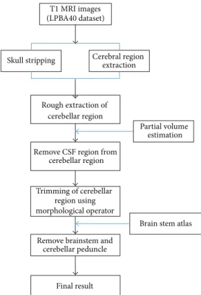
segmentation consists of several consecutive steps (Figure 1).
A preprocessing step includes intensity inhomogeneity cor-rection, skull stripping, tissue classification, and partial volume estimation. We removed the cerebral region from the skull-stripped image to define the cerebellar region
Skull stripping T1 MRI images (LPBA40 dataset) Partial volume estimation Cerebral region extraction Rough extraction of cerebellar region
Remove CSF region from cerebellar region
Trimming of cerebellar region using morphological operator
Brain stem atlas Remove brainstem and
cerebellar peduncle
Final result
Figure 1: The flowchart of the proposed cerebellar segmentation method.
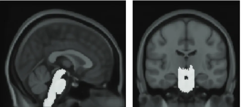
approximately and thresholded its partial volume image to remove the false positive. Morphological operators were then applied to eliminate the noise or nonconnected regions. The brain-stem template was generated by delineating the regions of brain stem and cerebellar peduncle manually on the International Consortium for Brain Mapping 152 (ICBM 152) template and aligned with the individual brain to remove the brain stem and cerebellar peduncle from the cerebellar
region (Figure 2).
2.2.1. Preprocessing and Extraction of Cerebellar Region. We corrected the intensity inhomogeneity, which involved varying the signal intensity slowly over the image caused
by magnetic field inhomogeneity [25]. Skull stripping was
performed using a Brain Extraction Tool (BET) that used a deformable model fitted to the brain surface using
optimiza-tion parameters [26]. Each brain was transformed separately
into a standardized stereotaxic space, that is, an ICBM 152
template, and resampled on a 1 mm3 voxel grid to account
for interindividual differences in absolute brain size [27].
An artificial neural network classifier was applied to identify
gray matter (GM), white matter (WM), and CSF [28].
Partial volume levels and MRI intensity mixing at the tissue interfaces due to the finite resolution of the imaging device were estimated and corrected using a trimmed minimum
covariance determinant method [29]. A cortical surface was
extracted automatically from each MR volume using the Constrained Laplacian-based Automated Segmentation with Proximities (CLASP) Algorithm to describe the cerebral
Figure 2: Brain-stem atlas image. The brain-stem atlas was defined manually on the International Consortium for Brain Mapping (ICBM) 152 template.
region without the cerebellum [30]. The cerebellar region was
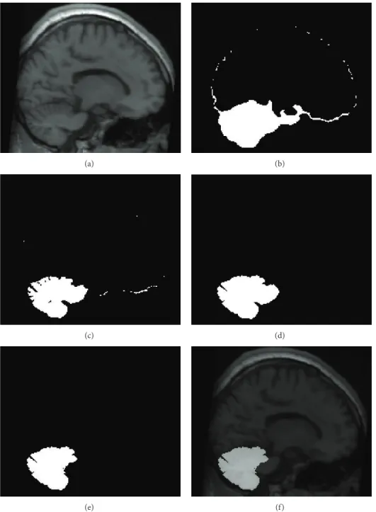
then defined approximately by subtracting the cerebral region generated by the cortical surface from the skull-stripped volume. This region was called cerebellar region candidate A (Figure 3(b)).
2.2.2. Morphological Operation and Template-Based Segmen-tation. It is important to separate the cerebellar tissue from nearby structures, such as the venous sinuses, cerebellar peduncle, and brain stem, which have a similar intensity of cerebellum. Because a thin CSF region divided the venous sinuses from the cerebellum, they were hard to be separated accurately because of the partial volume effect which was the amount of each tissue type within each voxel. Therefore, thresholding of the partial CSF volume image followed by morphological erosion was performed to remove the venous sinuses from the previously defined cerebellar region candi-date A. A connected component analysis was then applied to select the largest region as the true positive cerebellar region. We defined this region as cerebellar region candidate B. Finally, morphological dilation restored cerebellar region
candidate B to its original size (Figures3(c)and3(d)).
Since the brain stem is connected directly to the cere-bellum, it was not removed completely in the previous step. The template-based approach was applied to separate the brain stem from cerebellar region candidate B. The brain stem template was delineated manually on the ICBM 152 atlas which was generated by averaging anatomical MRI data of 152 healthy normal adults corrections for overall brain size and orientation. It was aligned to each subject using nonlinear registration to mask out the brain stem and cerebellar
pedun-cle from cerebellar region candidate B (Figures3(e)and3(f)).
Since noise might have been introduced inadvertently during the masking of the brain stem and cerebellar peduncle, a morphological opening operation, a serial combination of erosion and dilation, was applied to remove any noise from the final result.
2.3. Validation. FreeSurfer assigns a neuroanatomical label automatically to each voxel of an individual MRI volume
based on probabilistic information estimated from a man-ually labeled training set, and ITK-SNAP provides semiau-tomated segmentation using an active contour algorithm. In FreeSurfer, the “recon-all” command performed the intensity normalization, talairach registration, and labeling processes. In ITK-SNAP, the manually defined cerebellar region on the ICBM 152 template was registered to each subject using an affine transform and filled by an active contour algorithm. Differences between the gold standard and the segmenta-tion results from FreeSurfer, ITK-SNAP, and the proposed
method were examined with a paired𝑡-test.
The dice similarity index and precision and recall mea-sures were used for evaluating the proposed method. The dice similarity index, one of the most common methods for evaluating segmentation results, indicates a level of similarity
between the reference and segmented volumes [31]:
Dice Similarity Index= 𝑉𝑚∩ 𝑉𝑎
(𝑉𝑚+ 𝑉𝑎) /2, (1)
where𝑉𝑚and𝑉𝑎are the voxel sets segmented as cerebellum
in the manual delineation and each method, respectively. They range from 0 for sets that have no common elements to 1 for identical sets. The precision is the number of true positives (i.e., the number of items labeled correctly as belonging to the positive class) divided by the total number of elements labeled as belonging to the positive class (i.e., the sum of true positives and false positives). The recall is defined as the number of true positives divided by the total number of elements that actually belong to the positive class (i.e., the sum of true positives and
false negatives) [32]. They are defined as follows:
Precision= 𝑉𝑚∩ 𝑉𝑎
𝑉𝑎 ,
Recall= 𝑉𝑚∩ 𝑉𝑎
𝑉𝑚 .
(2)
A precision score of 1.0 for class 𝑉𝑚 means that every
item labeled as belonging to class𝑉𝑚belongs to class𝑉𝑎but
says nothing about the number of items from class𝑉𝑚 that
(a) (b)
(c) (d)
(e) (f)
Figure 3: Snapshot of each step in the proposed segmentation procedure. (a) Individual T1 image. (b) Roughly defined cerebellar region using skull mask and brain mask. Cerebellar region candidate A. (c) Improvement in defining the cerebellar region via the CSF partial volume estimation map. (d) Trimming of the cerebellar region using a morphological operator. Cerebellar region candidate B. (e) Removal of brain stem and cerebellar peduncle using template-based segmentation. (f) Final result.
means that every item from class𝑉𝑚 is labeled as belonging
to class𝑉𝑎but says nothing about how many other items are
incorrectly labeled as also belonging to class𝑉𝑚.
3. Result
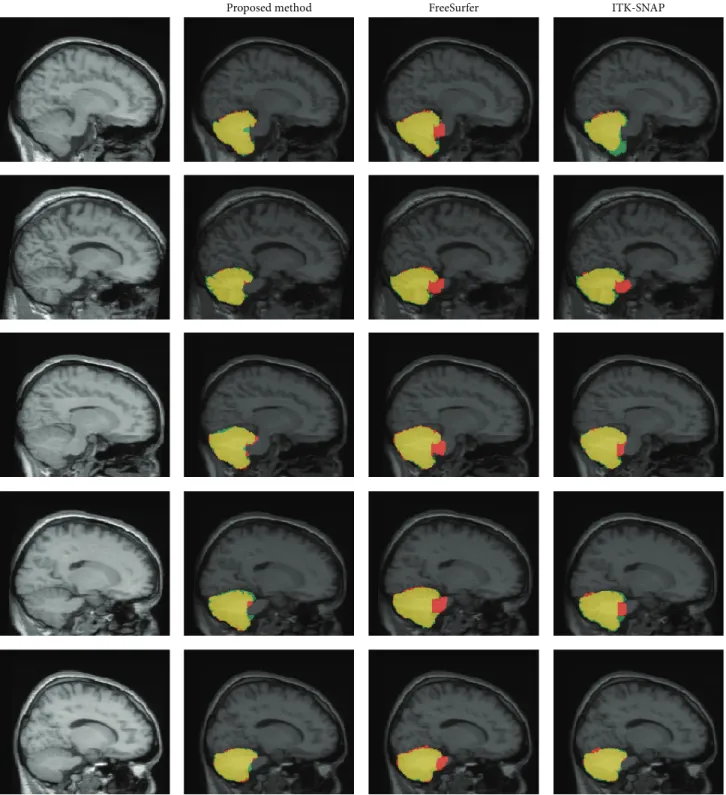
3.1. Qualitative Evaluation of Similarity and Comparison with FreeSurfer and ITK-SNAP. Figure 4 shows the segmenta-tion results from FreeSurfer, ITK-SNAP, and the proposed
method, and their differences from the manual gold standard. While FreeSurfer and ITK-SNAP exhibited over- or underes-timated results, the proposed method showed better results,
mainly from the removal of the brain stem.Figure 5shows
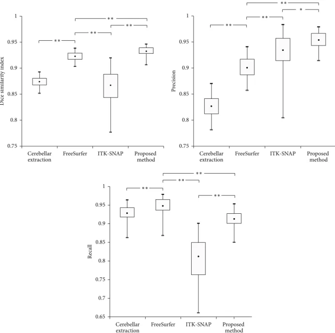
the dice similarity index for each method. The proposed method showed a significantly higher dice similarity index (0.932 ± 0.008) than FreeSurfer (0.923 ± 0.009) and ITK-SNAP (0.867 ± 0.033). This meant that the proposed method performed better than the other methods in terms of likeness.
Proposed method FreeSurfer ITK-SNAP
Figure 4: Comparison of each automatic segmentation method and manual definition. Yellow is an identical result; red is overestimation; green is underestimation.
We compared the results before and after removing the brain stem to investigate the effect of the template-based seg-mentation on the proposed method. The dice similarity index improved significantly after processing, as expected (0.874 ± 0.009 versus 0.932±0.008). For a better understanding, it was also applied to the results of FreeSurfer and ITK-SNAP. Even though there was no significant difference, indices for both
methods increased relatively (FreeSurfer:0.929 ± 0.009;
ITK-SNAP:0.879 ± 0.033) after removing the brain stem.
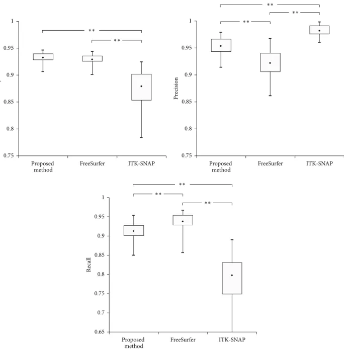
3.2. Qualitative Evaluation of Precision and Recall and Com-parison with FreeSurfer and ITK-SNAP. Figure 6shows dif-ferences in the recall and precision values among the three approaches. FreeSurfer exhibited significantly higher recall
Cerebellar
extraction FreeSurfer ITK-SNAP Proposed method Cerebellar
extraction FreeSurfer ITK-SNAP Proposed method
∗ 1 0.95 0.9 0.85 0.8 0.75 Dice simila ri ty index 1 0.95 0.9 0.85 0.8 0.75 P recisio n ∗∗ ∗∗ ∗∗ ∗∗ ∗∗ ∗∗ ∗∗ Cerebellar
extraction FreeSurfer ITK-SNAP Proposed method 1 0.95 0.9 0.85 0.8 0.7 0.65 0.75 Recall ∗∗ ∗∗ ∗∗ ∗∗
Figure 5: Box plot of quantitative indices (∗∗𝑝 < 0.001,∗𝑝 < 0.01, where 𝑝 indicates the statistical significance).
values (0.948±0.023) than ITK-SNAP (0.812±0.056) and the proposed method (0.913±0.023), while the proposed method showed significantly higher precision values (0.953 ± 0.017) than FreeSurfer (0.900 ± 0.022, 𝑝 < 0.0001) and ITK-SNAP (0.934 ± 0.037, 𝑝 = 0.004).
When the template-based segmentation of the brain stem was applied, the recall value decreased but the precision value increased after removing the brain stem from all the methods (Table 1).
4. Discussion
In this study, we propose a fully automated framework for cerebellum segmentation that consists of tissue classification, cortical surface extraction, template-based segmentation,
Table 1: Precision and recall indices before and after removing the brain stem.
Recall Precision
Before After Before After
Proposed method 0.928 (0.0226) 0.913 (0.0234) 0.827 (0.0210) 0.953 (0.0172)∗∗ FreeSurfer 0.948 (0.0225) 0.937 (0.0227) 0.900 (0.0234) 0.922 (0.0230)∗∗ ITK SNAP 0.812 (0.0562) 0.800 (0.0556) 0.934 (0.0366) 0.981 (0.0103)∗∗
∗∗𝑝 < 0.001, where 𝑝 indicates the statistical significance.
and morphological operations. The segmentation results of the proposed method were compared with FreeSurfer and
∗∗ ∗∗ 1 0.95 0.9 0.85 0.8 0.75 Dice simila ri ty index FreeSurfer ITK-SNAP Proposed
method FreeSurfer ITK-SNAP
∗∗ ∗∗ ∗∗ 1 0.95 0.9 0.85 0.8 0.75 Pre ci si on Proposed method FreeSurfer ITK-SNAP Proposed method ∗∗ ∗∗ ∗∗ 1 0.95 0.9 0.85 0.8 0.7 0.65 0.75 Recall
Figure 6: The box plot of quantitative indices after removing the brain stem and cerebellar peduncle (∗∗𝑝 < 0.001, where 𝑝 indicates the statistical significance).
ITK-SNAP, which are widely employed in brain segmenta-tion. The LPBA40 dataset with a manually defined cerebellum was used as the gold standard for an objective and righteous validation.
While the segmentation results seemed to show a rela-tively well-defined boundary for the cerebellum, the robust-ness and accuracy of each method were demonstrated using the quantitative evaluations of the dice similarity index,
and recall and precision values. As described inSection 3,
the proposed method showed better performance in the dice similarity index than FreeSurfer and ITK-SNAP. Most segmentation errors occurred in the cerebellar peduncle and venous sinuses, since the intensity of the cerebellar peduncle
was very similar to that of the cerebellar WM. FreeSurfer tended to fail in removing the cerebellar peduncle and venous
sinuses exactly [10]. This was because the atlas in FreeSurfer
excluded the venous sinuses and it could not distinguish
accurately between the brain stem and cerebellum (Figure 4)
[10, 23, 33, 34]. On the other hand, ITK-SNAP tended to
underestimate the cerebellar surface compared with
man-ual delineation (Figure 4). ITK-SNAP is a semiautomatic
approach using an active contour model where the seed point is extended to the image boundary. It showed relatively poor performance in the regions with inaccurate boundaries, since the propagation of the contour depends on an edge or
(a) (b)
(c) (d)
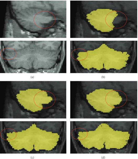
Figure 7: Comparison of the cerebellar segmentation performed with (b) proposed method (c), FreeSurfer, and (d) ITK-SNAP. Incorrect segmentation of venous sinuses (red circle) could be a problem when using the automated software.
Reliable separation of the brainstem and cerebellar peduncle from cerebellum is necessary to achieve an accurate
measurement of cerebellar volume [23]. For this reason, we
explored the effect of the brain stem including the cerebellar peduncle. After applying template-based segmentation for the brain stem to the results of FreeSurfer and ITK-SNAP, the cerebellar peduncle was separated more accurately than in the previous results and the similarity index and precision value
were significantly enhanced (Figure 6andTable 1). The
dif-ference in the similarity index between the proposed method and FreeSurfer or ITK-SNAP could be caused by incorrect discrimination of the cerebellar tissue from nearby structures,
such as the venous sinuses (Figure 7). To avoid this problem,
we eliminated the nonconnected region using a morphologi-cal operator and partial volume estimated images. As a result, our proposed method showed significantly higher indices of similarity than the other methods, even though they also removed the brain stem.
In conclusion, we propose a fully automated proce-dure for cerebellar segmentation including template-based segmentation and morphological operations. The proposed method showed accurate segmentation results when com-pared with manual delineation and removed the cerebellar peduncle from the cerebellum effectively.
Conflict of Interests
The authors declare that there is no conflict of interests regarding the publication of this paper.
Acknowledgments
This study was supported by a grant of the Korean Health Technology R&D Project, Ministry for Health, Welfare and Family Affairs, Republic of Korea (A120798), and by Basic Science Research Program through the National Research
Foundation of Korea (NRF) funded by the Ministry of Science, ICT & Future Planning (2014R1A1A1005536).
References
[1] E. J. Fine, C. C. Ionita, and L. Lohr, “The history of the devel-opment of the cerebellar examination,” Seminars in Neurology, vol. 22, no. 4, pp. 375–384, 2002.
[2] C. J. Stoodley and J. D. Schmahmann, “Functional topography in the human cerebellum: a meta-analysis of neuroimaging studies,” NeuroImage, vol. 44, no. 2, pp. 489–501, 2009. [3] H. Tiemeier, R. K. Lenroot, D. K. Greenstein, L. Tran, R.
Pierson, and J. N. Giedd, “Cerebellum development during childhood and adolescence: a longitudinal morphometric MRI study,” NeuroImage, vol. 49, no. 1, pp. 63–70, 2010.
[4] J. D. Schmahmann, “Disorders of the cerebellum: ataxia, dysme-tria of thought, and the cerebellar cognitive affective syndrome,” Journal of Neuropsychiatry and Clinical Neurosciences, vol. 16, no. 3, pp. 367–378, 2004.
[5] J. D. Schmahmann and D. Caplan, “Cognition, emotion and the cerebellum,” Brain, vol. 129, no. 2, pp. 290–292, 2006.
[6] V. S. Caviness Jr., D. N. Kennedy, C. Richelme, J. Rademacher, and P. A. Filipek, “The human brain age 7-11 years: a volumetric analysis based on magnetic resonance images,” Cerebral Cortex, vol. 6, no. 5, pp. 726–736, 1996.
[7] J. A. Scott, K. S. Hamzelou, V. Rajagopalan et al., “3D mor-phometric analysis of human fetal cerebellar development,” Cerebellum, vol. 11, no. 3, pp. 761–770, 2012.
[8] K. Ando, S. Obayashi, Y. Nagai et al., “PET analysis of dopamin-ergic neurodegeneration in relation to immobility in the MPTP-treated common marmoset, a model for Parkinson’s disease,” PLoS ONE, vol. 7, no. 10, Article ID e46371, 2012.
[9] J. Dukart, K. Mueller, H. Barthel, A. Villringer, O. Sabri, and M. L. Schroeter, “Meta-analysis based SVM classification enables accurate detection of Alzheimer’s disease across different clin-ical centers using FDG-PET and MRI,” Psychiatry Research— Neuroimaging, vol. 212, no. 3, pp. 230–236, 2013.
[10] J. Hwang, J. Kim, Y. Han, and H. Park, “An automatic cerebellum extraction method in T1-weighted brain MR images using an active contour model with a shape prior,” Magnetic Resonance Imaging, vol. 29, no. 7, pp. 1014–1022, 2011.
[11] Z. Y. Shan, Q. Ji, A. Gajjar, and W. E. Reddick, “A knowledge-guided active contour method of segmentation of cerebella on MR images of pediatric patients with medulloblastoma,” Journal of Magnetic Resonance Imaging, vol. 21, no. 1, pp. 1–11, 2005. [12] M.-E. Bolduc, A. J. du Plessis, N. Sullivan et al., “Regional
cerebellar volumes predict functional outcome in children with cerebellar malformations,” Cerebellum, vol. 11, no. 2, pp. 531–542, 2012.
[13] S. T. Camargos, W. Marques Jr., and A. C. dos Santos, “Brain stem and cerebellum volumetric analysis of Machado Joseph disease patients,” Arquivos de Neuro-Psiquiatria, vol. 69, no. 2, pp. 292–296, 2011.
[14] I. S. Gousias, A. D. Edwards, M. A. Rutherford et al., “Magnetic resonance imaging of the newborn brain: manual segmentation of labelled atlases in term-born and preterm infants,” NeuroIm-age, vol. 62, no. 3, pp. 1499–1509, 2012.
[15] N. Saeed and B. K. Puri, “Cerebellum segmentation employing texture properties and knowledge based image processing: applied to normal adult controls and patients,” Magnetic Res-onance Imaging, vol. 20, no. 5, pp. 425–429, 2002.
[16] R. Malladi and J. A. Sethian, “Image processing via level set curvature flow,” Proceedings of the National Academy of Sciences of the United States of America, vol. 92, no. 15, pp. 7046–7050, 1995.
[17] J. Diedrichsen, J. H. Balsters, J. Flavell, E. Cussans, and N. Ramnani, “A probabilistic MR atlas of the human cerebellum,” NeuroImage, vol. 46, no. 1, pp. 39–46, 2009.
[18] D. W. Shattuck, M. Mirza, V. Adisetiyo et al., “Construction of a 3D probabilistic atlas of human cortical structures,” NeuroIm-age, vol. 39, no. 3, pp. 1064–1080, 2008.
[19] F. van der Lijn, B. F. J. Verhaaren, M. A. Ikram et al., “Automated measurement of local white matter lesion volume,” NeuroImage, vol. 59, no. 4, pp. 3901–3908, 2012.
[20] F. S´egonne, A. M. Dale, E. Busa et al., “A hybrid approach to the skull stripping problem in MRI,” NeuroImage, vol. 22, no. 3, pp. 1060–1075, 2004.
[21] J. H. Kim, G. W. Astary, S. Kantorovich, T. H. Mareci, P. R. Carney, and M. Sarntinoranont, “Voxelized computational model for convection-enhanced delivery in the rat ventral hip-pocampus: comparison with in vivo MR experimental studies,” Annals of Biomedical Engineering, vol. 40, no. 9, pp. 2043–2058, 2012.
[22] M. J. Firbank, R. Barber, E. J. Burton, and J. T. O’Brien, “Validation of a fully automated hippocampal segmentation method on patients with dementia,” Human Brain Mapping, vol. 29, no. 12, pp. 1442–1449, 2008.
[23] K. Weier, A. Beck, S. Magon et al., “Evaluation of a new approach for semi-automatic segmentation of the cerebellum in patients with multiple sclerosis,” Journal of Neurology, vol. 259, no. 12, pp. 2673–2680, 2012.
[24] B. Fischl, D. H. Salat, E. Busa et al., “Whole brain segmentation: automated labeling of neuroanatomical structures in the human brain,” Neuron, vol. 33, no. 3, pp. 341–355, 2002.
[25] J. G. Sied, A. P. Zijdenbos, and A. C. Evans, “A nonparametric method for automatic correction of intensity nonuniformity in mri data,” IEEE Transactions on Medical Imaging, vol. 17, no. 1, pp. 87–97, 1998.
[26] S. M. Smith, “Fast robust automated brain extraction,” Human Brain Mapping, vol. 17, no. 3, pp. 143–155, 2002.
[27] J. C. Mazziotta, A. W. Toga, A. Evans, P. Fox, and J. Lancaster, “A probabilistic atlas of the human brain: theory and rationale for its development: the International Consortium for Brain Mapping (ICBM),” NeuroImage, vol. 2, no. 2, pp. 89–101, 1995. [28] A. P. Zijdenbos, R. Forghani, and A. C. Evans, “Automatic
‘pipeline’ analysis of 3-D MRI data for clinical trials: application to multiple sclerosis,” IEEE Transactions on Medical Imaging, vol. 21, no. 10, pp. 1280–1291, 2002.
[29] J. Tohka, A. Zijdenbos, and A. Evans, “Fast and robust param-eter estimation for statistical partial volume models in brain MRI,” NeuroImage, vol. 23, no. 1, pp. 84–97, 2004.
[30] S. K. June, V. Singh, K. L. Jun et al., “Automated 3-D extraction and evaluation of the inner and outer cortical surfaces using a Laplacian map and partial volume effect classification,” Neu-roImage, vol. 27, no. 1, pp. 210–221, 2005.
[31] L. R. Dice, “Measures of the amount of ecologic association between species,” Ecology, vol. 26, no. 3, pp. 297–302, 1945. [32] V. V. Raghavan, G. S. Jung, and P. Bollmann, “A critical
inves-tigation of recall and precision as measures of retrieval system performance,” ACM Transactions on Information Systems, vol. 7, no. 3, pp. 205–229, 1989.
[33] G. S´anchez-Benavides, B. G´omez-Ans´on, A. Sainz, Y. Vives, M. Delfino, and J. Pe˜na-Casanova, “Manual validation of FreeSurfer’s automated hippocampal segmentation in normal aging, mild cognitive impairment, and Alzheimer Disease subjects,” Psychiatry Research - Neuroimaging, vol. 181, no. 3, pp. 219–225, 2010.
[34] W. S. Tae, S. S. Kim, K. U. Lee, E. C. Nam, and K. W. Kim, “Validation of hippocampal volumes measured using a manual method and two automated methods (FreeSurfer and IBASPM) in chronic major depressive disorder,” Neuroradiology, vol. 50, no. 7, pp. 569–581, 2008.
[35] P. A. Yushkevich, J. Piven, H. C. Hazlett et al., “User-guided 3D active contour segmentation of anatomical structures: signifi-cantly improved efficiency and reliability,” NeuroImage, vol. 31, no. 3, pp. 1116–1128, 2006.
Submit your manuscripts at
http://www.hindawi.com
Stem Cells
International
Hindawi Publishing Corporationhttp://www.hindawi.com Volume 2014
Hindawi Publishing Corporation
http://www.hindawi.com Volume 2014
INFLAMMATION
Hindawi Publishing Corporation
http://www.hindawi.com Volume 2014
Behavioural
Neurology
Endocrinology
International Journal ofHindawi Publishing Corporation
http://www.hindawi.com Volume 2014
Hindawi Publishing Corporation
http://www.hindawi.com Volume 2014
Disease Markers
Hindawi Publishing Corporation
http://www.hindawi.com Volume 2014
BioMed
Research International
Oncology
Journal ofHindawi Publishing Corporation
http://www.hindawi.com Volume 2014
Hindawi Publishing Corporation
http://www.hindawi.com Volume 2014
Oxidative Medicine and Cellular Longevity
Hindawi Publishing Corporation
http://www.hindawi.com Volume 2014
PPAR Research
The Scientific
World Journal
Hindawi Publishing Corporation
http://www.hindawi.com Volume 2014
Immunology Research
Hindawi Publishing Corporation
http://www.hindawi.com Volume 2014
Journal of
Obesity
Journal ofHindawi Publishing Corporation
http://www.hindawi.com Volume 2014
Hindawi Publishing Corporation
http://www.hindawi.com Volume 2014
Computational and Mathematical Methods in Medicine
Ophthalmology
Journal ofHindawi Publishing Corporation
http://www.hindawi.com Volume 2014
Diabetes Research
Journal ofHindawi Publishing Corporation
http://www.hindawi.com Volume 2014
Hindawi Publishing Corporation
http://www.hindawi.com Volume 2014
Research and Treatment
AIDS
Hindawi Publishing Corporation
http://www.hindawi.com Volume 2014
Gastroenterology Research and Practice
Hindawi Publishing Corporation
http://www.hindawi.com Volume 2014