INTRODUCTION
Eosinophils are produced in the bone marrow along with oth-er white blood cells and circulate at relatively low levels in the bloodstream, making up 1-3% of white blood cells. Eosinophils also occur outside the bone marrow and blood vessels: in the medulla and the junction between the cortex and medulla of the thymus and in the lower gastrointestinal tract, ovary, uterus, spleen, and lymph nodes. In allergic conditions, they are found in the lung, skin, and esophagus. Eosinophils persist in the cir-culation for 8-12 hours and can survive in tissues for an addition-al 8-12 days in the absence of stimulation.1 Eosinophils are dis-tinguished based on their characteristic morphological features, namely bilobed nuclei and cytoplasmic granules of a distinctive granular pink,2 and they are about 12-17 μm in diameter (Fig. 1). While no cell surface proteins unique to eosinophils have as yet been recognized, they are armed with abundant specific cyto-plasmic granules with their structural packaging of cationic pro-teins, the eosinophils’ most characteristic morphologic feature.
Eosinophil Development, Regulation of Eosinophil-Specific Genes,
and Role of Eosinophils in the Pathogenesis of Asthma
Tae Gi Uhm,
†Byung Soo Kim,
†Il Yup Chung*
Division of Molecular and Life Sciences, College of Science and Technology, Hanyang University, Ansan, Korea
Eosinophil granules are composed mainly of cytotoxic cationic proteins and also harbor a multitude of cytokines and chemo-kines. Eosinophils are terminally differentiated cells that arise from hematopoietic CD34+ stem cells through commitment and differentiation and do not appear to multiply after leaving the bone marrow. An interplay of several key transcription factors dictates eosinophil lineage development and differentiation, and an almost identical set of factors activates transcription of eosinophil-specific genes encoding the major basic protein (MBP), eosinophil-derived neurotoxin (EDN), eosinophil per-oxidase (EPO), Charcot-Leyden crystal (CLC) protein, CC
che-Allergy Asthma Immunol Res. 2012 March;4(2):68-79. http://dx.doi.org/10.4168/aair.2012.4.2.68
pISSN 2092-7355 • eISSN 2092-7363
Eosinophils arise from hematopoietic CD34+ stem cells in the bone marrow. They acquire IL-5Rα on their surface at a very early stage during eosin-ophilopoiesis, and differentiate under the strong influence of interleukin (IL)-5. They then exit to the bloodstream, and enter the lung upon exposure to airway inflammatory signals, including eotaxins. In inflamed tissues, eosinophils act as key mediators of terminal effector functions and innate immunity and in linking to adaptive immune responses. Transcription factors GATA-1, CCAAT/enhancer-binding protein, and PU.1 play instructive roles in eosinophil specification from multipotent stem cells through a network of cooperative and antagonistic interactions. Not surprisingly, the in-terplay of these transcription factors is instrumental in forming the regulatory circuit of expression of eosinophil-specific genes, encoding eosinophil major basic protein and neurotoxin, CC chemokine receptor 3 eotaxin receptor, and IL-5 receptor alpha. Interestingly, a common feature is that the critical cis-acting elements for these transcription factors are clustered in exon 1 and intron 1 of these genes rather than their promoters. Elucidation of the mechanism of eosinophil development and activation may lead to selective elimination of eosinophils in animals and human subjects. Further-more, availability of a range of genetically modified mice lacking or overproducing eosinophil-specific genes will facilitate evaluation of the roles of eosinophils in the pathogenesis of asthma. This review summarizes eosinophil biology, focusing on development and regulation of eosinophil-spe-cific genes, with a heavy emphasis on the causative link between eosinophils and pathological development of asthma using genetically modified mice as models of asthma.
Key Words: Asthma; CCR3; eosinophils; eotaxin; GATA-1; IL-5
This is an Open Access article distributed under the terms of the Creative Commons Attribution Non-Commercial License (http://creativecommons.org/licenses/by-nc/3.0/) which permits unrestricted non-commercial use, distribution, and reproduction in any medium, provided the original work is properly cited.
Correspondence to: Il Yup Chung, PhD, Division of Molecular and Life Sciences, College of Science and Technology, Hanyang University, 1271 Sa-3-dong, Sangrok-gu, Ansan 426-791, Korea.
Tel: +82-31-400-5514; Fax: +82-31-419-1760; E-mail: [email protected] Received: July 30, 2011; Accepted: August 31, 2011
† These two authors contributed equally to this work.
mokine receptor 3 (CCR3), and interleukin-5 receptor alpha (IL-5Rα) chain. Eosinophils are multifunctional leukocytes im-plicated in the pathogenesis of inflammatory responses, nota-bly including allergic diseases and parasitic helminth infections. Much controversy exists as to the role of eosinophils in homeo-static and diseased conditions. Recent advances have allowed selective removal of eosinophils in rodents and asthmatic pa-tients through genetic manipulation and therapeutic agents. With these tools, we are now in a much better position to deter-mine the role of eosinophils in the pathophysiology of asthma and so develop novel therapeutic approaches.
Eosinophil development
Recent advances in the biology of cellular development/dif-ferentiation have highlighted the fact that any cell type can seemingly become any other, given the correct combinations of transcription factors and environment. Eosinophil develop-ment appears to faithfully conform to this notion. Eosinophil lineage fate is determined by the interplay of a few key transcrip-tion factors, including CCAAT/enhancer-binding protein (C/ EBP family member), GATA-1 (a zinc finger family member), PU.1 (an Ets family member), and friend of GATA (FOG). In par-ticular, C/EBPα/β and GATA-1, either individually or in concert, play a decisive role in eosinophil commitment from multipo-tent stem cells. C/EBPs are a family of transcription factors that contain a highly conserved, basic-leucine zipper domain at the C-terminus that is involved in dimerization and DNA binding. Six members of this family (α, β, γ, δ, ε, and ζ) have thus far been isolated and characterized.3 Expression of NF-M, the chicken homolog of C/EBPβ, fused to the ligand binding domain of es-trogen receptor induces the up-regulation of an eosinophil-specific surface marker EOS47 along with the down-regulation of a specific marker of a multipotent chicken progenitor cell line transformed by the Myb-Ets oncoprotein.4 Mice with a null mutation in C/EBPα fail to generate eosinophils and neutrophils, whereas other hematopoietic lineages, including monocytes, are not affected.5 The enforced expression of either C/EBPα or C/EBPβ induces eosinophil differentiation of the chicken-trans-formed cells.6 A dominant negative C/EBP that antagonizes all C/EBP members blocks granulocyte and monocyte
develop-ment from human cord blood CD34+ progenitors.7 There is a functional redundancy of C/EBPα and C/EBPβ family mem-bers for granulocyte development/differentiation, although C/ EBPβ-deficient mice do not show any defects in formation of myeloid lineage, unlike C/EBPα.8 Additionally, a dominant neg-ative C/EBPβ phenotype induces the formation of immature eosinophils, indicating that C/EBPβ also promotes eosinophil maturation.6
GATA-1 is a member of the GATA family of transcription fac-tors that contain two zinc finger motifs. GATA-1 is expressed in the hematopoietic system, including by erythroid cells, mega-karyocytes, eosinophils, and mast cells and in the Sertoli cells of the testis.9 GATA-1 reprograms avian myeloblastic cell lines to eosinophils, and its expression level fine tunes development of the eosinophil lineage.10 Human cord blood CD34+ cells that are transduced by GATA-1-expressing retrovirus exclusively give rise to eosinophils. The C-terminal zinc finger motif of GATA-1 is necessary for formation of eosinophils, and GATA-1-deficient fetal liver cells lack the ability to form eosinophils.11 Deletion of a high-affinity GATA-binding site in the GATA-1 promoter, ∆dblGATA, leads to selective loss of the eosinophil lineage, whereas development of platelet, mast cells, and red blood cells remains little-changed.12 When granulocyte/macrophage pro-genitors (GMPs) are isolated from bone marrow cells of trans-genic GATA-1 reporter-tagged GFP and grown in liquid culture, eosinophils are found only in the GFP-positive fraction, along with acquisition of surface IL-5Rα.13 C/EBPβ and GATA-1 syn-ergistically regulate activity of MBP promoter.14 The level of GATA-1 expression is an important element in establishing the eosinophil phenotype, as it activates an eosinophil-specific gene at low, and represses it at high, GATA-1 concentrations.10,15 Additionally, the timing of expression of these transcription fac-tors is critical. For instance, when GATA-2 acts on GMPs ex-pressing C/EBPα, it exclusively induces eosinophil formation, whereas it instructs GMPs to form basophils and/or mast cells if GMPs are not expressing C/EBPα.16 GATA-2 has an instructive capacity toward eosinophil lineage from human cord blood CD34+ progenitors comparable to that of GATA-1 and efficient-ly compensates for GATA-1-deficiency in terms of eosinophil development in vivo.11 GATA-2 also complements GATA-1 to activate EDN transcription.17 Nonetheless, the in vivo role of GATA-2 in eosinophil development remains to be determined, as GATA-2-deficient mice display a general reduction in hema-topoiesis, and a complete lack of mast cells.18
Given that the two transcription factors GATA-1 and C/EBP serve as the master regulators of eosinophil development, it is proper to mention how GATA-1 and C/EBP might induce eo-sinophil formation in CD34+ cells. Two models have been pro-posed.19 In the first, stochastic expression of either GATA-1 or C/EBPα in a common progenitor induces expression of the other, resulting in co-expression of both factors and ultimately eosinophil formation. In the second model, each of the factors Fig. 1. Eosinophils. (A) Peripheral blood eosinophils purified by negative
selec-tion. (B) and (C) Cord blood-derived eosinophils. Cord blood CD34+ cells were cultured for 18 days with a cytokine cocktail. Cultured cells were stained with Diff Quick (B) or probed with FITC-conjugated anti-MBP antibody (C). DAPI and MBP stains are shown in blue and green, respectively.
acts on a distinct type of CD34+ cell, leading to production of eosinophil lineage. These authors favor the second model, as several distinct subpopulations of CD34+ cells exist, and Myb-Ets-transformed multipotent progenitor cells can readily be converted to any cell type depending on the combination of transcription factors, including C/EBP, GATA-1, PU.1, and FOG (see below), to which they are exposed.
Other players also act in concert with C/EBP and GATA-1 in the process of eosinophil commitment. PU.1 is a transcription factor with a winged helix-turn-helix DNA binding domain that is a member of the Ets transcription family and is expressed in hematopoietic cells, including myeloid cells.20 Conditional acti-vation of PU.1 in Myb-Ets-transformed multipotent progenitor cells induces the formation of cells with properties of immature eosinophils after short-term culture.21 The mechanism by which PU.1 induces eosinophil commitment in transformed cells in-volves downregulation of GATA-1 expression,22 agreeing with the observation that an intermediate GATA-1 level is required for eosinophil commitment.10,15 When PU.1 is co-expressed with C/EBPε32 and GATA-1, however, it transactivates the MBP promoter.22 Hence, PU.1 differentially exerts its function de-pending on the context of available transcription factors. FOG contains nine zinc fingers, at least two of which are capable of binding to the N-terminal finger motif of GATA-1.23 Expression of FOG in eosinophils leads to a loss of eosinophil markers and the acquisition of a multipotent lineage, and constitutive ex-pression of FOG in multipotent progenitors inhibits activation of MBP gene transcription by GATA-1,14 C/EBPβ,24 or a combi-nation of GATA-1, C/EBPε32, and PU.1.22 Thus, FOG acts as a repressor of the eosinophil lineage. These results highlight the importance of both cooperative and antagonistic interactions of multiple transcription factors for eosinophil-lineage com-mitment from multipotent hematopoietic progenitors.
The involvement of two additional transcription factors in eo-sinophil development has been documented. IFN consensus sequence binding protein (Icsbp) is an IFN-γ-induced transcrip-tion factor that regulates IFN-responsive genes.25 Icsbp-deficient mice have reduced eosinophil developmental potential and eosinophil progenitors. Eosinophil progenitors from icsbp-de-ficient mice show reduced expression of GATA-1 and are un-able to respond to IL-5 in terms of eosinophil colony forma-tion.26 Therefore, Icsbp appears to play a critical role in the de-velopment of the eosinophil lineage, although little known about the underlying molecular mechanism. Id proteins are basic helix-loop-helix transcription factors that lack a basic DNA binding domain.27 Constitutive expression of Id1 inhibits eosinophil development, whereas Id2 accelerates the final mat-uration of eosinophils. The effects of Id factors do not seem to be restricted to eosinophils, however, because they also pro-mote neutrophil development and maturation.28 Notch is an evolutionarily conserved transmembrane protein that regu-lates a broad spectrum of cell-fate decisions and
differentia-tion.29 Notch signaling promotes eosinophil maturation30 as well as affecting eosinophil functionality.31,32 However, whether Notch signaling modulates the transcription factors or unidentified pathways key for deciding eosinophil fate or is itself affected by these factors remains to be determined.
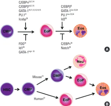
Once multipotent progenitor cells commit to becoming eo-sinophil progenitors, they go through several intermediate stag-es before becoming fully mature eosinophils that are seen in the circulation and tissues. These stages include promyeloblasts, promyelocytes, metamyelocytes, band form, and segmented form, based on morphological criteria.33 As readouts in most studies of hematopietic development are measured by the for-mation of fully mature cells, it is difficult to identify the exact development/differentiation stage at which a particular tran-scription factor exerts its function. Most of the aforementioned transcription factors positively or negatively act on the commit-ment stage. These include GATA-1, GATA-2, C/EBPα, C/EBPβ, C/EBPε, PU.1, Icsbp, FOG, and Id1, whereas fewer numbers of transcription factors are known to specifically act on the late or terminal stages (Fig. 2A). For instance, Id228 and moderate GATA-1 levels10,15 are required for progression through
matura-Mouse13 Human37 C/EBPα5,6,7,14 C/EBPβ4,6,7,15 GATA-110,11,12,13,14,15 PU.121 Icsbp26 FOG24 Id128 GATA-1high 10 C/EBPβ6 GATA-1low 22,34 PU.1low 22,34 Id228 C/EBPε34 Notch30
Fig. 2. Eosinophil development. (A) Transcription factors regulating eosinophil commitment and maturation. Eosinophil commitment is dictated largely by two transcription factors, CCAAT/enhancer-binding protein (C/EBP) and GATA-1, whose levels and functions are fine-tuned by interactions with the other tran-scription factors PU.1 and friend of GATA (FOG). Icsbp and Id1 individually regu-late eosinophil formation, although their relationship with C/EBP and GATA-1 are unknown. Eosinophil maturation is driven by a similar combination of tran-scription factors but is inhibited by C/EBPe. Notch signaling prevents eosinophil maturation by an unknown mechanism. (B) Different pathways of eosinophil development in the mouse and human. Human eosinophil progenitors arise di-rectly from a common myeloid progenitor, whereas mouse eosinophil progeni-tors arise from a common myeloid progenitor via a granulocyte/macrophage progenitor that is bipotent for eosinophils and neutrophils.
A
tion, whereas C/EBPε14/27 isoforms, which are highly ex-pressed on peripheral blood eosinophils34 and terminally dif-ferentiated eosinophils,35 block MBP transcription. Given that a wide spectrum of transcription factors is present in mature eo-sinophils, they may influence late-stage eosinophil differentia-tion and maturadifferentia-tion. These molecules could be novel targets for therapeutic approaches to eosinophil-associated inflamma-tion.
Despite sharing many features, such as transcription factors for eosinophil commitment and maturation, there are subtle differences in the lineage pathway through which eosinophils are generated in mice and humans (Fig. 2B). In mouse hemato-poiesis, eosinophil potential exists along with the granulocyte/ monocyte differentiation pathway from hematopoietic stem cells, and at least a fraction of granulocyte/macrophage pro-genitors (GMPs) are bipotent for the eosinophil and the neutro-phil lineages. Eosinoneutro-phil progenitors are found within cells ac-tivating GATA-1, whereas GMPs that do not express GATA-1 give rise to neutrophils and macrophages. Thus, eosinophil progenitors exist as a distinct population downstream of GMP.13 The mouse bipotent basophil/mast cell progenitor and the ba-sophil lineage-committed progenitor are also identified down-stream of the granulocyte/macrophage progenitor,36 suggesting that the commitment of eosinophil and basophil/mast lineages occurs independently after the multipotent progenitor has lost the megakaryocyte/erythroid lineage potential. In contrast, in human hematopoiesis, GMPs lack eosinophil potential, and eosinophil progenitors are instead found in common myeloid progenitors (CMPs) that both do and do not express surface IL-5Rα. Cells expressing IL-5Rα give rise exclusively to eosinophils
but never basophils or neutrophils.37 However, as cells possess-ing both basophil and eosinophil granules have been found in leukemia patients,38 it is possible that a distinct cell type exists that has deviated from the known lineage pathway.
Regulation of eosinophil-specific genes
Analysis of the transcription factors that control eosinophil-specific genes may offer insights, at the molecular level, into the mechanisms behind the commitment of multipotent progeni-tors into the eosinophil lineage. Relatively small numbers of genes are exclusively or predominantly transcribed in eosino-phil progenitors and fully differentiated eosinoeosino-phils.39 These in-clude genes encoding eosinophil granule proteins (MBP, ECP, EDN, EPO, CLC protein) and surface receptors (IL-5Rα and CCR3). Not surprisingly, almost the same set of transcription factors that dictate eosinophil commitment and differentiation are also involved in controlling transcription of eosinophil-spe-cific genes. These include C/EBP family proteins, GATA factors, and PU.1. The regulatory regions of these genes include known or putative binding sites for these transcription factors. Interest-ingly enough, these cis-acting control elements are clustered in the sequences flanking their exon 1 and intron 1 rather than the promoter (Fig. 3), although the implication of this for transcrip-tional regulation remains to be determined.
MBP is a granule protein localized in the crystalline core with no known enzymatic activity. Transcriptional regulation of the human MBP gene is the most thoroughly studied of all eosino-phil-specific genes14,22,34,40,41 because MBP is a representative marker of eosinophils, and MBP transcript accounts for up to 8.1% of the total cellular mRNAs of eosinophils.42 Two different
CCR3 59,60,61,62 IL-5Rα 65,66,67,68,69 MBP 14,22,34,41 EDN 17,45,46,47,48,49 ECP EPO 53
Proximal promoter regions of genes encoding eosinophil granule proteins Proximal promoter regions of CCR3 and IL-5Rα genes
A B
Fig. 3. Regulatory regions of eosinophil-specific genes. Transcription factor binding sites in the MBP (NM002728.4), EDN (NM002934.2), ECP (NM002935.2), EPO (NM000502.4), CCR3 (NM001837.3), and IL-5α genes (NM000564.3). Functional binding sites are indicated by dark figures, and putative binding sites that have not been confirmed as functional are indicated by light figures. Numbering is relative to the transcriptional start site of each gene.
transcripts arise from differential splicing of alternative MBP transcripts from promoters P1 and P2, respectively, located 32 kb apart in the genomic DNA. The P2 promoter is predomi-nantly responsible for MBP expression in eosinophil lineage cells.40 The P2 promoter of the MBP gene contains a functional GATA site and a C/EBP site (Fig. 3A). Binding of GATA-1 or C/ EBPα/β to its respective binding site transactivates the MBP P2 promoter.41 A subsequent study by the same group showed that GATA-1 and C/EBPβ interact physically to synergistically trans-activate the MBP P2 promoter. Furthermore, FOG acts as a neg-ative cofactor for GATA-1-, but not C/EBPβ-, mediated transac-tivation.14 The P2 promoter is activated by GATA-1 alone but is synergistically transactivated by low levels of PU.1 in the pres-ence of optimal GATA-1 levels. PU.1 and C/EBPε individually activate the P2 promoter.34 In addition to GATA-1 and C/EBPβ, the combination of GATA-1 and PU.1 transactivates the MBP P2 promoter.34 By contrast, C/EBPε14, which lacks the transactiva-tion domain and is expressed at high levels in terminally differ-entiated eosinophils,34,35 strongly inhibits the P2 promoter. C/ EBPε27 also represses P2 promoter activity via protein–protein interaction through the C/EBP and/or GATA-binding sites, but not the PU.1 sites.34 Thus, the active transcription complex con-sisting of well-known transcription factors is required for regu-lation of MBP P2 promoter activity. The complex includes inter-actions between GATA-1 and C/EBPα, GATA-1 and C/EBPβ, GATA-1 and C/EBPε isoforms, GATA-1 and PU.1, PU.1 and C/ EBPε isoforms, and GATA-1 and FOG.14,22,34,41 These findings es-tablish a combinatorial cooperation and antagonism through protein–protein interactions of the transcription factors that control eosinophil development.
EDN is a cationic granule protein synthesized in eosinophils,43 and it has ribonuclease activity that can degrade the RNA ge-nomes of some viruses. EDN also has an immunomodulatory function in terms of regulation of dendritic cell migration44. As seen in Fig. 3, the key regulatory sequence of EDN transcription resides in the promoter and intron, which contain GATA, C/ EBP, PU.1, NFAT, and AP-1 sites,17,45 most of which are function-al. PU.1,46 C/EBP isoforms α, β, and ε47, or NFAT binding48 to their respective binding sites in intron 1 of the gene induces transac-tivation. The promoter region also contains two GATA sites, which are 600 bp apart. GATA-1 and GATA-2 bind the two func-tional GATA sites in the EDN promoter. GATA-2 can replace the effect of GATA-1.17 Elsewhere, HNF4 interacts with Sp1 to stim-ulate EDN promoter activity.49
ECP is found in the matrix of the eosinophil-specific granule and has more potent anti-helminthic activity but less ribonu-clease activity than EDN.50 The ECP gene sequence is highly ho-mologous to that of EDN, in particular, with 92% identity in the upstream 1-kb sequence.51 Given that the ECP gene shares with the EDN gene all cis-acting elements at identical positions (Fig. 3), almost identical molecular cues appear to govern regulation of gene expression.
EPO is a heme-containing glycoprotein that possesses peroxi-dase activity. It is located in the matrix of the granule and has a sequence that is closely related to neutrophil myeloperoxidase.52 A number of positively and negatively cis-acting elements are mapped to the proximal promoter of this gene, including tran-scription factors Egr-1, H4TF-1, CTCF, UBP-1, and GaEII, al-though it is not known whether these potential binding sites are functional for EPO transcription.53 Additionally, sequence anal-ysis shows that binding sites for GATA factors, PU.1, and C/EBP are present in intron 1 and the promoter (Fig. 3), again suggest-ing that the transcription factors for eosinophil development are actively involved in the transcriptional regulation of the EPO gene.
CCR3 is constitutively expressed at high levels in eosinophils, with 16,000–60,000 receptors per cell; it serves as the primary chemokine receptor responsible for eosinophil trafficking to tissues in diseased and healthy conditions.54,55 CCR3 is also ex-pressed on prominent allergic inflammatory cells, including Th2 helper56 and mast cells.57 The restricted expression of CCR3 leads to a notion that it plays an integral role in the pathogene-sis of allergic diseases including asthma, allergic dermatitis, and allergic rhinitis. Furthermore, as airway epithelial cells ex-press functional CCR3, this protein is postulated to play roles beyond simple cell trafficking, such as in airway remodeling.58 A recent study revealed that CCR3 serves as an identification marker, along with IL-5Rα, in eosinophil progenitors at the very early stage of human eosinophil development.37 Hence, analy-sis of the transcription factors that control CCR3 expression may offer insights into the mechanisms behind the commit-ment of common myeloid progenitors to the eosinophil lin-eage. The key sequences for CCR3 gene transcription reside in exon 1 and intron 1 rather than in the promoter,59-61 (Fig. 2B). Multiple GATA binding sites are present in exon 1 and intron 1. Exon 1, in particular, has five GATA sites, each of which has a different GATA-1 binding affinity, with one of the five as a posi-tively acting element and two as negaposi-tively acting elements for transcription in vitro.62 C/EBP and PU.1 binding sites are locat-ed in the promoter and intron 1 regions, respectively, although their function remains to be determined. Additionally, AML-1 and CREB binding motifs are present in exon 1. We recently found evidence that protein binding to AML-1 and CREB sites contributes to transactivation of the CCR3 gene (our unpub-lished results), as much as does GATA binding. Therefore, it is plausible that these transcription factors participate in eosino-phil development and maturation.
IL-5R consists of heterodimer, a unique ligand-binding α chain and β chain shared with IL-3 and GM-CSF receptors that is linked to the Janus kinase/signal transducer and activator of transcription and phosphoinositol-3-kinase.63 IL-5R mediates their differentiation and maturation, survival, chemotaxis, and effector functions.64 Eosinophils, but not basophils or neutro-phils, possess a high level of IL-5Rα, and IL-5Rα is a key surface
molecule in sorting of murine eosinophil progenitors.13 Al-though IL-5Rα is expressed as a result of commitment to the eosinophilic lineage,13 human CMPs that express IL-5Rα give rise to only eosinophils.37 Therefore, IL-5Rα is presumably the earliest phenotypic marker that eosinophils acquire at the com-mitment step of the developmental pathway. Given that IL-5Rα-positive CMPs are derived from the CMPs that lack this surface marker, the signals and transcription factors that induce IL-5Rα transcription may play an integral role in eosinophil fate decision. An early study demonstrated that 34 bp of the proxi-mal region of the IL-5Rα promoter serves as the binding site for a myeloid- and eosinophil-specific transcription factor.65 These turned out to be RFX family transcription factors, although RFX family proteins are not expressed in a myeloid- or eosinophil-specific manner.66 An AP-1 site, located upstream of the RFX binding site, functionally cooperates with a neighboring EOS site to mediate IL-5Rα transcription. C-Jun, CREB, and CREM bind to the AP-1 site.67 There is a second promoter, designated P2, for the human IL-5Rα gene. Oct2 transactivates murine B cells’ IL-5α gene by binding to its promoter.68 A short sequence of 6 bp within the P2 promoter is responsible for the binding of an uncharacterized protein and is sufficient for promoter activ-ity in eosinophilic cells.69 In C/EBPα-null mice, expression of the IL-5Rα gene was greatly reduced.69 Moreover, sequence analysis of exon 1 and intron 1 as well as promoters shows that a number of GATA factors, C/EBP, and PU.1 binding sites are concentrated in these sequences (Fig. 2B). Nevertheless, the important transcription factors GATA-1 and C/EBP, which are believed to direct cells toward the eosinophil lineage, have not yet been reported as necessary and/or sufficient for transcrip-tion of the IL-5Rα gene.
Although the aforementioned transcription factors are pri-marily responsible for the regulation of eosinophil-specific gene expression, their mere presence even in combination is not sufficient for induction of gene transcription. Many eosino-phil-specific genes encoding eosinophil basic proteins, CCR3, and IL-5R are induced by modifiers of histone structure such as histone acetyltransferase inhibitors,14,46,53,60,65 and expression of many asthma-related inflammatory genes is affected by these agents.70 DNA methyltransferase inhibitors also have a high propensity to alter eosinophil-specific gene expression. More-over, regulation of these gene products by microRNAs has not yet been reported. As the relative importance of role of epigen-etic regulation has increasingly become evident, the study of epigenetic regulation of eosinophil-specific genes is vital. Tak-en together, these findings show that the deciphering of eosino-phil-specific gene expression will provide both a molecular ba-sis for eosinophil development and targets for novel therapies for the treatment of eosinophil-associated diseases.
Role of eosinophils in asthma
Eosinophils are associated with the pathogenesis of asthma,
and the presence of eosinophils in the airway lumen and lung tissues is often regarded as a defining feature of this disease.71 The role of eosinophils in the pathogenesis of asthma is due to their ability to mediate terminal effector functions and innate immune responses by secreting a wide variety of cationic pro-teins, lipid mediators, and cytokines/chemokines. Further-more, eosinophils are capable of bridging innate and adaptive immune responses by elaborating T cells, dendritic cells, and mast cells. The recent availability of genetically modified mice makes possible the elucidation of a causal relationship between eosinophil recruitment and the onset or progression of pulmo-nary pathologies associated with asthma and provides new in-sight into the role of eosinophils in the pathogenesis of the al-lergic disease. In these animals, eosinophils are depleted or overproduced by manipulating expression of transcription fac-tors regulating eosinophil development, production of IL-5 and eotaxins, and expression of receptors responding to these cyto-kines, through transgenic systems, gene disruption, and neu-tralizing antibodies. Thus, much information on the role of eo-sinophils roles has been accumulated using experimental mod-els. This section describes the contribution of eosinophils to the pathogenesis of allergic disease within the context of asthma. Eosinophil-deficient mice
Two strains of mice that lack eosinophils were engineered in different genetic backgrounds. Removal of a high-affinity dou-ble GATA site from the GATA-1 promoter (∆dbl-GATA) in a BALB/c background selectively ablates eosinophils.12 When ∆dbl-GATA mice are subjected to a standard experimental asthma protocol of sensitization and challenge with allergen, the absence of eosinophils does not protect the mice from AHR development, but are required for airway remodeling.72 How-ever, ∆dbl-GATA mice created in a C57BL/6 background show decreased allergen-induced AHR, T cell recruitment to the lung, and production of Th2 cytokines and chemokines (Table 1). Furthermore, adoptive transfer of eosinophils or CCL11/eo-taxin-1 delivery to ∆dbl-GATA BABL/c mice results in recruit-ment of lung T cells and restoration of airway inflammation.73 A second line of mice devoid of eosinophils, PHIL mice, was cre-ated in the C57BL/6 background by transgenic expression of diphtheria toxin A driven by the EPO promoter.74 In this line of mice, eosinophils are nearly completely deficient in all organs in which they occur under homeostatic conditions. Allergen challenge of these mice does not induce AHR or pulmonary mucus accumulation, suggesting a link between eosinophils and allergic pulmonary pathologies. The combined transfer of Th2-polarized OVA-specific transgenic T cells and eosinophils to PHIL mice, but not transgenic T cells alone, results in accu-mulation of the effector T cells and airway Th2 responses, sug-gesting that the primary role of pulmonary eosinophils is to elicit localized recruitment of effector T cells.75 These data sup-port the central hypothesis that eosinophils are required for the
recruitment of T cells to the lung and thus are not only terminal effector cells but also important modulators of allergic asthma. IL-5- or IL-5Rα-deficient mice
IL-5 plays a role in the pathogenesis of eosinophilic inflam-mation and asthma. Airway allergen challenge in asthmatics induces expression of IL-5 by T cells,76 whereas increased levels of IL-5 and MBP can be detected in the airway of symptomatic asthmatics.77 IL-5-deficient mice in a C57BL/6 background fail to develop AHR and airway eosinophilia upon aeroallergen challenge (Table 2), suggesting an essential role for IL-5 in in-duction of eosinophilia and development of AHR.78 Indeed, re-duced lung eosinophils and AHR are observed in mice treated with IL-5 antibodies.79 IL-5-deficient mice also show lesser al-terations in tissue remodeling events, including peribronchial fibrosis and thickness of the peribronchial muscle layer, along with a reduction in the production of TGF-β and MBP by eosin-ophils.80 In contrast, IL-5-deficient BABL/c mice develop aller-gen-induced AHR, as wild-type mice do, despite markedly re-duced blood and lung eosinophilia,81,82 suggesting dissociation of airway eosinophilia from AHR development. On the other hand, transgenic mice that constitutively express IL-5 in the lung epithelium develop an accumulation of eosinophils and pathologic changes including goblet cell hyperplasia, epithelial hypertrophy, and AHR even in the absence of antigen chal-lenge.83 Genetic IL-5Rα deficiency decreases antigen-induced airway eosinophilia and AHR.84 The confusion involving the ef-fect of IL-5 on lung functions is also observed in human clinical studies. An initial study using humanized anti-IL-5 antibody in patients with mild asthma demonstrated >90% lower blood and sputum eosinophilia but was not effective in improving lung function, as measured by FEV1.85 A subsequent study showed that anti-IL-5 did not reduce the level of MBP in the
airways, even in the presence of partially inhibited airway eo-sinophils (approximately 55%).86 In contrast, anti-IL-5 therapy was effective in treatment of a small group of patients with eo-sinophilic asthma.87,88 Thus, studies from both human subjects and murine models show that IL-5 is responsible for the induc-tion of pulmonary eosinophilia, but the role of IL-5-induced eosinophils in the pathogenesis of asthma remains unan-swered. Nevertheless, the association/dissociation of airway eosinophilia with lung function seen in some mouse strains and the differential clinical benefits of anti-IL-5 therapy have important implications for the treatment of asthma and testify to the complex pathogenesis of the disease.
Eotaxins (CCL11, CCL24, and CCL26)- and/or CCR3-deficient mice
Three eotaxin family proteins, eotaxin-1/CCL11, eotaxin-2/ CCL24, and eotaxin-3/CCL26 have been identified,89 all of which selectively bind to CCR3. Eotaxin-2 and -3 are distantly related to eotaxin-1, with ~30% sequence identity and different chromosomal locations. Gene disruption studies of eotaxins-1 and -2 have been published, and both eotaxin-1 and eotaxin-2 have not yet been characterized as a functional murine homo-logue of eotaxin-3 (Table 2). Targeted disruption of CCL11/eo-taxin 1 leads to partially reduced eosinophil counts in the blood and airways under baseline conditions without affecting eosin-ophil hematopoiesis in the bone marrow. Upon exposure to aeroallergen, eotaxin-1-deficient mice show ~70% reductions in eosinophil numbers in the airway compared with un-sensi-tized wild-type mice,90 but they retain substantial levels of pul-monary eosinophils. The same knock-out mice have a selective reduction (approximately 95%) in eosinophil counts in the jeju-num and thymus, indicating that eotaxin-1 is a fundamental regulator of the physiological trafficking of eosinophils in the Table 1. Mice lacking eosinophils and their phenotypes compared with the wild type
Disrupted gene
(or transgene) Mouse strain Immunization protocol Major phenotypes
∆dbl GATA BALB/c Standard* Fail to develop eosinophilia in airways and bone marrow.72IIDevelop AHR and goblet cell metaplasia.72,73 Reduced airway remodeling72
Fungus† Reduced eosinophilia in airways and lung.99 Reduced goblet cell metaplasia ∆dbl GATA C57BL/6 Standard Reduced recruitment of CD4+ T cells.4 Fail to develop lung inflammation and AHR73
Injection of eosinophils or eotaxin-1‡ Restore recruitment of CD4+ T cells4 and lung inflammation73 (PHIL: EPO promoter-
driven diphtheria toxin A)
C57BL/6 Baseline Eosinophils are absent in bone marrow, uterus, small intestine, and thymus74 Standard Fail to develop airways eosinophilia2 and Th2 cytokine production in airways.75
Partial-ly reduced goblet cell metaplasia74
Transfer of eosinophils§ Recruitment of effector T cells in airways75 (Restore Th2 cytokine production in air-ways)
*A protocol in which mice are sensitized via a peritoneal injection followed by intranasal administration of ovalbumin. †Intranasal challenge with Aspergillus fumigatus.
‡Eosinophils and/or eotaxin-1 are delivered via intravenous and intranasal routes, respectively.
§Eosinophils are injected with ovalbumin-specific T cells via the intratracheal and intravenous routes, respectively. IIPhenotype identical to that of the wild type.
body during health.91 However, eotaxin 1-deficient mice, whose eotaxin gene had been replaced with a transgenic Escherichia coli β-galactosidase gene, developed lung eosinophilia in re-sponse to allergen challenge and had no histologic or hemato-logic abnormalities,92 contradicting two earlier studies.90,91 An-other study suggested that eotaxin 1-deficient mice in a BALB/ c background possessed no defect in the development of aller-gen-induced AHR and blood eosinophila, with partially re-duced airway eosinophilia,82 suggesting that incomplete elimi-nation of lung eosinophils is not sufficient to abolish AHR. Eo-taxin-2-deficient mice have normal baseline eosinophil levels in the hematopoietic tissues and gastrointestinal tract. Howev-er, these mice do not develop airway eosinophilia in response to IL-13. Additionally, IL-13 induces eotaxin-2, but not eotax-in-1, expression by macrophages in BALF. These results suggest non-redundant roles for these two CCR3 ligands in response to inflammatory airway environments.93 Eotaxin-1/2
double-defi-cient mice exhibit a profound decrease in eosinophils in BALF and peribronchial tissue compared with mice carrying a single deletion, comparable to the effect in CCR3-deficient mice.94 Additionally, eotaxin-1 and eotaxin-2 contribute to lung pa-thology differently: eotaxin-1 is important in the development and maintenance of peribronchial eosinophilia,95 whereas eo-taxin-2 is primarily responsible for IL-13-induced airway ep-sonophilia.93 Another type of double knock-out BALB/c mice that are deficient in both IL-5 and eotaxin-1 fail to develop al-lergen-induced AHR and completely lack eosinophils in the blood and lungs, whereas either IL-5 or eotaxin-1-deficient mice develop AHR, as do wild-type mice, suggesting that com-plete removal of airway eosinophils is required to impede AHR development. Additionally, Th2 cells in these mice produce re-duced IL-13 levels, a critical regulator of pathologic changes in the asthmatic lung, indicating that eosinophils can link to adap-tive immune responses by modulating CD4+ T cell functions.82 Table 2. Transgenic and gene knockout mice: phenotypes compared with the wild type
Disrupted gene (or transgene) Mouse strain Immunization protocol Major phenotypes
IL-5 KO C57BL/6 Standard Fail to develop airway and blood eosinophilia and AHR78
Standard Reduced eosinophilia in airways and epithelium,80 reduced airway remodeling80 IL-5 KO BALB/c Standard Reduce or fail to develop airway and blood eosinophilia.81,82 Develop AHR (the same as
wild type)*.81,82 Develop normal levels of specific IgE in serum*81 (IL-5 TG lung-specific) C57BL/6 Baseline Eosinophilia in blood and bone marrow.8 Slight airway eosinophilia83 IL-5α KO BALB/c Standard Reduced airway eosinophilia and specific IgE in levels.84 Fail to develop AHR84 Eot-1 KO 129SvEv Baseline Reduced blood eosinophilia.90 Eosinophils are absent in jejunum and thymus91
Standard Reduced airway eosinophilia91
Eot-1 KO BALB/c Standard Develop AHR and blood eosinophili*.82
Eot-1 promoter-driven β–gal TG ICR Standard Reduced eosinophilia82
Eot-2 KO 129SvEv Baseline Fail to develop in airway eosinophilia92
IL-13† NO change in eosinophils numbers in bone marrow, blood, spleen, and jejunum93 Standard No change in AHR93
Eot-2 KO (IL-13 TG lung) ? Baseline Reduced eosinophils in airways but not in peribronchi94
Eot-1/2 KO 129SvEv Baseline Reduced airway eosinophilia93
Standard Increased in bone marrow and blood sinophilia.94 Eosinophils are absent jejunum94 CCR3 KO BALB/c Baseline Reduced airway eosinophilia,16,18 Reduced goblet cell metaplasia99
Standard Increased spleen eosinophilia.96 Reduced small intestine eosinophilia96 Epicutaneous
sensitization‡ Reduced lung eosinophilia
94 (eosinophils do not enter lung parenchyma). Develop exac-erbation of AHR96. Increased mast cells in airways96
Fungus§ Fail to develop skin eosinophilia.97 Develop splenocyte Th2 cytokine production. Fail to develop air way and lung eosinophilia and AHR97
CCR3 KO C57BL/6 Baseline Reduced airway and lung eosinophilia.99 Reduced goblet cell metaplasia99 Standard Reduced blood and spleen eosinophilia.94 Eosinophils are absent in jejunum94 Eot-1/IL-5 KO BALB/c Standard Fail to develop airway and peribrochial eosinophilia94
Fail to develop airway and blood eosinophilia and AHR82 *Phenotype identical to that of the wild type.
†IL-13 administered via the intratracheal route.
‡Ovalbumin on a patch of gauze is introduced three times into the skin. §Intranasal challenge with Aspergillus fumigatus.
Analysis of CCR3-deficient mice shows that a lack of CCR3 re-sults in markedly reduced eosinophil recruitment to the lung, with the majority of eosinophils trapped in the subendothelial space. However, CCR3-deficient mice unexpectedly exhibit greater airway responses to methacholine than do wild-type mice when subjected to systemic sensitization followed by re-spiratory antigen challenge, indicating that CCR3 disruption confers no protection, but rather exacerbates AHR.96 However, allergen-challenged CCR3-deficient mice fail to develop AHR upon epicutaneous sensitization.97 Therefore, it is not clear whether CCR3 is the dominant pathway in chronic models of allergic airway inflammation. Moreover, CCR3-deficient mice have more mast cells in the airways after antigen challenge,96 reflecting a more complex role for CCR3 in the pathological events of asthma. In contrast to the conflicting findings from CCR3-deficient mice, administration of anti-CCR3 antibody via both systemic and local routes abolishes antigen-induced lung eosinophilia and AHR.98
To summarize the gene-ablation studies, two axes, IL-5/IL-5R and eotaxins/CCR3, play dominant roles in allergen-induced pulmonary eosinophilia. However, the contribution of eosino-phils to the pathogenesis of this allergic disease has been con-troversial, depending on the rodent strain (largely C57BL/6 vs. BALB/c mice), experimental protocol (e.g. aerosol versus cuta-neous routes, chronic cytokine exposure versus allergen chal-lenges, and severity of antigen challenge), and pathological conditions (e.g. the milieu of Th2 cytokines present in the lung). Moreover, conflicting effects of eosinophil depletion are ob-served in human diseases. Nevertheless, the discrepancies in the pathological phenotypes reflect the heterogeneous nature of asthma in humans and have important implications for se-lection of therapeutic targets and designing therapeutic agents. CONCLUSIONS
Understanding of eosinophil development, trafficking, and effector function may lead to the development of a core experi-mental instrument, reduction or elimination of eosinophils in asthma model and human subjects. The anti-eosinophil ap-proaches allow intense testing of the link of eosinophils to the lung functions and pathologies of asthmatic lungs, prove useful to identify critical pathways involved in the recruitment and ac-tivation of eosinophils in the asthmatic lung, and draw atten-tion to the potential of anti-eosinophil-directed therapeutics. Despite increasing knowledge in eosinophil’s role by the use of eosinophil-deficient mice in models of disease, none of these models fully reflects the human disease. Furthermore, these models might not be predictive of the role played by the eosin-ophil in the human disease. This is at least in part due to the fact that the causative relationship between eosinophil activities and the onset/progression of allergic respiratory pathology is affected by a variety of pathologic conditions and
inflammato-ry microenvironments in the lung and system. Further studies are needed to clarify role of eosinophils in diverse disease set-tings and to identify the downstream mechanism, such as co-operation with resident lung cells. Such analyses will help to es-tablish pathophysiological paradigms and to uncover the mo-lecular insight into disease pathogenesis.
ACKNOWLEDGMENTS
This work was supported by the National Research Founda-tion (2011-0014580 to IYC), Republic of Korea, and NaFounda-tional Re-search Foundation (2009-0072520 to IYC), Republic of Korea. BSK was supported from BK21, National Research Foundation. The authors have no financial conflicts of interest.
REFERENCES
1. Young B, Lowe JS, Stevens A, Heath JW, Heath JW. Wheater’s func-tional histology: a text and colour atlas. 5th ed. Edinburgh:Elsevier; 2006.
2. Weller PF, Dvorak AM. Human eosinophils-development, matura-tion and funcmatura-tional morphology. In: Busse W, Holgate ST, editors. Asthma and rhinitis. Boston: Blackwell Scientific; 1994. 225-274. 3. Ramji DP, Foka P. CCAAT/enhancer-binding proteins: structure,
function and regulation. Biochem J 2002;365:561-75.
4. Müller C, Kowenz-Leutz E, Grieser-Ade S, Graf T, Leutz A. NF-M (chicken C/EBP beta) induces eosinophilic differentiation and apoptosis in a hematopoietic progenitor cell line. EMBO J 1995;14: 6127-35.
5. Zhang DE, Zhang P, Wang ND, Hetherington CJ, Darlington GJ, Te-nen DG. Absence of granulocyte colony-stimulating factor signal-ing and neutrophil development in CCAAT enhancer bindsignal-ing pro-tein alpha-deficient mice. Proc Natl Acad Sci U S A 1997;94:569-74. 6. Nerlov C, McNagny KM, Döderlein G, Kowenz-Leutz E, Graf T. Dis-tinct C/EBP functions are required for eosinophil lineage commit-ment and maturation. Genes Dev 1998;12:2413-23.
7. Iwama A, Osawa M, Hirasawa R, Uchiyama N, Kaneko S, Onodera M, Shibuya K, Shibuya A, Vinson C, Tenen DG, Nakauchi H. Recip-rocal roles for CCAAT/enhancer binding protein (C/EBP) and PU.1 transcription factors in Langerhans cell commitment. J Exp Med 2002;195:547-58.
8. Tanaka T, Akira S, Yoshida K, Umemoto M, Yoneda Y, Shirafuji N, Fujiwara H, Suematsu S, Yoshida N, Kishimoto T. Targeted disrup-tion of the NF-IL6 gene discloses its essential role in bacteria killing and tumor cytotoxicity by macrophages. Cell 1995;80:353-61. 9. Ferreira R, Ohneda K, Yamamoto M, Philipsen S. GATA1 function,
a paradigm for transcription factors in hematopoiesis. Mol Cell Biol 2005;25:1215-27.
10. Kulessa H, Frampton J, Graf T. GATA-1 reprograms avian myelo-monocytic cell lines into eosinophils, thromboblasts, and erythro-blasts. Genes Dev 1995;9:1250-62.
11. Hirasawa R, Shimizu R, Takahashi S, Osawa M, Takayanagi S, Kato Y, Onodera M, Minegishi N, Yamamoto M, Fukao K, Taniguchi H, Nakauchi H, Iwama A. Essential and instructive roles of GATA fac-tors in eosinophil development. J Exp Med 2002;195:1379-86. 12. Yu C, Cantor AB, Yang H, Browne C, Wells RA, Fujiwara Y, Orkin
GATA-1 promoter leads to selective loss of the eosinophil lineage in vivo. J Exp Med 2002;195:1387-95.
13. Iwasaki H, Mizuno S, Mayfield R, Shigematsu H, Arinobu Y, Seed B, Gurish MF, Takatsu K, Akashi K. Identification of eosinophil lineage-committed progenitors in the murine bone marrow. J Exp Med 2005;201:1891-7.
14. Yamaguchi Y, Nishio H, Kishi K, Ackerman SJ, Suda T. C/EBPbeta and GATA-1 synergistically regulate activity of the eosinophil gran-ule major basic protein promoter: implication for C/EBPbeta activ-ity in eosinophil gene expression. Blood 1999;94:1429-39. 15. McNagny KM, Sieweke MH, Döderlein G, Graf T, Nerlov C.
Regula-tion of eosinophil-specific gene expression by a C/EBP-Ets com-plex and GATA-1. EMBO J 1998;17:3669-80.
16. Iwasaki H, Mizuno S, Arinobu Y, Ozawa H, Mori Y, Shigematsu H, Takatsu K, Tenen DG, Akashi K. The order of expression of tran-scription factors directs hierarchical specification of hematopoietic lineages. Genes Dev 2006;20:3010-21.
17. Qiu Z, Dyer KD, Xie Z, Rådinger M, Rosenberg HF. GATA transcrip-tion factors regulate the expression of the human eosinophil-de-rived neurotoxin (RNase 2) gene. J Biol Chem 2009;284:13099-109. 18. Tsai FY, Orkin SH. Transcription factor GATA-2 is required for
pro-liferation/survival of early hematopoietic cells and mast cell for-mation, but not for erythroid and myeloid terminal differentiation. Blood 1997;89:3636-43.
19. McNagny K, Graf T. Making eosinophils through subtle shifts in transcription factor expression. J Exp Med 2002;195:F43-7. 20. Klemsz MJ, McKercher SR, Celada A, Van Beveren C, Maki RA. The
macrophage and B cell-specific transcription factor PU.1 is related to the ets oncogene. Cell 1990;61:113-24.
21. Nerlov C, Graf T. PU.1 induces myeloid lineage commitment in multipotent hematopoietic progenitors. Genes Dev 1998;12:2403-12.
22. Gombart AF, Kwok SH, Anderson KL, Yamaguchi Y, Torbett BE, Koeffler HP. Regulation of neutrophil and eosinophil secondary granule gene expression by transcription factors C/EBP epsilon and PU.1. Blood 2003;101:3265-73.
23. Tsang AP, Visvader JE, Turner CA, Fujiwara Y, Yu C, Weiss MJ, Cross-ley M, Orkin SH. FOG, a multitype zinc finger protein, acts as a co-factor for transcription co-factor GATA-1 in erythroid and megakaryo-cytic differentiation. Cell 1997;90:109-19.
24. Querfurth E, Schuster M, Kulessa H, Crispino JD, Döderlein G, Or-kin SH, Graf T, Nerlov C. Antagonism between C/EBPbeta and FOG in eosinophil lineage commitment of multipotent hematopoietic progenitors. Genes Dev 2000;14:2515-25.
25. Weisz A, Marx P, Sharf R, Appella E, Driggers PH, Ozato K, Levi BZ. Human interferon consensus sequence binding protein is a nega-tive regulator of enhancer elements common to interferon-induc-ible genes. J Biol Chem 1992;267:25589-96.
26. Milanovic M, Terszowski G, Struck D, Liesenfeld O, Carstanjen D. IFN consensus sequence binding protein (Icsbp) is critical for eo-sinophil development. J Immunol 2008;181:5045-53.
27. Biggs J, Murphy EV, Israel MA. A human Id-like helix-loop-helix protein expressed during early development. Proc Natl Acad Sci U S A 1992;89:1512-6.
28. Buitenhuis M, van Deutekom HW, Verhagen LP, Castor A, Jacob-sen SE, Lammers JW, Koenderman L, Coffer PJ. Differential regu-lation of granulopoiesis by the basic helix-loop-helix transcription-al inhibitors Id1 and Id2. Blood 2005;105:4272-81.
29. Artavanis-Tsakonas S, Rand MD, Lake RJ. Notch signaling: cell fate
control and signal integration in development. Science 1999;284: 770-6.
30. Kang JH, Lee DH, Lee JS, Kim HJ, Shin JW, Lee YH, Lee YS, Park CS, Chung IY. Eosinophilic differentiation is promoted by blockage of Notch signaling with a gamma-secretase inhibitor. Eur J Immunol 2005;35:2982-90.
31. Kang JH, Lee da H, Seo H, Park JS, Nam KH, Shin SY, Park CS, Chung IY. Regulation of functional phenotypes of cord blood derived eo-sinophils by gamma-secretase inhibitor. Am J Respir Cell Mol Biol 2007;37:571-7.
32. Radke AL, Reynolds LE, Melo RC, Dvorak AM, Weller PF, Spencer LA. Mature human eosinophils express functional Notch ligands mediating eosinophil autocrine regulation. Blood 2009;113:3092-101.
33. Diggs LW, Sturm D, Bell A. The morphology of human blood cells. Chicago: Abbott Laboratories; 1975.
34. Du J, Stankiewicz MJ, Liu Y, Xi Q, Schmitz JE, Lekstrom-Himes JA, Ackerman SJ. Novel combinatorial interactions of GATA-1, PU.1, and C/EBPepsilon isoforms regulate transcription of the gene en-coding eosinophil granule major basic protein. J Biol Chem 2002; 277:43481-94.
35. Bedi R, Du J, Sharma AK, Gomes I, Ackerman SJ. Human C/EBP-epsilon activator and repressor isoforms differentially reprogram myeloid lineage commitment and differentiation. Blood 2009;113: 317-27.
36. Arinobu Y, Iwasaki H, Gurish MF, Mizuno S, Shigematsu H, Ozawa H, Tenen DG, Austen KF, Akashi K. Developmental checkpoints of the basophil/mast cell lineages in adult murine hematopoiesis. Proc Natl Acad Sci U S A 2005;102:18105-10.
37. Mori Y, Iwasaki H, Kohno K, Yoshimoto G, Kikushige Y, Okeda A, Uike N, Niiro H, Takenaka K, Nagafuji K, Miyamoto T, Harada M, Takatsu K, Akashi K. Identification of the human eosinophil lin-eage-committed progenitor: revision of phenotypic definition of the human common myeloid progenitor. J Exp Med 2009;206:183-93.
38. Boyce JA, Friend D, Matsumoto R, Austen KF, Owen WF. Differen-tiation in vitro of hybrid eosinophil/basophil granulocytes: auto-crine function of an eosinophil developmental intermediate. J Exp Med 1995;182:49-57.
39. Byström J, Wynn TA, Domachowske JB, Rosenberg HF. Gene mi-croarray analysis reveals interleukin-5-dependent transcriptional targets in mouse bone marrow. Blood 2004;103:868-77.
40. Li MS, Sun L, Satoh T, Fisher LM, Spry CJ. Human eosinophil major basic protein, a mediator of allergic inflammation, is expressed by alternative splicing from two promoters. Biochem J 1995;305:921-7. 41. Yamaguchi Y, Zon LI, Ackerman SJ, Yamamoto M, Suda T. Forced
GATA-1 expression in the murine myeloid cell line M1: induction of c-Mpl expression and megakaryocytic/erythroid differentiation. Blood 1998;91:450-7.
42. Plager DA, Adolphson CR, Gleich GJ. A novel human homolog of eosinophil major basic protein. Immunol Rev 2001;179:192-202. 43. Gomolin HI, Yamaguchi Y, Paulpillai AV, Dvorak LA, Ackerman SJ,
Tenen DG. Human eosinophil Charcot-Leyden crystal protein: cloning and characterization of a lysophospholipase gene promot-er. Blood 1993;82:1868-74.
44. Yang D, Chen Q, Su SB, Zhang P, Kurosaka K, Caspi RR, Michalek SM, Rosenberg HF, Zhang N, Oppenheim JJ. Eosinophil-derived neurotoxin acts as an alarmin to activate the TLR2-MyD88 signal pathway in dendritic cells and enhances Th2 immune responses. J
Exp Med 2008;205:79-90.
45. Tiffany HL, Handen JS, Rosenberg HF. Enhanced expression of the eosinophil-derived neurotoxin ribonuclease (RNS2) gene requires interaction between the promoter and intron. J Biol Chem 1996; 271:12387-93.
46. van Dijk TB, Caldenhoven E, Raaijmakers JA, Lammers JW, Koen-derman L, de Groot RP. The role of transcription factor PU.1 in the activity of the intronic enhancer of the eosinophil-derived neuro-toxin (RNS2) gene. Blood 1998;91:2126-32.
47. Baltus B, Buitenhuis M, van Dijk TB, Vinson C, Raaijmakers JA, Lammers JW, Koenderman L, de Groot RP. C/EBP regulates the promoter of the eosinophil-derived neurotoxin/RNS2 gene in hu-man eosinophilic cells. J Leukoc Biol 1999;66:683-8.
48. Dyer KD, Nitto T, Moreau JM, McDevitt AL, Rosenberg HF. Identifi-cation of a purine-rich intronic enhancer element in the mouse eosinophil-associated ribonuclease 2 (mEar 2) gene. Mamm Ge-nome 2004;15:126-34.
49. Wang HY, Ho PC, Lan CY, Chang MD. Transcriptional regulation of human eosinophil RNase2 by the liver-enriched hepatocyte nucle-ar factor 4. J Cell Biochem 2009;106:317-26.
50. Slifman NR, Loegering DA, McKean DJ, Gleich GJ. Ribonuclease activity associated with human eosinophil-derived neurotoxin and eosinophil cationic protein. J Immunol 1986;137:2913-7.
51. Wang HY, Chang HT, Pai TW, Wu CI, Lee YH, Chang YH, Tai HL, Tang CY, Chou WY, Chang MD. Transcriptional regulation of hu-man eosinophil RNases by an evolutionary- conserved sequence motif in primate genome. BMC Mol Biol 2007;8:89.
52. Sakamaki K, Tomonaga M, Tsukui K, Nagata S. Molecular cloning and characterization of a chromosomal gene for human eosino-phil peroxidase. J Biol Chem 1989;264:16828-36.
53. Yamaguchi Y, Zhang DE, Sun Z, Albee EA, Nagata S, Tenen DG, Ackerman SJ. Functional characterization of the promoter for the gene encoding human eosinophil peroxidase. J Biol Chem 1994; 269:19410-9.
54. Daugherty BL, Siciliano SJ, DeMartino JA, Malkowitz L, Sirotina A, Springer MS. Cloning, expression, and characterization of the hu-man eosinophil eotaxin receptor. J Exp Med 1996;183:2349-54. 55. Ponath PD, Qin S, Post TW, Wang J, Wu L, Gerard NP, Newman W,
Gerard C, Mackay CR. Molecular cloning and characterization of a human eotaxin receptor expressed selectively on eosinophils. J Exp Med 1996;183:2437-48.
56. Sallusto F, Mackay CR, Lanzavecchia A. Selective expression of the eotaxin receptor CCR3 by human T helper 2 cells. Science 1997; 277:2005-7.
57. Forsythe P, Befus AD. CCR3: a key to mast cell phenotypic and functional diversity? Am J Respir Cell Mol Biol 2003;28:405-9. 58. Beck LA, Tancowny B, Brummet ME, Asaki SY, Curry SL, Penno
MB, Foster M, Bahl A, Stellato C. Functional analysis of the chemo-kine receptor CCR3 on airway epithelial cells. J Immunol 2006;177: 3344-54.
59. Zimmermann N, Daugherty BL, Kavanaugh JL, El-Awar FY, Moult-on EA, Rothenberg ME. Analysis of the CC chemokine receptor 3 gene reveals a complex 5’ exon organization, a functional role for untranslated exon 1, and a broadly active promoter with eosino-phil-selective elements. Blood 2000;96:2346-54.
60. Scotet E, Schroeder S, Lanzavecchia A. Molecular regulation of CC-chemokine receptor 3 expression in human T helper 2 cells. Blood 2001;98:2568-70.
61. Vijh S, Dayhoff DE, Wang CE, Imam Z, Ehrenberg PK, Michael NL.
Transcription regulation of human chemokine receptor CCR3: evi-dence for a rare TATA-less promoter structure conserved between drosophila and humans. Genomics 2002;80:86-95.
62. Kim BS, Uhm TG, Lee SK, Lee SH, Kang JH, Park CS, Chung IY. The crucial role of GATA-1 in CCR3 gene transcription: modulated bal-ance by multiple GATA elements in the CCR3 regulatory region. J Immunol 2010;185:6866-75.
63. Kouro T, Takatsu K. IL-5- and eosinophil-mediated inflammation: from discovery to therapy. Int Immunol 2009;21:1303-9.
64. Rothenberg ME, Hogan SP. The eosinophil. Annu Rev Immunol 2006;24:147-74.
65. Sun Z, Yergeau DA, Tuypens T, Tavernier J, Paul CC, Baumann MA, Tenen DG, Ackerman SJ. Identification and characterization of a functional promoter region in the human eosinophil IL-5 receptor alpha subunit gene. J Biol Chem 1995;270:1462-71.
66. Iwama A, Pan J, Zhang P, Reith W, Mach B, Tenen DG, Sun Z. Di-meric RFX proteins contribute to the activity and lineage specifici-ty of the interleukin-5 receptor alpha promoter through activation and repression domains. Mol Cell Biol 1999;19:3940-50.
67. Baltus B, van Dijk TB, Caldenhoven E, Zanders E, Raaijmakers JA, Lammers JW, Koenderman L, de Groot RP. An AP-1 site in the pro-moter of the human IL-5R alpha gene is necessary for propro-moter activity in eosinophilic HL60 cells. FEBS Lett 1998;434:251-4. 68. Emslie D, D’Costa K, Hasbold J, Metcalf D, Takatsu K, Hodgkin PO,
Corcoran LM. Oct2 enhances antibody-secreting cell differentia-tion through reguladifferentia-tion of IL-5 receptor alpha chain expression on activated B cells. J Exp Med 2008;205:409-21.
69. Zhang J, Kuvelkar R, Cheewatrakoolpong B, Williams S, Egan RW, Billah MM. Evidence for multiple promoters of the human IL-5 re-ceptor alpha subunit gene: a novel 6-base pair element determines cell-specific promoter function. J Immunol 1997;159:5412-21. 70. Adcock IM, Tsaprouni L, Bhavsar P, Ito K. Epigenetic regulation of
airway inflammation. Curr Opin Immunol 2007;19:694-700. 71. Bousquet J, Chanez P, Lacoste JY, Barnéon G, Ghavanian N,
En-ander I, Venge P, Ahlstedt S, Simony-Lafontaine J, Godard P, Michel FB. Eosinophilic inflammation in asthma. N Engl J Med 1990;323: 1033-9.
72. Humbles AA, Lloyd CM, McMillan SJ, Friend DS, Xanthou G, McK-enna EE, Ghiran S, Gerard NP, Yu C, Orkin SH, Gerard C. A critical role for eosinophils in allergic airways remodeling. Science 2004; 305:1776-9.
73. Walsh ER, Sahu N, Kearley J, Benjamin E, Kang BH, Humbles A, August A. Strain-specific requirement for eosinophils in the re-cruitment of T cells to the lung during the development of allergic asthma. J Exp Med 2008;205:1285-92.
74. Lee JJ, Dimina D, Macias MP, Ochkur SI, McGarry MP, O’Neill KR, Protheroe C, Pero R, Nguyen T, Cormier SA, Lenkiewicz E, Colbert D, Rinaldi L, Ackerman SJ, Irvin CG, Lee NA. Defining a link with asthma in mice congenitally deficient in eosinophils. Science 2004; 305:1773-6.
75. Jacobsen EA, Ochkur SI, Pero RS, Taranova AG, Protheroe CA, Col-bert DC, Lee NA, Lee JJ. Allergic pulmonary inflammation in mice is dependent on eosinophil-induced recruitment of effector T cells. J Exp Med 2008;205:699-710.
76. Robinson DS, Hamid Q, Ying S, Tsicopoulos A, Barkans J, Bentley AM, Corrigan C, Durham SR, Kay AB. Predominant TH2-like bron-choalveolar T-lymphocyte population in atopic asthma. N Engl J Med 1992;326:298-304.
Eosino-philic inflammation is associated with elevation of interleukin-5 in the airways of patients with spontaneous symptomatic asthma. J Allergy Clin Immunol 1995;96:661-8.
78. Foster PS, Hogan SP, Ramsay AJ, Matthaei KI, Young IG. Interleukin 5 deficiency abolishes eosinophilia, airways hyperreactivity, and lung damage in a mouse asthma model. J Exp Med 1996;183:195-201.
79. Hamelmann E, Oshiba A, Loader J, Larsen GL, Gleich G, Lee J, Gel-fand EW. Antiinterleukin-5 antibody prevents airway hyperrespon-siveness in a murine model of airway sensitization. Am J Respir Crit Care Med 1997;155:819-25.
80. Cho JY, Miller M, Baek KJ, Han JW, Nayar J, Lee SY, McElwain K, McElwain S, Friedman S, Broide DH. Inhibition of airway remod-eling in IL-5-deficient mice. J Clin Invest 2004;113:551-60. 81. Hogan SP, Matthaei KI, Young JM, Koskinen A, Young IG, Foster PS.
A novel T cell-regulated mechanism modulating allergen-induced airways hyperreactivity in BALB/c mice independently of IL-4 and IL-5. J Immunol 1998;161:1501-9.
82. Mattes J, Yang M, Mahalingam S, Kuehr J, Webb DC, Simson L, Ho-gan SP, Koskinen A, McKenzie AN, Dent LA, Rothenberg ME, Mat-thaei KI, Young IG, Foster PS. Intrinsic defect in T cell production of interleukin (IL)-13 in the absence of both IL-5 and eotaxin pre-cludes the development of eosinophilia and airways hyperreactivi-ty in experimental asthma. J Exp Med 2002;195:1433-44.
83. Lee JJ, McGarry MP, Farmer SC, Denzler KL, Larson KA, Carrigan PE, Brenneise IE, Horton MA, Haczku A, Gelfand EW, Leikauf GD, Lee NA. Interleukin-5 expression in the lung epithelium of trans-genic mice leads to pulmonary changes pathognomonic of asth-ma. J Exp Med 1997;185:2143-56.
84. Tanaka H, Kawada N, Yamada T, Kawada K, Takatsu K, Nagai H. Al-lergen-induced airway inflammation and bronchial responsive-ness in interleukin-5 receptor alpha chain-deficient mice. Clin Exp Allergy 2000;30:874-81.
85. Leckie MJ, ten Brinke A, Khan J, Diamant Z, O’Connor BJ, Walls CM, Mathur AK, Cowley HC, Chung KF, Djukanovic R, Hansel TT, Holgate ST, Sterk PJ, Barnes PJ. Effects of an interleukin-5 blocking monoclonal antibody on eosinophils, airway hyper-responsive-ness, and the late asthmatic response. Lancet 2000;356:2144-8. 86. Flood-Page PT, Menzies-Gow AN, Kay AB, Robinson DS.
Eosino-phil’s role remains uncertain as anti-interleukin-5 only partially depletes numbers in asthmatic airway. Am J Respir Crit Care Med 2003;167:199-204.
87. Nair P, Pizzichini MM, Kjarsgaard M, Inman MD, Efthimiadis A, Pizzichini E, Hargreave FE, O’Byrne PM. Mepolizumab for predni-sone-dependent asthma with sputum eosinophilia. N Engl J Med 2009;360:985-93.
88. Haldar P, Brightling CE, Hargadon B, Gupta S, Monteiro W, Sousa A, Marshall RP, Bradding P, Green RH, Wardlaw AJ, Pavord ID. Mepo-lizumab and exacerbations of refractory eosinophilic asthma. N Engl J Med 2009;360:973-84.
89. Pease JE. Asthma, allergy and chemokines. Curr Drug Targets 2006; 7:3-12.
90. Rothenberg ME, MacLean JA, Pearlman E, Luster AD, Leder P. Tar-geted disruption of the chemokine eotaxin partially reduces anti-gen-induced tissue eosinophilia. J Exp Med 1997;185:785-90. 91. Matthews AN, Friend DS, Zimmermann N, Sarafi MN, Luster AD,
Pearlman E, Wert SE, Rothenberg ME. Eotaxin is required for the baseline level of tissue eosinophils. Proc Natl Acad Sci U S A 1998; 95:6273-8.
92. Yang Y, Loy J, Ryseck RP, Carrasco D, Bravo R. Antigen-induced eo-sinophilic lung inflammation develops in mice deficient in che-mokine eotaxin. Blood 1998;92:3912-23.
93. Pope SM, Fulkerson PC, Blanchard C, Akei HS, Nikolaidis NM, Zimmermann N, Molkentin JD, Rothenberg ME. Identification of a cooperative mechanism involving interleukin-13 and eotaxin-2 in experimental allergic lung inflammation. J Biol Chem 2005;280: 13952-61.
94. Pope SM, Zimmermann N, Stringer KF, Karow ML, Rothenberg ME. The eotaxin chemokines and CCR3 are fundamental regula-tors of allergen-induced pulmonary eosinophilia. J Immunol 2005; 175:5341-50.
95. Pope SM, Brandt EB, Mishra A, Hogan SP, Zimmermann N, Mat-thaei KI, Foster PS, Rothenberg ME. IL-13 induces eosinophil re-cruitment into the lung by an IL-5- and eotaxin-dependent mech-anism. J Allergy Clin Immunol 2001;108:594-601.
96. Humbles AA, Lu B, Friend DS, Okinaga S, Lora J, Al-Garawi A, Mar-tin TR, Gerard NP, Gerard C. The murine CCR3 receptor regulates both the role of eosinophils and mast cells in allergen-induced air-way inflammation and hyperresponsiveness. Proc Natl Acad Sci U S A 2002;99:1479-84.
97. Ma W, Bryce PJ, Humbles AA, Laouini D, Yalcindag A, Alenius H, Friend DS, Oettgen HC, Gerard C, Geha RS. CCR3 is essential for skin eosinophilia and airway hyperresponsiveness in a murine model of allergic skin inflammation. J Clin Invest 2002;109:621-8. 98. Justice JP, Borchers MT, Crosby JR, Hines EM, Shen HH, Ochkur SI,
McGarry MP, Lee NA, Lee JJ. Ablation of eosinophils leads to a re-duction of allergen-induced pulmonary pathology. Am J Physiol Lung Cell Mol Physiol 2003;284:L169-78.
99. Fulkerson PC, Fischetti CA, McBride ML, Hassman LM, Hogan SP, Rothenberg ME. A central regulatory role for eosinophils and the eotaxin/CCR3 axis in chronic experimental allergic airway inflam-mation. Proc Natl Acad Sci U S A 2006;103:16418-23.