65
-Vol. 12, No. 2(December), 2015 The Journal of Medicine and Life Science
The pulmonary artery catheter(PAC) is widely used to monitor the hemodynamic status for patients of cardiac surgery and intensive care unit(ICU) [1]. However, use of catheter is associated with complication and some are fatal. These complications can occur anytime from the moment of insertion until after its removal [2,3]. Right ventricle(RV) perforation by PAC is rare complication [4], and cardiac surgeon usually detects the perforation during heart manipulation at open cardiac surgery. However, the diagnosis of RV perforation may be difficult when it occurs in the ICU. This case, the perforation of RV was caused by PAC at ICU after double valve replacement. It was incidentally diagnosed at reoperation to control of postoperative bleeding.
A 68-year-old woman with rheumatic aortic stenosis(AS) and mitral stenosis(MS) was planned double valve replacement. Preoperative echocardiography showed
rheumatic severe AS (Vmax 4.1ml/sec, pressure gradient 67/37 mmHg, aortic valve area 0.6/0.7cm2) with aortic regurgitation grade 2, moderate MS (pressure gradient 18/7 mmHg, mitral valve area 1.2/1.4cm2), mild tricuspid regurgitation and mild resting pulmonary hypertension. Preoperative medication included beta blocker, calcium channel blocker, digoxin, aspirin and furosemide, because she has hypertension and congestive heart failure.
Standard monitors were applied, such as ECG(lead II, V5), non-invasive blood pressure(BP), pulse oximetry, cerebral oximeter, and bispectral index (BIS). After arterial catheterization was performed at left radial artery, anesthesia was induced with etomidate, vecuronium and fentanyl. Anesthesia was maintained with intermittent dose of midazolam and vecuronium, and continuous infusion of remifentanil. Swan-Ganz catheter and central venous catheter was assessed in right internal jugular vein assisted by ultrasonography. The PAC was difficult to pass into RV, because of tricuspid regurgitation. Despite several attempts though, PAC was failed to advance RV before cardiopulmonary bypass(CPB). Transesophageal echocardiography was performed to get the information of cardiac status.
Surgery was uneventfully done with routine procedure. After CPB weaning, PAC could not advance through tricuspid valve, and surgeon incidentally detected the RV free wall tear. It was not related to the surgical incision site or PAC tip. Surgeon closed the RV tear site with direct
Right ventricular perforation caused by pulmonary artery catheter at intensive care unit
InYoung Huh, DaeYoung Kim, MinHa Sung, MinHyun Lee, SunEun Park
Ulsan University Hospital
(Received November 30, 2015; Revised December 7, 2015; Accepted December 14, 2015)
Pulmonary artery catheter (PAC) is considered the useful hemodynamic monitoring tool in cardiac surgery and intensive care unit (ICU). Placement of PAC has potential risks of complications, though. A various type of complications have been reported. However, right ventricular perforation by PAC is rare finding. In this case, a 68-year-old woman with rheumatic aortic stenosis and mitral stenosis was planned double valve replacement. PAC was not advanced in pulmonary artery during surgery. After transferred ICU, PAC was advanced into right ventricle (RV) with ballooning. A large amount of bleeding and hemodynamic instability was developed, and then, reoperation was decided. After median sternotomy, surgeon detected the protruding PAC tip from the free wall of RV. Direct suture was performed, and catheter tip was withdrawn back into the RV cavity. It is important to keep in mind that RV perforation could arise after PAC insertion and PAC should be gently handled. (J Med Life Sci 2015;12(2):65-68)
Key Words
: Complication, Intensive care unit, Pulmonary artery catheter, Right ventricle perforation.Introduction
Case Report
Correspondence to : MinHa Sung
Ulsan University Hospital, 877, Bangeojinsunhwando-ro, Dong-gu, Ulsan, 44033, Rep. of KOREA
E-mail : [email protected]
Abstract
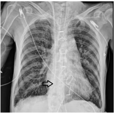
suture, and patient was transferred to ICU. Chest x-ray showed that PAC tip was placed at right atrium(Fig. 1A). After patient’s hemodynamic stabilization, PAC was advanced through the tricuspid valve with ballooning, but PA pressure waveform and cardiac output were not monitored. Chest X-ray showed that PAC tip was advanced through tricuspid valve into RV, but we could not determine the exact location of PAC tip in retrospective review(Fig. 1B). At ICU, 2,400 ml blood was drained through chest tube during the first six hours. Blood pressure gradually decreased, and then blood product and vasopressor(norepinephrine) were administered. Subsequently, chest tube drainage was not decreased(200 ml/hr), surgeon decided to re-operation to control of the bleeding. So far, chest tube drainage was totally 3,870ml before reoperation, and packed red blood cell 6U, fresh frozen plasma 6U, platelet concentrate 25U and cryoprecipitate 10U were transfused. Arterial blood gas analysis was shown that pH 7.36, PaCO248mmHg, PaO2283 mmHg, hematocrit 31% and lactate 6.5 mEq/L.
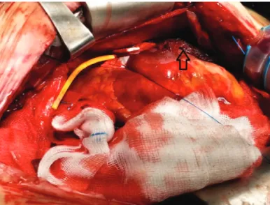
After opening of sternum, blood was evacuated from mediastinum, and surgeon did not find the definite bleeding focus. During cardiac manipulation, protruding PAC tip was detected at RV free wall, 5 cm remote from the previous RV closure site(Fig. 2A). The catheter tip was withdrawn and the perforation site was closed with direct suture at the same time(Fig. 2B). After surgery was uneventfully undertaken, patient was transferred to the ICU with stable vital sign. On follow-up, the patient was stable.
66
-InYoung Huh, DaeYoung Kim, MinHa Sung, MinHyun Lee, SunEun Park
Figure 1A. Chest X-ray finding. (A) Immediately
postoperative chest X-ray shows that pulmonary artery catheter (PAC) tip is placed at right atrium. (B) PAC is advanced into the right ventricle through tricuspid valve, but we can not determine the exact location of catheter tip in retrospective review. Black arrow indicates the tip of PAC.
Figure 1B. Intraoperative finding. (A) Protruding
pulmonary artery catheter (PAC) tip is visualized in the free wall of right ventricle (RV). Black arrow indicates the previous RV tear site during double valve replacement. (B) PAC tip is withdrawn into the right ventricle cavity with direct suture of perforating site. White arrow indicates the direct suture site of RV of PAC tip.
67
-Right ventricular perforation caused by pulmonary artery catheter at intensive care unit
In this case, postoperative bleeding after open heart surgery is related to RV perforation by PAC. Namely, our case was manifested a large amount of bleeding at immediate postoperative period. Surgeon decided to re-operation in order to control of the bleeding. And then, surgeon found the PAC tip protruding from the RV during cardiac manipulation.
The incidence of serious complication related to Swan-Ganz catheter is reported 0.03 to 1% and its mortality rate is 42 to 70% [5,6]. Risk factors for complication of PAC could be: age over 60 years, female gender, pulmonary hypertension, hypothermia(increase the rigidity of catheter), coagulation abnormalities, medication of anticoagulant, and surgical manipulation, etc [4]. However, most of cardiac surgical patients have above mentioned risk factors. Perforations of RV by PACs are well-known complications, and there was only one fatal case in the largest retrospective study [2]. Perforation of RV by PAC has been reported less than pulmonary artery perforation, because it occasionally does not present definitive symptoms [2,7]. This is because myocardium of RV can wrap around the perforating devices [8]. In most of cases, it can be waited the spontaneous hemostasis while serial echocardiography will be checked [4]. However, if cardiac tamponade occurs in severe case, it can be treated by drainage the blood from the pericardium through surgical intervention. Rarely, it will be required median sternotomy. In this case, median sternotomy was performed to control of postoperative bleeding, and RV perforation by PAC was incidentally found. Bleeding into the pericardial space by protruding PAC occurred progressively during postoperative period, and might be enhanced by postoperative coagulation abnormality, such as increased activated clotting time.
RV perforation by PAC is directly diagnosed by surgeon during open heart surgery, but it is difficult to detect in the ICU. There are different signs depending on the position of the perforating catheter, such as change of the PA pressure waveform, or bleeding. The PA pressure waveform is not useful for detection of perforation on occasion [9]. In our case, PA pressure waveform and cardiac output were not monitored, although Swan-Ganz catheter was advanced through the tricuspid valve with ballooning at ICU. During advancing, there was no exact record of pressure waveform or its change. We should be suspected the abnormal position of PAC, when waveform or cardiac output was not monitored after advancing the PAC. Echocardiography and
Discussion
Figure 2A. Intraoperative finding. (A) Protruding
pulmonary artery catheter (PAC) tip is visualized in the free wall of right ventricle (RV). Black arrow indicates the previous RV tear site during double valve replacement. (B) PAC tip is withdrawn into the right ventricle cavity with direct suture of perforating site. White arrow indicates the direct suture site of RV of PAC tip.
Figure 2B. Intraoperative finding. (A) Protruding
pulmonary artery catheter (PAC) tip is visualized in the free wall of right ventricle (RV). Black arrow indicates the previous RV tear site during double valve replacement. (B) PAC tip is withdrawn into the right ventricle cavity with direct suture of perforating site. White arrow indicates the direct suture site of RV of PAC tip.
computed tomography(CT) are considered the useful diagnostic tools for RV perforation [9,10]. The perforating catheter tip is detected in pericardial space, through the enhanced CT scan [11]. Echocardiography is also useful tool of serial examination of pericardial bleeding, especially if surgery is not required [4]. In this case, she checked chest X-ray and echocardiography after advancing the catheter, but RV perforation was not diagnosed before the re-operation. We did not determine the exact location of PAC tip in retrospective review of chest X-ray(Fig. 1B). It is helpful to check lateral view of chest X-ray, but we did not. Echocardiography showed poor window and only evaluated the cardiac function. Thus, we did not suspect the abnormal position of PAC tip before reoperation, because of massive bleeding and hemodynamic instability.
Factors which can induce ventricular perforation during cardiac catheterization include the small ventricular chamber, myocardial infarction, outflow tract obstruction, or stiff catheter etc [5]. In this case, RV tear was detected regardless of the surgical site after weaning of CPB during double valve replacement. We assumed the fragility of the RV myocardium to some degree. However, there was no abnormality of RV myocardium in the preoperative echocardiography. Accordingly, the thickness of RV wall is not guaranteed against RV perforation [12]. Although the PAC was advanced with ballooning, it was repeatedly inflated and deflated in RV and placed in RV, because of difficulty to advance in PA, and consequently it was induced perforation of RV. If PAC is difficult to advance into pulmonary artery, we should avoid locating PAC tip in RV and regressing into right atrium. In addition, it might be related to excessive manipulation, because they are not familiar with handling of PAC at ICU.
In conclusion, RV perforation is one of the serious complications of PAC. RV wall damage may be self-limited, but pericardial bleeding can happen that is required surgical intervention. In this case, reoperation was performed in order to solve the postoperative bleeding, that was induced by RV perforation of PAC. After advancing the PAC, we should check chest X-ray and PA pressure waveform. If it is suspicious, echocardiography and CT can be helpful to confirm the PAC location.
1) Swan HJ, Ganz W, Forrester J, Marcus H, Diamond G, Chonette D. Catheterization of the heart in man with use of a flow-directed balloon-tipped catheter. N Engl J
Med 1970; 283: 447-51.
2) Shah KB, Rao TL, Laughlin S, El-Etr AA. A review of pulmonary artery catheterization in 6,245 patients. Anesthesiology 1984; 61: 271-5.
3) Damen J, Bolton D. A prospective analysis of 1,400 pulmonary artery catheterizations in patients undergoing cardiac surgery. Acta Anaesthesiol Scand 1986; 30: 386-92.
4) Bossert T, Gummert JF, Bittner HB, Barten M, Walther T, Falk V, et al. Swan-Ganz catheter-induced severe complications in cardiac surgery: right ventricular perforation, knotting, and rupture of a pulmonary artery. J Card Surg 2006; 21: 292-5.
5) Kearney TJ, Shabot MM. Pulmonary artery rupture associated with the Swan-Ganz catheter. Chest 1995; 108: 1349-52.
6) Sirivella S, Gielchinsky I, Parsonnet V. Management of catheter-induced pulmonary artery perforation: a rare complication in cardiovascular operations. Ann Thorac Surg 2001; 72: 2056-9.
7) Domaingue CM, White AL. Right ventricular perforation in a patient with a pulmonary artery catheter. J Cardiothorac Anesth 1988; 2: 223-4.
8) Cohn PF, Braunwald E. Traumatic heart disease. In: Heart Disease: A Textbook of Cardiovascular Medicine. Edited by Braunwald E. Philadelphia, WB Saunders. 1992, pp 1517-27.
9) Chuang KC, Lan AK, Luk HN, Wang CS, Huang CJ, Cheng KW, et al. Perforation of the right ventricle by a pulmonary artery catheter that continues to measure cardiac output and mixed venous saturation. J Clin Anesth 2005; 17: 124-7.
10) Goodman A, Perera P, Mailhot T, Mandavia D. The role of bedside ultrasound in the diagnosis of pericardial effusion and cardiac tamponade. J Emerg Trauma Shock 2012; 5: 72-5.
11) Ohira S, Matsushita T, Masuda S, Ishise T. Right ventricular perforation caused by pulmonary artery catheter three days after insertion in a patient with acute pulmonary embolism. Heart Lung Circ 2013; 22: 1040-2.
12) Lyew MA, Bacon DR, Nesarajah MS. Right ventricular perforation by a pulmonary artery catheter during coronary artery bypass surgery. Anesth Analg 1996; 82: 1089-90.
68
-InYoung Huh, DaeYoung Kim, MinHa Sung, MinHyun Lee, SunEun Park