© 2018 Korean Breast Cancer Society. All rights reserved. http://ejbc.kr | pISSN 1738-6756
INTRODUCTION
Improvements in breast reconstruction technique have sig-nificantly enhanced the quality of life for most patients with breast cancer. Therapeutic standards have evolved from radi-cal intervention to skin-sparing mastectomy (SSM) or most recently nipple-sparing mastectomy (NSM), advancing recon-structive and aesthetic outcomes incrementally [1,2]. Al-though NSM has raised safety issues, this technique is gener-ally considered an acceptable oncologic option in properly se-lected patients [3-5].
The demand for immediate breast reconstruction following
conservative mastectomy has increased, with implant-based reconstruction most commonly pursued. According to Albornoz et al. [6], approximately 80% of all breast reconstructions per-formed in the United States involve prosthetic devices, and the vast majority are implanted immediately, placed at the time of mastectomy. When performing immediate implant-based breast reconstruction, it is still debatable whether a sin-gle-stage, direct-to-implant (DTI) strategy or a two-stage (tis-sue expander/implant) solution is best.
NSM with immediate prosthetic reconstruction may be performed using either a one-stage or two-stage method. Pro-ponents of the one-stage DTI approach emphasize its low re-vision rate, fewer operations, reduced overall cost, and excel-lent patient outcomes [7-9]. Those supporting the two-stage method cite the opportunity to recontour and select an ideal device at second stage, reduced capsular contracture risk in the setting of postmastectomy radiation, lower unplanned re-vision rate, as well as excellent patient outcomes [10,11].
When performing NSM, the choice of incision may be
Comparison of Outcomes between Direct-to-Implant Breast Reconstruction
Following Nipple-Sparing Mastectomy through Inframammary Fold Incision
versus Noninframammary Fold Incision
Tai Suk Roh, Jae Yoon Kim, Bok Ki Jung, Joon Jeong1, Sung Gwe Ahn1, Young Seok Kim
Departments of Plastic & Reconstructive Surgery and 1Surgery, Gangnam Severance Hospital, Yonsei University College of Medicine, Seoul, Korea
ORIGINAL ARTICLE
Purpose: In properly selected patients with breast cancer, nipple-sparing mastectomy (NSM) is generally considered safe by on-cologic standards. We examined two groups of patients who underwent direct-to-implant (DTI) reconstruction after NSM, comparing complications encountered, revision rates, and aes-thetic outcomes. The patients were stratified based on type of surgical incision and assigned to inframammary fold (IMF) and non-IMF groups. Methods: We investigated 141 patients (145 breasts) subjected to NSM and immediate DTI reconstruction between 2013 and 2016. A total of 62 breasts (in 58 patients) were surgically removed via IMF incisions, with the other 83 breasts (in 83 patients) removed by non-IMF means. Results: Complications associated with IMF (n=62) and non-IMF (n=83) incisions were as follows: skin necrosis (IMF, 9; non-IMF, 18); he-matoma (IMF, 3; non-IMF, 4); seroma (IMF, 8; non-IMF, 4); mild
capsular contracture (IMF, 4; non-IMF, 7); and tumor recurrence (IMF, 2; non-IMF, 8). Surgical revisions were counted as dupli-cates (IMF, 18; non-IMF, 38). Aesthetic outcomes following IMF incisions were rated as very good (44.2%), good (23.1%), fair (23.1%), or poor (9.6%). Conclusion: IMF incision enables com-plete preservation of the nipple-areolar complex, yielding superi-or aesthetic results in immediate DTI breast reconstruction after NSM. The nature of incision used had no significant impact on postoperative complications or reoperation rates and had com-parable oncologic safety to that of non-IMF incisions. IMF inci-sions produced the least visible scarring and did not affect breast shape. Most patients were satisfied with the aesthetic outcomes.
Key Words: Breast implants, Mammaplasty, Mastectomy
Correspondence to: Young Seok Kim
Department of Plastic and Reconstructive Surgery, Gangnam Severance Hospital, Yonsei University College of Medicine, 211 Eonju-ro, Gangnam-gu, Seoul 06273, Korea
Tel: +82-2-2019-3420, Fax: +82-2-577-4914 E-mail: [email protected]
Received: January 20, 2018 Accepted: May 17, 2018
Cancer
problematic, given that numerous variations have reportedly shown merit. Alternatives include radial, transverse periareo-lar, lateral, inferolateral, double concentric periareoperiareo-lar, vertical infra-areolar, and inframammary fold (IMF) incisions, with or without an axillary extension [12-15]. Endara et al. [16] have comprehensively addressed the process of choosing an appropriate incision for NSM. A radial or IMF incision is known to minimize scarring.
Although no incision is ideal, some certainly should be avoided. Endara et al. [16] found similar rates of nipple necro-sis for radial and IMF incisions (8.8% and 9.1%, respectively), whereas this rate was much higher (17.8%) for periareolar/ circumareolar incision lines. The highest incidence of nipple necrosis (81.8%) was recorded for a transareolar approach [16]. Using IMF incisions, the Beth Israel Deaconess group achieved an 82.0% nipple-areolar complex (NAC) survival rate in 17 breasts [17]. Blechman et al. [18] also reported a 94.0% NAC survival rate; in our series, the NAC survival rate was 95.1%.
Both oncologic and plastic surgeons would nevertheless agree on the convenience of a non-IMF incision in perform-ing NSM. Moreover, a clearer intraoperative view is enabled. The drawback is a potential for visible scarring and nipple de-formity or positional changes as likely consequences of scar contractures.
In this study, we examined two groups of patients who un-derwent DTI reconstruction after NSM, comparing complica-tions encountered, revision rates, and aesthetic outcomes. The patients were stratified by the nature of the surgical incision, assigned to IMF and non-IMF groups.
METHODS
Patient populationWe investigated 141 patients (145 breasts) subjected to NSM and immediate DTI reconstruction at Gangnam Severance Hospital, Seoul, Korea between 2013 and 2016. A total of 62 breasts (in 58 patients) were operated upon using IMF incisions, with the remaining 83 breasts (in 83 patients) involving non-IMF incisions. Detailed patient chart reviews were conducted retrospectively. The Yonsei University College of Medicine Institutional Review Board approved this study (number: 2017-08352014-001), and all participating patients granted informed consent.
Prior to 2016, mastectomy and breast reconstruction at our facility entailed non-IMF incisions. Since then, IMF incision has been the method of choice. NSM is also our surgical pref-erence and is typically reserved for smaller tumors (<3 cm across) situated >3 cm from the NAC. If undertaking SSM or
confronting likely postmastectomy radiotherapy for nodal positivity, a tissue expander (rather than DTI) would be in-serted.
Surgical technique
Preoperative markings were discussed and agreed upon by the oncologic and reconstructive teams. A lateral IMF inci-sion was our preferred method. Inciinci-sions were made along the curvilinear skin crease, extending as far lateral as the 3 o’clock position for the left breast (9 o’clock for the right breast) and extending as far inferior as the 6 o’clock position. Sentinel lymph node biopsies or axillary dissections were also attempt-ed through such incisions. If broader exposure was neattempt-edattempt-ed, counter-incisions of the axilla or along previous scars were made.
Acellular dermal matrices (DermACELL® [LifeNet Health, Virginia Beach, USA]; MegaDerm® [L&C BIO, Seoul, Korea]; or AlloDerm® [LifeCell Corp., Branchburg, USA]) were regu-larly used as inferolateral slings to expand inferior pockets and gain better IMF control. All permanent breast implants (Mentor Worldwide LLC, Santa Barbara, USA) were placed using dual plane technique (subpectoral and acellular dermal matrix positioning).
Skin incisions were routinely sutured to deep tissue (3-0 Vicryl; Ethicon, Bridgewater, USA), thereafter using 4-0 V-LocTM (Medtronic, Minneapolis, USA) and 5-0 Monosof (Medtronic) layer by layer. Two drains were placed at the time of mastectomy and removed after ~2–3 weeks. In the interim, patients were maintained on intravenous or oral antibiotics to protect against indigenous skin flora.
Postoperative care
Until the day of operation and on postoperative days 1 and 2, methylprednisolone sodium succinate (125 mg Solu-Medrol®; Pfizer, New York, USA) was injected twice daily. This regimen served to promote skin flap perfusion and pre-vent postoperative edema.
As early as 2002, reports had surfaced on use of leukotriene inhibitors to treat capsular contractures [19,20]. The benefits of montelukast (Singulair®; Merck, Kenilworth, USA) in soft-ening breasts and mitigating capsular contractures have since been documented in a number of studies. Our facility allows once-daily montelukast administration, starting on postoper-ative day 7.
Surgical complications
Complications were defined as necrosis of mastectomy skin and/or NAC, necessitating surgical intervention; seroma; cap-sular contracture; infection requiring intravenous antibiotics;
implant loss; and tumor recurrence. Incidences of various re-operations in both groups were analyzed as well. To assess the impact of postoperative radiation on various complication and reoperation rates, subgroup analyses were performed, separating patients into postmastectomy radiation therapy (PMRT) and non-PMRT groups.
Aesthetic outcomes
All patients were photographed pre- and postoperatively by the plastic surgeon. Aesthetic outcomes were gauged through subjective evaluation of certain factors, including NAC posi-tion, breast shape, and symmetry (Table 1), as suggested by Salibian et al. [21] Using a three-point scale, results were rated as very good, good, fair, or poor. Clinical examinations and reviews of photo documentation were carried out for this pur-pose by two plastic surgeons and one oncologic surgeon. Bi-lateral mastectomies in two patients were excluded. Subgroup analysis (PMRT vs. non-PMRT) was also conducted.
Statistical analysis
Complication and reoperation data were analyzed statisti-cally. Descriptive statistics and measures of central tendency were used to describe absolute and mean results, respectively, with analysis of binary datasets via t-test and invoking chi-square analysis for proportional response comparisons. Fisher exact test was applied to small values. All computations relied on standard software (SPSS version 15.0.1 for Windows; SPSS Inc., Chicago, USA), setting significance at p<0.05.
RESULTS
Therapeutic considerationsWe analyzed 145 NSM procedures (unilateral, 141; bilateral, 2). The IMF group accounted for 62 breasts in 58 patients (mean age, 46.4±5.9 years; mean body mass index [BMI], 22.7±2.3 kg/m2). Neoadjuvant and adjuvant chemotherapy was given to six and 28 patients, respectively; 47 patients re-ceived hormonal therapy; and 16 breasts were irradiated after reconstruction. The mean size of implanted prosthetics was 219.9±57.4 mL. In 62 DTI reconstructions, our oncologic breast surgeons performed sentinel lymph node biopsy in 46, axillary lymph node dissection in 13, and neither procedure
in two.
The non-IMF group accounted for 83 breasts in 83 patients (mean age, 45.2±8.5 years; mean BMI, 22.6±3.2 kg/m2). Neoadjuvant and adjuvant chemotherapy was given to three and 25 patients, respectively; 65 patients received hormonal therapy; and 14 breasts were irradiated after reconstruction. The mean size of implanted prosthetics was 255.3±76.9 mL. In 83 DTI reconstructions, our oncologic breast surgeons per-formed sentinel lymph node biopsy in 74 and axillary lymph node dissection in nine (Table 2).
Complications and reoperations
Procedural complications are summarized in Table 3. In the IMF group, skin necrosis (9/62, 14.5%), hematoma (3/62, 4.8%), seroma (8/62, 12.9%), capsular contracture (4/62, 6.5%) and infection (4/62, 6.5%) were encountered. Severe compli-cations requiring breast implant removal were limited to three instances (4.8%). One of these patients acquired a postopera-tive infection and another developed severe capsular contrac-ture after adjuvant radiotherapy. The third patient experienced extensive skin necrosis, which we debrided. The implant was then exchanged for a smaller one, anticipating a later reduc-tion mammoplasty on the other side. There were two
instanc-Table 1. Aesthetic outcome categories
Aesthetic evaluation factor Very good Good Fair Poor
NAC location Symmetrically positioned Mild displacement Moderate displacement Severe displacement Breast shape No irregularity Mildly irregular Moderate irregularities Severely distorted
Symmetry Symmetric Mild asymmetry Moderate asymmetry Severe asymmetry
NAC=nipple-areolar complex.
Table 2. Comparative demographics for direct-to-implant breast recon-structions performed with IMF incisions and non-IMF incisions
Characteristic IMF incision (n=58) No. (%) Non-IMF incision (n=83) No. (%) p-value No. of breasts 62 83 0.001 Unilateral 54 (87.1) 83 (100) Bilateral 8 (12.9) 0 Age (yr)* 46.4±5.9 45.2±8.5 0.804 BMI (kg/m²)* 22.7±2.3 22.6±3.2 0.874 Neoadjuvant chemotherapy 6 (10.3) 3 (3.6) 0.105 Adjuvant chemotherapy 28 (48.3) 25 (30.1) 0.122 Hormone therapy 47 (81.0) 65 (78.3) 0.431 Postmastectomy radiotherapy 16 (27.6) 14 (16.9) 0.094 Implant volume (mL)* 219.9±57.4 255.3±76.9 <0.001 Node dissection
Sentinel lymph node biopsy 46 (79.3) 74 (89.2) 0.085 Axillary lymph node dissection 13 (22.4) 9 (10.8) 0.053
No procedure 2 (3.4) 0 0.167
IMF=inframammary fold; BMI=body mass index. *Mean±SD.
es of tumor recurrence (3.2%), each adequately managed by oncologic surgeons.
In the non-IMF group, skin necrosis (18/83, 21.7%), hema-toma (4/83, 4.8%), seroma (4/83, 4.8%), capsular contracture (7/83, 8.4%), infection (10/83, 12.0 %), implant removal (7/83, 8.4%), and tumor recurrence (8/83, 9.6%) were recorded. In the IMF (vs. non-IMF) group, capsular contracture (p= 0.656), infection (p=0.259), implant loss (p=0.398), and tu-mor recurrence (p=0.132) rates tended to be lower, and the risk of skin necrosis (p=0.030) was significantly less. Howev-er, there were no significant between-group differences in terms of complications (Table 3, Figures 1 and 2).
We also investigated reoperations for secondary correc-tions, which were counted as duplicates. Results are shown in
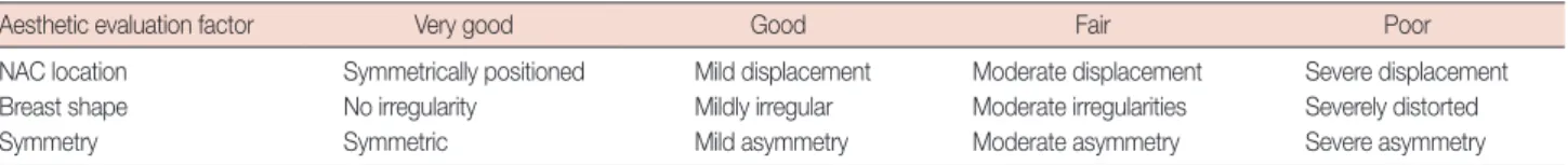
Figure 1. Representative reconstructive outcome: a 43-year-old woman with ductal carcinoma in situ of left breast. (A) Preoperative view. (B) Symmetric nipple-areolar complex position and breast shape in immediate postoperative state.
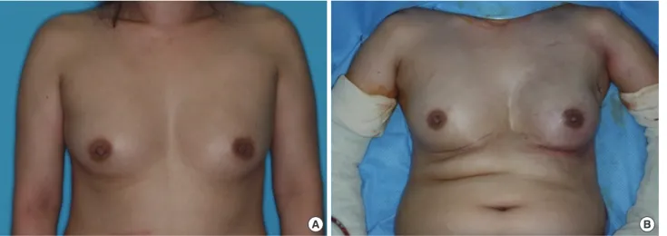
Figure 2. Representative reconstructive outcome: a 40-year-old woman with mucinous carcinoma of left breast. (A) Preoperative status, prior to nipple-sparing mastectomy (via inframammary incision) removing 464 g of tissue. (B) Outcome 6 months after immediate direct-to-implant recon-struction.
A
A
B
B Table 3. Postoperative complications in patients undergoing DTI breast
reconstruction following conservative mastectomy through IMF incision and non-IMF incision
Complication Total DTI (n=145) No. (%) IMF incision (n=62) No. (%) Non-IMF incision (n=83) No. (%) p-value Skin breakdown/necrosis 27 (18.6) 9 (14.5) 18 (21.7) 0.030 Hematoma 7 (4.8) 3 (4.8) 4 (4.8) 0.067 Seroma 12 (8.3) 8 (12.9) 4 (4.8) 0.080 Capsular contracture 11 (7.6) 4 (6.5) 7 (8.4) 0.656 Infection 14 (9.7) 4 (6.5) 10 (12.0) 0.259 Implant loss 10 (6.9) 3 (4.8) 7 (8.4) 0.398 Recurrence 10 (6.9) 2 (3.2) 8 (9.6) 0.132 Each complication was counted in duplicate.
Table 4. In the IMF group, there were six skin flap revisions (9.7%), three hematoma evacuations (4.8%), two capsular contracture corrections (3.2%), three contralateral augmenta-tion mammoplasties (4.8%), three implant extracaugmenta-tions (4.8%), and one implant exchange (1.6%). In the non-IMF group, there were 13 skin flap revisions (15.7%), four hematoma evacuations (4.8%), one capsular contracture correction (1.2%), three scar revisions (3.6%), three contralateral aug-mentation mammoplasties (3.6%), four implant extractions (4.8%), and 10 implant exchanges (12.0%). The rate of implant exchange owing to size mismatch was significantly lower in the IMF (vs. non-IMF) group (p=0.019).
Subgroup analyses
In subgroup analysis, a higher incidence of complications was evident in the PMRT (vs. non-PMRT) group across all fields, including skin necrosis (17/30, 56.7%), hematoma (4/30, 13.3%), seroma (9/30, 30.0%), capsular contracture (9/30, 30.0%), infection (10/30, 33.3%), implant loss (8/30, 26.7%) and tumor recurrence (7/30, 23.3%). Again, skin ne-crosis in the IMF (vs. non-IMF) group was significantly less (p=0.024), but no other significant differences emerged (Ta-ble 5).
PMRT had no apparent impact on reoperation outcomes, and there were no significant differences according to type of incision (Table 6).
Aesthetic outcomes
To evaluate aesthetic outcomes, we compared pre- and postoperative photos. In the IMF group (n=58), two patients (four mastectomies) were excluded, and postoperative follow-up photos were lacking in four patients. In the non-IMF group (n=83), 28 patients also lacked follow-up photos. Ulti-mately, 107 patients (IMF, 52; non-IMF, 55) were eligible for review and comparison (Table 7).
Aesthetic outcomes in the IMF group were rated as follows: very good, 44.2% (23/52); good, 23.1% (12/52); fair, 23.1% (12/52); and poor, 9.6% (5/52). Of the 12 patients with fair re-sults, six had received postoperative radiotherapy and two had experienced intractable seromas, leading to deep wound in-fections. The other four patients were free of comorbidities. One of the five patients with poor results showed breast asym-metry, having undergone implant removal for postoperative infection. Skin flap necrosis occurring in another three pa-tients required surgical revision, and a capsular contracture necessitated capsulotomy and implant exchange.
Aesthetic outcomes in the non-IMF incision group were
Table 4. Reoperation outcomes in patients undergoing DTI breast re-construction following conservative mastectomy through IMF incision and non-IMF incision
Reoperation Total DTI (n=145) No. (%) IMF incision (n=62) No. (%) Non-IMF incision (n=83) No. (%) p-value Skin flap revision 19 (13.1) 6 (9.7) 13 (15.7) 0.291 Hematoma evacuation 7 (4.8) 3 (4.8) 4 (4.8) 0.996 Capsular contracture
correction
3 (2.1) 2 (3.2) 1 (1.2) 0.398 Scar revision 3 (2.1) 0 3 (3.6) 0.130 Incision and drainage
due to infection 0 0 0 Contralateral augmentation mammoplasty due to size mismatch 6 (4.1) 3 (4.8) 3 (3.6) 0.714 Implant extraction 7 (4.8) 3 (4.8) 4 (4.8) 0.996 Implant change 11 (7.6) 1 (1.6) 10 (12.0) 0.019 Liposuction 0 0 0 Fat graft 0 0 0
Each complication was counted in duplicate. DTI=direct-to-implant; IMF=inframammary fold.
Table 5. Subgroup analysis of postoperative complications based on PMRT
Complication PMRT (n=30) Non-PMRT (n=115) Total DTI No. (%) IMF incision (n=16) No. (%) Non-IMF incision (n=14) No. (%) p-value Total DTI No. (%) IMF incision (n=46) No. (%) Non-IMF incision (n=69) No. (%) p-value Skin breakdown/necrosis 17 (56.7) 6 (37.5) 11 (78.6) 0.024 10 (8.7) 3 (6.5) 7 (10.1) 0.499 Hematoma 4 (13.3) 2 (12.5) 2 (14.3) 0.886 3 (2.6) 1 (2.2) 2 (2.9) 0.811 Seroma 9 (30.0) 6 (37.5) 3 (21.4) 0.338 3 (2.6) 2 (4.3) 1 (1.4) 0.339 Capsular contracture 9 (30.0) 3 (18.8) 6 (42.9) 0.151 2 (1.7) 1 (2.2) 1 (1.4) 0.771 Infection 10 (33.3) 3 (18.8) 7 (50.0) 0.070 4 (3.5) 1 (2.2) 3 (4.3) 0.533 Implant loss 8 (26.7) 3 (18.8) 5 (35.7) 0.295 2 (1.7) 0 2 (2.9) 0.244 Recurrence 7 (23.3) 1 (6.3) 6 (42.9) 0.222 3 (2.6) 1 (2.2) 2 (2.9) 0.811
Each complication was counted in duplicate.
rated as follows: very good, 16.4% (9/55); good, 32.7% (18/55); fair, 29.1% (16/55); and poor, 21.8% (12/55). Of the 16 pa-tients with fair results, most developed capsular contracture or implant malposition. In those rated as poor, some showed capsular contracture or mismatched implant size. There were also instances of implant malposition or skin necrosis needing reoperation. Outcomes rated as very good were significantly more numerous in the IMF (vs. non-IMF) group (p=0.002).
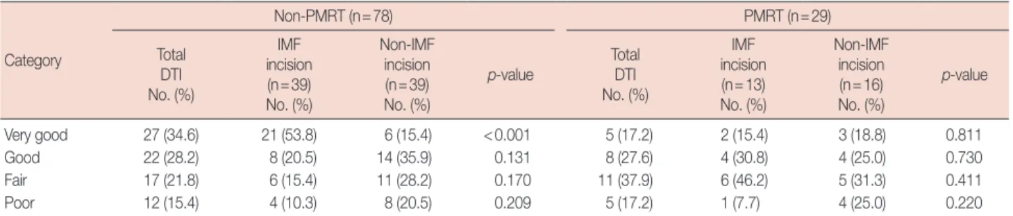
In subgroup analysis, the aesthetic superiority of the IMF incision was upheld in the non-PMRT group (n=78), with
significantly more patients rated very good (p<0.001) and fewer rated as poor (p=0.209). However, in breast reconstruc-tions performed after PMRT (n=29), there were no signifi-cant between-group differences (IMF vs. non-IMF) (Table 8).
DISCUSSION
Presently, NSM is a valid surgical option for treating breast cancer in selected patients. The combination of NSM and im-mediate breast reconstruction has been gaining traction as a
Table 6. Subgroup analysis of reoperation outcomes based on PMRT
Reoperation PMRT (n=30) Non-PMRT (n=115) Total DTI No. (%) IMF incision (n=16) No. (%) Non-IMF incision (n=14) No. (%) p-value Total DTI No. (%) IMF incision (n=46) No. (%) Non-IMF incision (n=69) No. (%) p-value Skin flap revision 5 (16.7) 1 (6.3) 4 (28.6) 0.102 11 (9.6) 2 (4.3) 9 (13.0) 0.120
Hematoma evacuation 0 1 (6.3) 2 (14.3) 0.464 4 (3.5) 2 (4.3) 2 (2.9) 0.678
Capsular contracture correction 2 (6.7) 1 (6.3) 1 (7.1) 0.922 1 (0.9) 1 (2.2) 0 0.219
Scar revision 1 (3.3) 0 1 (7.1) 0.277 2 (1.7) 0 2 (2.9) 0.244
Incision and drainage due to infection 0 0 0 0 0 0
Contralateral augmentation mammoplasty
due to size missmatch 2 (6.7) 1 (6.3) 1 (7.1) 0.922 4 (3.5) 2 (4.3) 2 (2.9) 0.678 Implant extraction 4 (13.3) 2 (12.5) 2 (14.3) 0.886 3 (2.6) 1 (2.2) 2 (2.9) 0.811
Implant change 4 (13.3) 1 (6.3) 3 (21.4) 0.222 7 (6.1) 0 7 (10.1) 0.026
Liposuction 0 0 0 0 0 0
Fat graft 0 0 0 0 0 0
Each complication was counted in duplicate.
PMRT=postmastectomy radiation therapy; DTI=direct-to-implant; IMF=inframammary fold.
Table 7. Aesthetic outcomes of the reconstructions
Category Total DTI (n=107) No. (%) IMF incision (n=52) No. (%) Non-IMF incision (n=55) No. (%) p-value Very good 32 (29.9) 23 (44.2) 9 (16.4) 0.002 Good 30 (28.0) 12 (23.1) 18 (32.7) 0.267 Fair 28 (26.2) 12 (23.1) 16 (29.1) 0.479 Poor 17 (15.9) 5 (9.6) 12 (21.8) 0.084
DTI=direct-to-implant; IMF=inframammary fold.
Table 8. Comparisons of the aesthetic outcomes of the breast reconstruction after PMRT status
Category Non-PMRT (n=78) PMRT (n=29) Total DTI No. (%) IMF incision (n=39) No. (%) Non-IMF incision (n=39) No. (%)
p-value Total DTI No. (%) IMF incision (n=13) No. (%) Non-IMF incision (n=16) No. (%) p-value Very good 27 (34.6) 21 (53.8) 6 (15.4) <0.001 5 (17.2) 2 (15.4) 3 (18.8) 0.811 Good 22 (28.2) 8 (20.5) 14 (35.9) 0.131 8 (27.6) 4 (30.8) 4 (25.0) 0.730 Fair 17 (21.8) 6 (15.4) 11 (28.2) 0.170 11 (37.9) 6 (46.2) 5 (31.3) 0.411 Poor 12 (15.4) 4 (10.3) 8 (20.5) 0.209 5 (17.2) 1 (7.7) 4 (25.0) 0.220
preferred surgical strategy [22-24], Furthermore, NSM tech-niques now include removal of glandular tissue from the nip-ple. This is a critical point of differentiation from prior subcu-taneous mastectomy methods, where significant residual breast tissue was retained within and beneath the nipple. Benediktsson and Perbeck [25] have subsequently confirmed the oncologic safety of NSM, showing survival rates compara-ble to those of conventional mastectomy, given a 13-year me-dian follow-up period.
One- and two-stage prosthetic reconstructions have been performed at a number of institutions using an array of inci-sions, including periareolar, inferior radial, inframammary, lateral, lateral radial, and inferolateral variations. The selection of a one- or two-stage approach in the setting of NSM is a complex process. Two-stage reconstruction yields better re-sults, improving symmetry and implant position. Moreover, this strategy allows greater control over final breast volume and shape. However, cost is clearly a factor in that surgery is carried out at least twice. On the other hand, one-stage pros-thetic reconstruction has few complications and confers rela-tively good aesthetic outcomes. At our facility, two-stage pros-thetic reconstruction is elected if skin flap volume is scant or postoperative radiotherapy is anticipated.
As Endara et al. [16] have maintained, choosing the right incision for NSM facilitates both therapeutic and reconstruc-tive efforts, preserving nipple-areola blood flow and produc-ing an aesthetically favorable scar. Our oncologic surgeons generally relied upon non-IMF incisions until 2016. There-after, IMF incisions were introduced, supported by ideal inci-sion criteria (referenced earlier). Since this transition, patient satisfaction has improved, and surgeons are pleased with the results, hoping to even further improve postoperative aesthet-ics.
Herein, we examined patients undergoing one-stage DTI breast reconstruction via non-IMF incisions and those recon-structed through IMF incisions, comparing complications. Skin necrosis in the IMF (n=9, 14.5%) vs. non-IMF (n=18, 21.7%) group was significantly less prevalent (p=0.030). There were nine instances of IMF-related skin necrosis in ear-ly 2016, suggesting a learning curve for this this new method. By preserving 3rd and 4th intercostal perforators to fortify the blood supply, our oncologic surgeon successfully enhanced remnant skin flap thickness and viability, and skin necrosis was seldom seen later that year. Nevertheless, skin necrosis did remain more problematic in non-IMF (vs. IMF) incisions, especially at the lower pole of the breast. One potential expla-nation is the gravitational pressure of implants against the lower reaches of skin flaps.
In the evolution of NSM procedures, the advantage of
im-mediate breast reconstruction has brought to bear the impor-tance of skin flap thickness in maintaining flap viability. Using 4 to 5 mm flaps, some sources [26] have confined necrosis to ~17%; through even thicker (10-mm) flaps, others have achieved rates <5% [27,28]. Although thicker remnant flaps may indeed limit necrosis, no standard method is yet available for calibration, so consistency may be difficult to ensure. On average, our team generates 8-mm skin flaps. We are con-vinced that such relatively thick flaps may contribute to the lower rates of flap necrosis presently achieved. This also ex-plains the lesser mean implant size in our IMF group, com-pared with the non-IMF group. The thin skin flaps formerly left behind were needed to cover larger sized implants. How-ever, current (i.e., less aggressive) mastectomy trends leave rel-atively thick skin flaps, restricting the size of breast implants.
Rates of hematoma and seroma did not differ significantly by incision type, nor did the rate of capsular contractures, al-beit somewhat premature. The follow-up period of ~1 year in the IMF group is possibly insufficient to reasonably assess contracture rates. Extended follow-up monitoring in future studies is truly needed. Rates of infection, implant loss, and tumor recurrence similarly showed no between-group differ-ences.
In a subgroup analysis comparing postoperative tions in PMRT and non-PMRT groups, the various complica-tions encountered did not differ significantly. As for reopera-tion outcomes, implant extracreopera-tion was significantly more like-ly in the non-IMF (vs. IMF) group. This may be related to the higher rate of skin necrosis shown by the non-IMF group. Our data further indicated that PMRT had no impact on re-operation outcomes, regardless of incision type.
A multiplicity of criteria has been developed for assessing the aesthetic outcomes of NSM, including patient self-assess-ment, nipple sensitivity, postoperative scarring, nipple place-ment, breast contour, and overall symmetry [17,29,30]. We used three criteria in our evaluations: NAC position, breast shape, and symmetry. IMF incisions concealed postoperative scarring and also resulted in fewer patient complaints.
Bilateral reconstruction (IMF group: four mastectomies) was excluded in our analysis of aesthetic outcome, owing to an inherent advantage in terms of symmetry. Accordingly, 35 patients (67.3%) in the IMF group were rated as having very good or good results, surpassing the corresponding rate (49.1%) in the non-IMF group. In the IMF (vs. non-IMF) group, significantly more patients were rated as very good (p=0.002).
Of note, we found no significant differences in aesthetic outcomes of IMF and non-IMF incisions after PMRT, regard-less of aesthetic rating achieved. It may be that PMRT effects
offset any aesthetic benefit conferred by IMF incisions. How-ever, the aesthetic superiority of IMF (vs. non-IMF) incisions was upheld in non-PMRT subgroup analysis. Finally, because aesthetic outcomes were evaluated by our staff plastic sur-geons, bias cannot be ruled out.
In conclusion, the use of IMF incisions for DTI breast re-construction after NSM allows complete preservation of the NAC, with superior aesthetic results. We found that related postoperative complications, secondary corrective operations, and oncologic safety all compared favorably with non-IMF incisions. Moreover, IMF incisions produced the least visible scars and did not affect breast shape. Most patients expressed satisfaction with their aesthetic outcomes.
CONFLICT OF INTEREST
The authors declare that they have no competing interests.
REFERENCES
1. Didier F, Radice D, Gandini S, Bedolis R, Rotmensz N, Maldifassi A, et al. Does nipple preservation in mastectomy improve satisfaction with cosmetic results, psychological adjustment, body image and sexuality? Breast Cancer Res Treat 2009;118:623-33.
2. Didier F, Arnaboldi P, Gandini S, Maldifassi A, Goldhirsch A, Radice D, et al. Why do women accept to undergo a nipple sparing mastectomy or to reconstruct the nipple areola complex when nipple sparing mas-tectomy is not possible? Breast Cancer Res Treat 2012;132:1177-84. 3. Jensen JA, Orringer JS, Giuliano AE. Nipple-sparing mastectomy in 99
patients with a mean follow-up of 5 years. Ann Surg Oncol 2011;18: 1665-70.
4. Jensen JA. Breast cancer: is nipple sparing mastectomy safe? Ann Surg 2009;250:657-8.
5. Crile G Jr, Esselstyn CB Jr, Hermann RE, Hoerr SO. Partial mastectomy for carcinoma of the breast. Surg Gynecol Obstet 1973;136:929-33. 6. Albornoz CR, Bach PB, Mehrara BJ, Disa JJ, Pusic AL, McCarthy CM,
et al. A paradigm shift in U.S. breast reconstruction: increasing implant rates. Plast Reconstr Surg 2013;131:15-23.
7. Salzberg CA, Ashikari AY, Koch RM, Chabner-Thompson E. An 8-year experience of direct-to-implant immediate breast reconstruction using human acellular dermal matrix (AlloDerm). Plast Reconstr Surg 2011;127:514-24.
8. Colwell AS, Damjanovic B, Zahedi B, Medford-Davis L, Hertl C, Austen WG Jr. Retrospective review of 331 consecutive immediate single-stage implant reconstructions with acellular dermal matrix: indications, com-plications, trends, and costs. Plast Reconstr Surg 2011;128:1170-8. 9. Colwell AS. Current strategies with 1-stage prosthetic breast
recon-struction. Gland Surg 2015;4:111-5.
10. Pusic AL, Cordeiro PG. Breast reconstruction with tissue expanders and implants: a practical guide to immediate and delayed reconstruc-tion. Semin Plast Surg 2004;18:71-7.
11. Spear SL, Seruya M, Rao SS, Rottman S, Stolle E, Cohen M, et al.
Two-stage prosthetic breast reconstruction using AlloDerm including out-comes of different timings of radiotherapy. Plast Reconstr Surg 2012;130:1-9.
12. Regolo L, Ballardini B, Gallarotti E, Scoccia E, Zanini V. Nipple sparing mastectomy: an innovative skin incision for an alternative approach. Breast 2008;17:8-11.
13. Boneti C, Yuen J, Santiago C, Diaz Z, Robertson Y, Korourian S, et al. Oncologic safety of nipple skin-sparing or total skin-sparing mastecto-mies with immediate reconstruction. J Am Coll Surg 2011;212:686-93. 14. Colwell AS, Gadd M, Smith BL, Austen WG Jr. An inferolateral
approach to nipple-sparing mastectomy: optimizing mastectomy and reconstruction. Ann Plast Surg 2010;65:140-3.
15. Maxwell GP, Storm-Dickerson T, Whitworth P, Rubano C, Gabriel A. Advances in nipple-sparing mastectomy: oncological safety and inci-sion selection. Aesthet Surg J 2011;31:310-9.
16. Endara M, Chen D, Verma K, Nahabedian MY, Spear SL. Breast recon-struction following nipple-sparing mastectomy: a systematic review of the literature with pooled analysis. Plast Reconstr Surg 2013;132:1043-54.
17. Yueh JH, Houlihan MJ, Slavin SA, Lee BT, Pories SE, Morris DJ. Nipple-sparing mastectomy: evaluation of patient satisfaction, aesthetic results, and sensation. Ann Plast Surg 2009;62:586-90.
18. Blechman KM, Karp NS, Levovitz C, Guth AA, Axelrod DM, Shapiro RL, et al. The lateral inframammary fold incision for nipple-sparing mastectomy: outcomes from over 50 immediate implant-based breast reconstructions. Breast J 2013;19:31-40.
19. Schlesinger SL, Ellenbogen R, Desvigne MN, Svehlak S, Heck R. Zafirlukast (Accolate): a new treatment for capsular contracture. Aesthet Surg J 2002;22:329-36.
20. Schlesinger SL, Desvigne MN, Ellenbogen R, Svehlak S, Heck R. Results of using zafirlukast (Accolate) and montelukast (Singulair) for treat-ment of capsular contracture. Aesthet Surg J 2003;23:101-2.
21. Salibian AH, Harness JK, Mowlds DS. Inframammary approach to nipple-areola-sparing mastectomy. Plast Reconstr Surg 2013;132:700e-8e.
22. Rusby JE, Smith BL, Gui GP. Nipple-sparing mastectomy. Br J Surg 2010;97:305-16.
23. de Alcantara Filho P, Capko D, Barry JM, Morrow M, Pusic A, Sacchini VS. Nipple-sparing mastectomy for breast cancer and risk-reducing surgery: the Memorial Sloan-Kettering Cancer Center experience. Ann Surg Oncol 2011;18:3117-22.
24. Spear SL, Willey SC, Feldman ED, Cocilovo C, Sidawy M, Al-Attar A, et al. Nipple-sparing mastectomy for prophylactic and therapeutic indi-cations. Plast Reconstr Surg 2011;128:1005-14.
25. Benediktsson KP, Perbeck L. Survival in breast cancer after nipple-sparing subcutaneous mastectomy and immediate reconstruction with implants: a prospective trial with 13 years median follow-up in 216 patients. Eur J Surg Oncol 2008;34:143-8.
26. Verheyden CN. Nipple-sparing total mastectomy of large breasts: the role of tissue expansion. Plast Reconstr Surg 1998;101:1494-500. 27. Newman LA, Kuerer HM, Hunt KK, Kroll SS, Ames FC, Ross MI, et al.
Presentation, treatment, and outcome of local recurrence after skin-sparing mastectomy and immediate breast reconstruction. Ann Surg Oncol 1998;5:620-6.
of skin preservation at mastectomy when combined with immediate reconstruction of the breast. Surg Gynecol Obstet 1991;172:17-20. 29. Djohan R, Gage E, Gatherwright J, Pavri S, Firouz J, Bernard S, et al.
Patient satisfaction following nipple-sparing mastectomy and immedi-ate breast reconstruction: an 8-year outcome study. Plast Reconstr Surg
2010;125:818-29.
30. Mosahebi A, Ramakrishnan V, Gittos M, Collier J. Aesthetic outcome of different techniques of reconstruction following nipple-areola-pre-serving envelope mastectomy with immediate reconstruction. Plast Reconstr Surg 2007;119:796-803.