INTRODUCTION
In recent years, soft tissue esthetics has been one of the major issues in implant dentistry.1-5The main influencing factors in soft tissue esthetics are the height of interproximal papilla and the level of facial gingiva.6The level of facial gingiva is influenced by several factors such as thickness of labial bony wall, the position and orientation of implant, the gingival biotypes6-9
whereas the height of interproximal papilla around a single implant restoration is mainly dependent on the bone level of adjacent teeth.10
Recently, excellent soft tissue esthetic outcomes were reported by delivering a provisional restoration on the day of immediate implant placement.11,12In the esthetic point of view, the greatest benefit of immediate provisionalization following immediate implant placement is that the interproximal soft tissue height could be maintained by supporting the overlying soft tissue during healing period.12,13As the presence and height of the interproximal papilla is attributable to the presence of adjacent teeth attachment and the size of the gingival embrasure formed
by these teeth,14immediate support of interproximal papilla by provisional restoration could minimize the collapse of interproximal soft tissue following the extraction of a tooth.13,15-17
Instead of delivering a provisional restoration, connecting a healing abutment still can be an option following an immediate implant placement. By gradually increasing the diameter of the healing abutment, clinicians can manipulate soft tissue around the implants.18 In such cases, however, clinicians may be concerned about the loss of papilla height due to the absence of immediate papilla support. Numerous studies have been performed to assess the soft tissue esthetics around implant restorations.19,20However, still less information is available on the soft tissue esthetics of single implant restorations which were immediately placed and immediately provisionalized.
The purpose of the current study was to evaluate and compare the soft tissue esthetic outcomes of immediately placed single tooth implant restorations with or without immediate provisional restorations.
IMPACT OF IMMEDIATE AND NON-IMMEDIATE
PROVISIONALIZATION ON THE SOFT TISSUE ESTHETICS OF
FINAL RESTORATIONS ON IMMEDIATELY PLACED IMPLANTS
Chong-Hyun Han1, DDS, PhD, Jeong-Won Paik2, DDS, PhD, Keun-Woo Lee3, DDS, PhD, Dong-Hoo Han3, DDS, PhD,
Moon-Kyu Chung3, DDS, PhD, Sunjai Kim4*, DDS, PhD
1Professor, Department of Prosthodontoics, YongDong Severance Dental Hospital, College of Dentistry, Yonsei University 2Clinical Assistant Professor, Department of Periodontics, SMC dental clinic, Samsng Medical Center, Sungkyunkwan University
3Professor, Department of Prosthodontics, Yonsei University Dental Hospital, College of Dentistry, Yonsei University 4Assistant Professor, Department of Prosthodontoics, YongDong Severance Dental Hospital,
College of Dentistry, Yonsei University
Corresponding Author: Sunjai Kim
Department of Prosthodontics, YongDong Severance Dental Hospital, College of Dentistry, Yonsei University 146-92 Dogok-dong, Kangnam-gu, Seoul, 135-720, Korea +82 2 2019 3568: e-mail, [email protected]
MATERIALS AND METHODS
A total of 10 patients, each with a hopeless maxillary anterior tooth, were selected between Sep 2005 and Oct 2006. The patients were comprised of 6 women and 4 men, aged between 30 to 68 (mean 42.2). The hopeless teeth included 6 central incisors, 2 lateral incisors and 2 canines. Under specific patient inclusion/exclusion criteria, the placement of implant into fresh extraction socket was selected as a treatment plan for each patient.
Inclusion criteria were as follows
1. a hopeless single maxillary anterior tooth with adequate and similar level of the gingival and underlying bony architecture as the contralateral natural tooth
2. reasons of extractions were root fractures and endodontic failures.
3. good oral hygiene
4. adequate bone volume to place an implant with a minimum dimension of 4.3×11.5 mm without necessity of bone grafting.
5. primary stability with 35 Ncm of insertion torque.
Fig. 1. Initial frontal view of the hopeless left maxillary central
incisor.
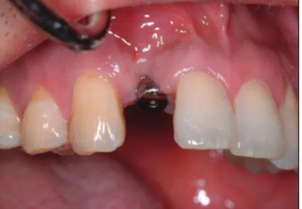
Fig. 2. An immediate implant was placed without flap elevation.
Fig. 3. A screw retained provisional restoration was delivered on
the day of implant placement (picture was taken at 2 weeks after surgery).
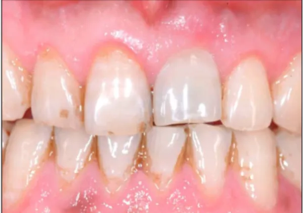
Fig. 4. Frontal view of the final restoration. Five soft tissue esthetic
variables. 1; mesial papilla, 2; distal papilla, 3; soft tissue level, 4; soft tissue contour, 5; facial soft tissue prominence.
Exclusion criteria were as follows
1. active or acute infection around the hopeless tooth. 2. uncontrolled diabetes, coagulation disorders,
psychologic disorders. 3. alcohol or drug abuse. 4. parafunctional habit.
5. heavy smoker (more than 10 cigarettes a day)
6. perforation, dehiscence or loss of labial bony plate following tooth extraction or implant osteotomy. Five patients were randomly assigned to the immediate provisionalization group (IP), and screw retained fixed provisional restorations were delivered on the day of implant surgery. The mean provisionalization period until delivery of final restorations for IP group was 155 days.
immediate provisionalization group (NIP). Healing abutments were connected to the implants on the day of implant surgery and were maintained for 75 to 154 (mean 100) days. The healing abutments were replaced with screw retained fixed provisional restorations and were maintained for an additional 61 to 86 days (mean 77 days) until delivery of final restorations.
The implants placed were nine 4.3 mm diameter implants (FIT43115, Warantec, Seoul, Korea) and one 5.3 mm diameter implant (FIT53115, Warantec, Seoul, Korea). The abutments used for the final restorations were 7 castable gold abutments (IOCA37GE, Warantec, Seoul, Korea) and 3 preparable titanium abutments (IOTA4536, Warantec, Seoul, Korea). Intraoral photographs of the restoration and
Fig. 5. A hopeless maxillary right lateral incisor due to endodontic
failure.
Fig. 6. A healing abutment was placed on the day of surgery
(pic-ture was taken at 1 month after surgery).
Fig. 7. A provisional restoration was delivered about 3 month after
implant placement.
camera (FinePix S2Pro, Fujifilm, Japan) on the delivery of the final restoration.
A total of 20 dentists (5 prosthodontists, 5 periodontists, 5 orthodontists and 5 dental students) were chosen to assess five soft tissue variables around implants in comparison with their contralateral teeth. The variables assessed were the height of mesial papilla (MP), the height of distal papilla (DP), the level of facial gingival margin (Level), the contour of facial gingival margin (Contour), and the facial soft tissue prominence of alveolar process (Prominence). Frontal view photographs of each final restoration and its contralateral tooth were shown to each dentist in randomized order, and five esthetic variables were scored. The scores were rated in numbers 0 (lack of esthetics), 1 (esthetic but incomplete), and 2 (perfect esthetic match to the contralateral tooth). The scores were not the absolute values but the relative values compared to the contralateral natural tooth.
Student t-test was used in order to evaluate the statistic significance of the scores rated for IP and NIP. To evaluate
the statistic significance of the five esthetic variables and the specialty of the four dentist groups, one-way ANOVA was used. Tukey grouping was chosen for post hoc test in both analyses (α= 0.05).
RESULTS
Table I presented the soft tissue esthetic scores of IP and NIP
The assessment resulted in significantly higher esthetic scores of the IP group in two variables; Level and Prominence.
Table II presents the esthetic scores rated by each dentist group.
Considering different specialty areas in dentistry, there was no notable disagreement among the dentists groups in assessing the esthetic variables.
Table I. Means and standard deviations of esthetic scores of IP and NIP
MP DP Level Contour Prominence
IP 1.56±0.52 1.56±0.52 1.41±0.55 1.49±0.56 1.60±0.59
NIP 1.51±0.56 1.44±0.56 1.23±0.71 1.38±0.69 1.40±0.67
P value 0.459 0.064 0.046* 0.146 0.002*
IP; immediate provisionalization group, NIP; non-immediate provisionalization group, MP; the height of mesial papilla, DP; the height of distal papilla, Level; the level of facial gingival marginl, Contour; the contour of facial gingival margin, Prominence; the facial soft tissue prominence of alveolar process. *; statistical significance was noted between immediate and non-immediate provisionalization groups.
Table II. The means and standard deviations of esthetic scores rated according to each dentist group
MP DP Level Contour Prominence
Prosthodontists 1.60±0.50 a 1.60±0.50 a 1.48±0.59 a 1.52±0.51 a 1.72±0.46 a Students 1.64±0.57 a 1.56±0.51 a 1.44±0.51 a 1.64±0.49 a 1.68±0.63 a IP Orthodontists 1.56±0.51 a 1.6±0.58 a 1.52±0.51 a 1.56±0.51 a 1.64±0.57 a Periodontists 1.44±0.51 a 1.48±0.51 a 1.2±0.58 a 1.24±0.66 a 1.36±0.64 a P value 0.562 0.832 0.166 0.062 0.119 Prosthodontists 1.48±0.59 a 1.44±0.58 a 1.32±0.69 a 1.48±0.71 a 1.56±0.51 a Students 1.68±0.56 a 1.52±0.59 a 1.2±0.71 a 1.52±0.65 a 1.44±0.65 a NIP Orthodontists 1.44±0.51 a 1.44±0.51 a 1.24±0.72 a 1.32±0.63 a 1.36±0.76 a Periodontists 1.44±0.58 a 1.36±0.57 a 1.16±0.75 a 1.2±0.76 a 1.24±0.72 a P value 0.372 0.798 0.877 0.336 0.387
DISCUSSION
Successful clinical results were achieved by submerging an immediate implant or connecting a healing abutment following immediate implant placement.18,21,22Along with these approaches, excellent esthetic outcomes were reported with immediate provisional restorations following immediate implant placements.11,12,23One of the advantages of immediate provisionalization following immediate implant placement is that it better maintains the interproximal soft tissue height around the implant restorations.13,24It is reported that 3 mm was measured in the facial dimension of normal dentogingival complex whereas 5 mm was measured in the interproximal dimension.23Only when the interproximal soft tissue was supported by adjacent teeth, the interproximal dimension of 5 mm could be maintained.25 Once the tooth is extracted, the interproximal soft tissue would lose its support from the tooth, resulting in decrease of soft tissue dimension to 3 mm, consequently compromising the gingival esthetics. Therefore, immediate support by a provisional restoration is essential to maintain the height of interproximal soft tissue and eventually produce desired esthetic outcome.11,14,25
Few studies have investigated the soft tissue change around immediate implant restorations. Schropp et al.19 reported that no significant difference was noted between the early and delayed placement groups at either final restorations delivery or 1.5 year follow up. However, the risk of negative or no papilla at final restorations delivery was 7 times greater in the delayed placement group.19 Cornelini and co-workers26
evaluated the soft tissue changes of 22 single implant restorations which were placed into fresh extraction socket and provisionalized within 24 hours. They reported that the mean recession of midfacial gingiva was 0.75 mm at 1 year follow up. As immediate provisional restorations were delivered to support the interproximal soft tissue, 100% of subjects showed esthetically acceptable papilla height (Jem t papilla index 2 or 3).26Kan and colleagues12
assessed the soft tissue changes around single implant restorations which were immediately placed and provisionalized following extraction of hopeless teeth. They reported that the mean amount of recession for midfacial, mesial and distal soft tissue were 0.55, 0.53 and 0.39 mm,
soft tissue changes on single implant restorations, which were immediately placed and immediately provisionalized, and thereby reported complete fill of papilla in 82 out of 86 implants.27One notable finding was recently published by Evans and Chen28. They reported that the bucco-lingual position of implant head influenced significantly on the level of facial gingiva. The mean recession of midfacial gingiva was 3 times greater the implants with a buccally positioned implant head rather than a lingually placed implant head.28In the current study, however, each screw hole of provisional restoration was around the cingulum, therefore, the influence of implant head position was not a critical factor for the level of facial gingiva.
Until now, immediate support of interproximal soft tissue was considered as a prerequisite in maintaining the preexisting papilla height.11-13,16,24However, current study showed that the preexisting papilla height can be also achieved by connecting a healing abutment following immediate implant placement. Immediate or early implant placement resulted early papilla regeneration on final restoration delivery even though the implants were submerged following implant surgery. It is assumed that the interproximal soft tissue around immediate implant might have more potential to regenerate the papilla.
Contrast to the level of papilla, significant difference was noted in the level of facial gingiva between IP and NIP groups. The difference in the level of facial gingiva between IP and NIP groups could be due to the following reason; immediate provisional restorations give support not only to the interproximal papillae but also to the facial soft tissue. The dimension of healing abutments used was smaller than the dimension of corresponding extracted tooth whereas the dimension of provisional restoration was similar to the extracted tooth. Furthermore, the implant system used in current study has a so-called narrow neck design. The diameter of abutment is about 1 mm narrower than that of implant platform at abutment/implant interface. The narrow healing abutment alone may not provide adequate support for the maintenance of facial soft tissue and eventually result in slight collapse of the facial soft tissue. Once the healing abutment was replaced with a provisional restoration, the greater facial-lingual dimension of provisional restoration would push the immature facial soft
gingival margin. The difference in facial soft tissue prominence between IP and NIP may come from the same reason. Considering the result of the current study, achieving esthetic facial gingival margin is more demanding rather than restoring the height of the papilla and the level of facial gingiva can be better maintained when facial soft tissue is immediately supported. However, clinicians should bear in mind that greater risk of failure is present when immediate provisional restorations are delivered on immediately placed implants. Careful patient selection and higher level of surgical and restorative skills are required for successful outcomes.
Four dentist groups with different specialties have participated in the current study to assess the esthetic variables. These dental specialties were selected based on previous studies.29In the previous report, orthodontists were more critical than other dentist groups. In the current study, however, the overall scores showed no significant differences in opinion between the dentist groups. The special characteristic of the current study was that the soft tissue esthetic variables of implant restoration were compared to those of the contralateral natural tooth and this guideline for esthetic assessment may have diluted the esthetic concerns of different specialties.
CONCLUSION
Within the limitation of this study, immediate provisionalization on immediately placed implants could be a superior treatment option to non-immediate provisionalization approaches to achieve more esthetic facial gingival margin and facial soft tissue prominence.
REFERENCES
1. Jemt T. Restoring the gingival contour by means of provisional resin crowns after single-implant treatment. Int J Periodontics Restorative Dent 1999;19:20-9. 2. McAllister BS. Scalloped implant designs enhance
in-terproximal bone levels. Int J Periodontics Restorative Dent 2007;27:9-15.
3. Tarnow D, Elian N, Fletcher P, Froum S, Magner A, Cho SC, Salama M, Salama H, Garber DA. Vertical dis-tance from the crest of bone to the height of the inter-proximal papilla between adjacent implants. J Periodontol 2003;74:1785-88.
4. Tarnow DP, Cho SC, Wallace SS. The effect of inter-implant distance on the height of inter-inter-implant bone crest. J Periodontol 2000;71:546-9.
5. Zetu L, Wang HL. Management of inter-dental/inter-implant papilla. J Clin Periodontol 2005;32:831-9. 6. Buser D, Martin W, Belser UC. Optimizing esthetics
for implant restorations in the anterior maxilla: anatom-ic and surganatom-ical considerations. Int J Oral Maxillofac Implants 2004;19 Suppl:43-61.
7. Saadoun AP, Touati B. Soft tissue recession around im-plants: is it still unavoidable?--Part I. Pract Proced Aesthet Dent 2007;19:55-62; quiz 64.
8. Saadoun AP, LeGall M, Touati B. Selection and ideal tridimensional implant position for soft tissue aesthet-ics. Pract Periodontics Aesthet Dent 1999;11:1063-72; quiz 1074.
9. Kois JC. Predictable single tooth peri-implant esthetics: five diagnostic keys. Compend Contin Educ Dent 2001;22:199-206; quiz 208.
10. Choquet V, Hermans M, Adriaenssens P, Daelemans P, Tarnow DP, Malevez C. Clinical and radiographic eval-uation of the papilla level adjacent to single-tooth den-tal implants. A retrospective study in the maxillary an-terior region. J Periodontol 2001;72:1364-71.
11. Kan JY, Rungcharassaeng K. Immediate placement and provisionalization of maxillary anterior single implants: a surgical and prosthodontic rationale. Pract Periodontics Aesthet Dent 2000;12:817-24; quiz 826. 12. Kan JY, Rungcharassaeng K, Lozada J. Immediate
placement and provisionalization of maxillary anterior single implants: 1-year prospective study. Int J Oral Maxillofac Implants 2003;18:31-9.
13. Harvey BV. Optimizing the esthetic potential of plant restorations through the use of immediate im-plants with immediate provisionals. J Periodontol 2007;78:770-6.
14. Spear FM. Maintenance of the interdental papilla fol-lowing anterior tooth removal. Pract Periodontics Aesthet Dent 1999;11:21-8; quiz 30.
15. Kan JY, Rungcharassaeng K, Umezu K, Kois JC. Dimensions of peri-implant mucosa: an evaluation of maxillary anterior single implants in humans. J Periodontol 2003;74:557-62.
16. Kan JY, Rungcharassaeng K. Interimplant papilla preservation in the esthetic zone: a report of six consec-utive cases. Int J Periodontics Restorative Dent 2003;23:249-59.
17. Mankoo T. Contemporary implant concepts in aesthetic dentistry--Part 2: Immediate single-tooth implants. Pract Proced Aesthet Dent 2004;16:61-8; quiz 70. 18. Touati B, Guez G, Saadoun A. Aesthetic soft tissue
in-tegration and optimized emergence profile: provisional-ization and customized impression coping. Pract
Periodontics Aesthet Dent 1999;11:305-14; quiz 316. 19. Schropp L, Isidor F, Kostopoulos L, Wenzel A.
Interproximal papilla levels following early versus de-layed placement of single-tooth implants: a controlled clinical trial. Int J Oral Maxillofac Implants 2005;20:753-61.
20. Juodzbalys G, Wang HL. Soft and hard tissue assess-ment of immediate implant placeassess-ment: a case series. Clin Oral Implants Res 2007;18:237-43.
21. Bianchi AE, Sanfilippo F. Single-tooth replacement by immediate implant and connective tissue graft: a 1-9-year clinical evaluation. Clin Oral Implants Res 2004;15:269-77.
22. Nemcovsky CE, Artzi Z, Moses O. Rotated palatal flap in immediate implant procedures. Clinical evaluation of 26 consecutive cases. Clin Oral Implants Res 2000;11:83-90.
23. Kois JC. Altering gingival levels : The restorative con-nection. Part 1 : Biologica variables. J Esthet Dent 1994;6:3-9.
24. Barone A, Rispoli L, Vozza I, Quaranta A, Covani U. Immediate restoration of single implants placed imme-diately after tooth extraction. J Periodontol 2006;
77:1914-20.
25. Kan JY, Rungcharassaeng K. Site development for an-terior single implant esthetics: the dentulous site. Compend Contin Educ Dent 2001;22:221-6, 228, 230-1; quiz 232.
26. Cornelini R, Cangini F, Covani U, Wilson TG, Jr. Immediate restoration of implants placed into fresh ex-traction sockets for single-tooth replacement: a prospective clinical study. Int J Periodontics Restorative Dent 2005;25:439-47.
27. Groisman M, Frossard WM, Ferreira HM, de Menezes Filho LM, Touati B. Single-tooth implants in the maxil-lary incisor region with immediate provisionalization: 2-year prospective study. Pract Proced Aesthet Dent 2003;15:115-22, 124; quiz 126.
28. Evans CD, Chen ST. Esthetic outcomes of immediate implant placements. Clin Oral Implants Res 2008;19:73-80.
29. Furhauser R, Florescu D, Benesch T, Haas R, Mailath G, Watzek G. Evaluation of soft tissue around single-tooth implant crowns: the pink esthetic score. Clin Oral Implants Res 2005;16:639-44.
IMPACT OF IMMEDIATE AND NON-IMMEDIATE PROVISIONALIZATION
ON THE SOFT TISSUE ESTHETICS OF FINAL RESTORATIONS ON
IMMEDIATELY PLACED IMPLANTS
Chong-Hyun Han1, DDS, PhD, Jeong-Won Paik2, DDS, PhD, Keun-Woo Lee3, DDS, PhD, Dong-Hoo Han3, DDS, PhD,
Moon-Kyu Chung3
, DDS, PhD, Sunjai Kim4
*, DDS, PhD
1Professor, Department of Prosthodontoics, YongDong Severance Dental Hospital, College of Dentistry, Yonsei University 2Clinical Assistant Professor, Department of Periodontics, SMC dental clinic, Samsng Medical Center, Sungkyunkwan University
3Professor, Department of Prosthodontics, Yonsei University Dental Hospital, College of Dentistry, Yonsei University 4Assistant Professor, Department of Prosthodontoics, YongDong Severance Dental Hospital,
College of Dentistry, Yonsei University
STATEMENT OF PROBLEM: Interpoximal papilla could be re-established without immeidate support with a provisional resotration
lowing an immdiate implant placement. PURPOSE: Successful esthetic outcomes were reported utilizing immediate provisionalization fol-lowing immediate implant placements. The aim of this study was to evaluate the soft tissue esthetics around immediately placed single tooth implant restorations with or without immediate provisional restorations. METHODS: A total of ten patients, who had a hopeless maxillary anterior tooth, were enrolled in this study. Screw retained provisional restorations were delivered to the randomly chosen 5 patients (imme-diate provisionalization group) on the day of imme(imme-diate implant placement and maintained for about 5 months. For the remaining five pa-tients (non-immediate provisionalization group), healing abutments were delivered on the day of surgery, replaced with screw retained pro-visional restorations approximately 3 months afterwards, and the propro-visional restorations were maintained for about 3 months. Digital pho-tographs were taken at the delivery of final restorations in order to assess following variables; mesial papilla, distal papilla, soft tissue level, soft tissue contour and facial soft tissue prominence. The variables were compared to those of the contralateral natural tooth and scored by prosthodontists, periodontists, orthodontists and dental students. RESULTS: The immediate provisionalization group marked significantly higher scores on the following variables; soft tissue level and facial soft tissue prominence. In evaluating each variable, there were no no-table differences in opinion between four dentist groups. CONCLUSION: Immediate provisionalization can be a treatment option to achieve superior soft tissue esthetics around immediately placed single implant restorations rather than non-immediate provisionalization approaches.
KEY WORDS: Soft tissue esthetics, Immediate implant, Immediate provisionalization
Corresponding Author: Sunjai Kim
Department of Prosthodontics, YongDong Severance Dental Hospital, College of Dentistry, Yonsei University 146-92 Dogok-dong, Kangnam-gu, Seoul, 135-720, Korea +82 2 2019 3568: e-mail, [email protected] Received March 28, 2008 Last Revison April 11, 2008 Accepted June 20, 2008.