저작자표시-비영리-변경금지 2.0 대한민국 이용자는 아래의 조건을 따르는 경우에 한하여 자유롭게 l 이 저작물을 복제, 배포, 전송, 전시, 공연 및 방송할 수 있습니다. 다음과 같은 조건을 따라야 합니다: l 귀하는, 이 저작물의 재이용이나 배포의 경우, 이 저작물에 적용된 이용허락조건 을 명확하게 나타내어야 합니다. l 저작권자로부터 별도의 허가를 받으면 이러한 조건들은 적용되지 않습니다. 저작권법에 따른 이용자의 권리는 위의 내용에 의하여 영향을 받지 않습니다. 이것은 이용허락규약(Legal Code)을 이해하기 쉽게 요약한 것입니다. Disclaimer 저작자표시. 귀하는 원저작자를 표시하여야 합니다. 비영리. 귀하는 이 저작물을 영리 목적으로 이용할 수 없습니다. 변경금지. 귀하는 이 저작물을 개작, 변형 또는 가공할 수 없습니다.
Ph.D. Dissertation of Agriculture
Characteristics of food irradiated by
7.5 MeV X-ray
7.5 MeV 엑스선 조사처리 식품의 특성 규명에 관한 연구
August, 2016
Graduate School
Seoul National University
Interdisciplinary Program in Agricultural Biotechnology
Characteristics of food irradiated by
7.5 MeV X-ray
Advisor: Prof. Sangryeol Ryu, Ph.D.
Submitting a Ph.D. Dissertation of Agriculture
June, 2016
Graduate School
Seoul National University
Interdisciplinary Program in Agricultural Biotechnology
Beom-Seok Song
Confirming the Ph.D. Dissertation written by
Beom-Seok Song
7.5 MeV 엑스선 조사처리 식품의 특성
규명에 관한 연구
Characteristics of food irradiated by 7.5 MeV X-ray
지도 교수 유상렬
이
논문을 농학박사학위논문으로 제출함
2016년 6월
서울대학교
대학원
협동과정
농업생물공학전공
송
범 석
송범석의
박사학위논문을 인준함
2016년 8월
위
원 장 장 판 식 (인)
부위원장
유 상 렬 (인)
위
원 권 대 영 (인)
위
원 권 중 호 (인)
위 원 조 철 훈 (인)
i
Abstract
Characteristics of food irradiated by
7.5 MeV X-ray
Beom-Seok Song
Interdisciplinary Program in Agricultural Biotechnology
Graduate School
Seoul National University
Food irradiation, which exploits the microbicidal effect of ionizing radiation, is used in many countries to improve the microbiological safety and to extend the shelf-life of food. Among different ionizing radiations including X-rays, gamma rays, and electron-beams (e-beams), 7.5 MeV X-rays generated when electrons with 7.5 MeV energy collide with a metal target have been newly suggested as an alternative food pasteurization process to overcome the application limitation from disadvantages of conventional irradiation sources such as low energy efficiency of 5 MeV X-rays, low consumer acceptance for gamma rays, and low penetration power of e-beams. To evaluate the availability of food irradiation using 7.5 MeV X-rays,
ii
characteristics of 7.5 MeV X-ray-irradiated foods must be investigated. Therefore, this study was conducted to evaluate the microbiological, physicochemical, and toxicological characteristics of model foods (red pepper powder, chicken breast meat, and ground beef) irradiated with 7.5 MeV X-rays, which have greater conversion efficiency than 5 MeV X-rays and higher penetration power than both gamma rays and e-beams.
The D10-values, which are the irradiation doses required achieving a decimal reduction in the initial bacterial population, of 7.5 MeV X-rays were ranged from 0.11 to 0.21 kGy for pathogens including Escherichia coli, Salmonella Typhimurium,
Listeria monocytogenes, and Staphylococcus aureus suspended in a buffer solution
and 0.22–0.41 kGy for beef-inoculated bacteria. Moreover, no significant differences in bactericidal efficiency of 7.5 MeV X-rays, gamma rays, and 10 MeV e-beams were observed for the bacterial strains inoculated on ground beef. In addition, 7.5 MeV X-ray irradiation showed the inactivation efficiency of 3 log reductions in total aerobic bacteria in red pepper powder irradiated at approximately 6 kGy. The total aerobic bacteria in chicken breast meat and ground beef were below the detection limit of 1 log CFU/g when they were irradiated at more than 4 kGy.
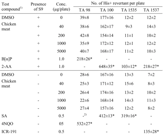
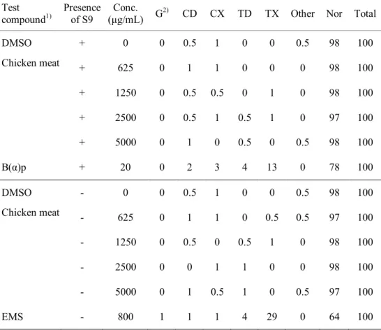
In the bacterial reverse-mutagen, in vitro chromosomal aberration, and in vivo micronucleus assays, the irradiated chicken breast meat with 7.5 MeV X-rays at 30 kGy exhibited dose-independent responses similar to those shown by the negative control. These results suggested that the irradiated samples were not genotoxic. In
iii
acute and sub-chronic toxicity studies, mortality or any abnormal clinical signs of ICR mice were not observed during the test periods. Several hematological and serum biochemical parameters of ICR mice showed significant differences from the values in the control group; however, those values were within the normal range for hematological and serum biochemical parameters of ICR mice. No specific toxic effects were observed in male and female ICR mice upon single oral administration of X-ray-irradiated (30 kGy) chicken breast at up to 2000 mg/kg body weight. Furthermore, daily intake of X-ray-irradiated chicken breast at 2500 mg/kg body weight for 90 days did not cause in any toxicological effects on the male or female mice. Therefore, these results revealed that chicken breast irradiated with 7.5 MeV X-rays at 30 kGy was not toxic to mice under the tested conditions.
Red pepper powder samples X-ray-irradiated at more than 8 kGy exhibited significantly more off-odor than did non-irradiated sample; however, there was no significant difference between non-irradiated and irradiated samples at less than 7 kGy, which is the maximum irradiation dose permitted in Korea. Characteristics of red pepper powder irradiated with 7.5 MeV X-rays including color, contents of capsaicinoids and capsanthin, and organoleptic properties except for off-odor exhibited no significant change upon an absorbed dose (p < 0.05). 2-thiobabituroinic acid reactive substance values of chicken breast meat and ground beef irradiated with 7.5 MeV X-rays increased as absorbed dose increased, whereas pH of meats did not exhibited significant changes, regardless of absorbed dose. Therefore, it is considered that 7.5 MeV X-ray-irradiated model foods at less than
iv
the upper dose limit did not exhibit quality deterioration induced by irradiation. The photo-stimulated luminescence photon counts for a minute (PCs/60 s) of red pepper powder irradiated at a dose less than 2 kGy were 700–5000 PCs/60 s, indicating the need for further confirmative analysis. In contrast, all samples irradiated at more than 4 kGy were correctly identified as irradiated (> 5000 PCs/60 s). In thermoluminescence (TL) analysis, TL ratios (TL1/TL2), which were calculated from the TL signal intensity of silicate mineral separated from red pepper powder irradiated with 7.5 MeV X-rays at 0.49–9.27 kGy (TL1) and the TL signal intensity of the TL1 mineral normalized with gamma rays at 1 kGy (TL2), of all the irradiated samples were over 0.1, resulting in positive results of all irradiated samples with 7.5 MeV at various doses. In X-ray-irradiated chicken breast meat, specific radiolytic hydrocarbons such as C16:2 and C17:1, which are used as markers for irradiated meat, were detected in the samples irradiated at > 4 kGy. Furthermore, 2-dodecylcyclobutanone and 2-tetradecylcyclobutanone, unique radiolytic products derived from lipids, were detected in all samples 7.5 MeV X-ray-irradiated at various doses and increased with a function of absorbed dose, whereas these were not detected in the non-irradiated sample.
From the result, 7.5 MeV X-ray irradiation can be used as a food pasteurization process not providing the physicochemical quality deterioration induced by irradiation within the allowed upper limit dose. Moreover, it is elucidated that 7.5 MeV X-ray-irradiated food is toxicologically safe and can be supervised by using physical and chemical identification methods.
v
Key words: X-ray, Gamma ray, Electron-beam, Bactericidal efficiency, Food quality, Identification of irradiated food, Toxicological safety
vi
Contents
Abstract ... i
Contents ...vi
List of Figures ... x
List of Tables ...xi
Chapter I. Literature review ... 1
I-1. Food irradiation by X-ray ... 1
I-2. Bacterial inactivation by X-ray irradiation ... 8
I-3. Physicochemical change by X-ray irradiation ... 11
I-4. Safety of irradiated food by 7.5 MeV X-ray ... 15
I-5. Food irradiation in Korea ... 17
Chapter II. Bacterial inactivation characteristics of 7.5 MeV X-ray irradiation ... 20
II-1. Introduction ... 20
II-2. Material and methods ... 22
II-2-1. Test strains and sample preparation ... 22
II-2-2. Irradiation of samples ... 25
II-2-3. Dosimetry ... 26
vii
II-2-5. Statistical analysis... 28
II-3. Results ... 29
II-3-1. Inactivation of foodborne pathogens by 7.5 MeV X-ray ... 29
II-3-2. Radiation sensitivity of bacteria in model foods for 7.5 MeV X-ray .. 37
II-4. Discussion ... 42
Chapter III. Toxicological safety of irradiated chicken breast meat by 7.5 MeV X-ray... 44
III-1. Introduction ... 44
III-2. Materials and Methods... 45
III-2-1. Genotoxicity test ... 45
III-2-2. Acute oral toxicity test ... 49
III-2-3. Sub-acute (90 days) oral toxicity test ... 51
III-2-4. Statistical analysis ... 53
III-3. Results ... 55
III-3-1. Genotoxicity of irradiated chicken breast meat by 7.5 MeV X-ray... 55
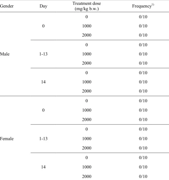
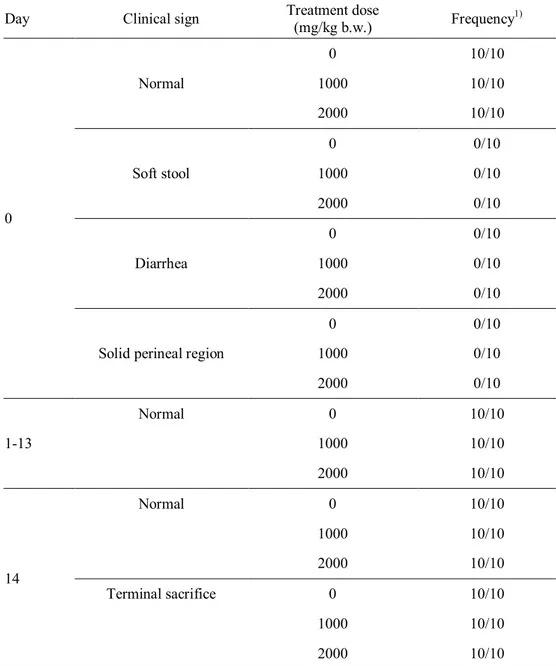
III-3-2. Acute toxicity of irradiated chicken breast meat by 7.5 MeV X-ray . 60 III-3-3. Sub-chronic toxicity of irradiated chicken breast meat by 7.5 MeV X-ray ... 71
viii
Chapter IV. Physicochemical quality characteristics of irradiated food by 7.5
MeV X-ray ... 86
IV-1. Introduction ... 86
IV-2. Materials and Methods ... 88
IV-2-1. Preparation of samples ... 88
IV-2-2. Irradiation of samples ... 88
IV-2-3. Dosimetry ... 89
IV-2-4. Hunter's color values ... 90
IV-2-5. Analysis of capsaicinoids and capsanthin ... 90
IV-2-6. pH and TBARS analysis ... 91
IV-2-7. Sensory evaluation ... 92
IV-2-8. Statistical analysis ... 93
IV-3. Results ... 94
IV-3-1. Quality characteristics of irradiated red pepper powder by 7.5 MeV X-ray ... 94
IV-3-2. Quality characteristics of irradiated chicken breast meat by 7.5 MeV X-ray ... 100
IV-3-3. Quality characteristics of irradiated ground beef by 7.5 MeV X-ray ... 108
ix
Chapter V. Identification characteristics of irradiated food by 7.5 MeV X-ray
... 116
V-1. Introduction ... 116
V-2. Materials and Methods ... 118
V-2-1. Preparation of samples ... 118
V-2-2. Irradiation of samples... 118
V-2-3. Dosimetry ... 119
V-2-4. Photostimulated luminescence analysis ... 120
V-2-5. Thermoluminescence analysis ... 121
V-2-6. Hydrocarbons and 2-ACBs analyses... 122
V-2-7. Statistical analysis ... 123
V-3. Results ... 124
V-3-1. Identification properties of irradiated red pepper powder by 7.5 MeV X-ray ... 124
V-3-2. Identification properties of irradiated meats by 7.5 MeV X-ray... 131
V-4. Discussion ... 135
References ... 138
x
List of Figures
Fig. 1. Growth curve of different bacteria incubated in tryptic soy broth at 37°C. .. 24 Fig. 2. Viability of bacterial pathogens inoculated on ground beef following treatment with different types of radiation... 35 Fig. 3. Histopathological examination of the liver of ICR mice administered with
X-ray-irradiated chicken meat at 30 kGy for 90 days. ... 82 Fig. 4. Histopathological examination of the kidney of ICR mice administered with X-ray-irradiated chicken meat at 30 kGy for 90 days. ... 83 Fig. 5. Organoleptic comparison in terms of off-odor intensities of red pepper powder irradiated by 7.5 MeV X-rays, gamma rays, or 10 MeV e-beams at 6 kGy. ... 99 Fig. 6. Comparison in TBARS value of chicken breast meat irradiated by 7.5 MeV X-rays, gamma rays, or 10 MeV e-beams at 2 kGy. ... 103 Fig. 7. Comparison in color of chicken breast meat irradiated by 7.5 MeV X-rays, gamma rays, or 10 MeV e-beams at 2 kGy. ... 107 Fig. 8. The first glow curves of red pepper powder irradiated by 7.5 MeV X-rays at different doses. ... 126 Fig. 9. TL signal intensity of quartz (A) and K-feldspar (B) irradiated by 7.5 MeV and 160 keV X-rays, gamma rays, and 10 MeV e-beams at 1 kGy. ... 130 Fig. 10. Chromatogram of hydrocarbons detected from chicken breast meat irradiated by 7.5 MeV X-ray. ... 132
xi
List of Tables
Table 1. Characteristics of three irradiation sources for food treatment ... 7 Table 2. List of food items permitted for gamma and e-beam irradiation in Korea . 19 Table 3. Viable cell count of Escherichia coli irradiated by 7.5 MeV X-rays at various doses ... 30 Table 4. Viable cell count of Salmonella Typhimurium irradiated by 7.5 MeV
X-rays at various doses ... 31 Table 5. Viable cell count of Listeria monocytogenes irradiated by 7.5 MeV X-rays at various doses ... 32 Table 6. Viable cell count of Staphylococcus aureus irradiated by 7.5 MeV X-rays at various doses ... 33 Table 7. Radiation sensitivity of foodborne pathogens for 7.5 MeV X-rays ... 34 Table 8. D10-values (kGy) for four strains inoculated on ground beef by radiation type ... 36 Table 9. Total aerobic bacteria count in red pepper powder irradiated by 7.5 MeV
X-rays at various doses ... 39 Table 10. Total aerobic bacteria count in chicken breast meat irradiated by 7.5 MeV X-rays at various doses during storage at 30°C for 7 days ... 40 Table 11. Total aerobic bacteria count in ground beef irradiated by 7.5 MeV X-rays at various doses during storage at 30°C for 7 days ... 41 Table 12. Formula of experimental diets ... 54
xii
Table 13. Salmonella Typhimurium reversion assay with X-ray-irradiated chicken meat at 30 kGy ... 57 Table 14. Chromosomal aberration test on X-ray-irradiated chicken meat at 30 kGy using a Chinese hamster lung cell line ... 58 Table 15. Frequency of micronuclei from marrow in mice treated with
X-ray-irradiated chicken meat at 30 kGy ... 59 Table 16. Mortality of ICR mice administered during acute toxicity test for
X-ray-irradiated chicken meat at 30 kGy ... 61 Table 17. Clinical signs of male ICR mouse during acute toxicity test for
X-ray-irradiated chicken meat at 30 kGy ... 62 Table 18. Clinical signs of female ICR mouse during acute toxicity test for
X-ray-irradiated chicken meat at 30 kGy ... 63 Table 19. Necropsy findings of ICR mice administered with X-ray-irradiated chicken meat (30 kGy) at various doses ... 64 Table 20. Body weight change of male ICR mice during acute toxicity test for
X-ray-irradiated chicken meat at 30 kGy ... 65 Table 21. Body weight change of female ICR mice during acute toxicity test for
X-ray-irradiated chicken meat at 30 kGy ... 66 Table 22. Hematological test of male ICR mice administered with X-ray-irradiated chicken (30 kGy) at various doses ... 67 Table 23. Hematological test of female ICR mice administered with
xiii
Table 24. Serum biochemistry test of male ICR mice administered with X-ray-irradiated chicken (30 kGy) at various doses ... 69 Table 25. Serum biochemistry test of female ICR mice administered with
X-ray-irradiated chicken (30 kGy) at various doses ... 70 Table 26. Mortality of ICR mice administered with X-ray-irradiated chicken meat at 0 and 30 kGy (2500 mg/ kg b.w./day) for 90 days ... 73 Table 27. Clinical signs of male ICR mice administered with X-ray-irradiated chicken at 0 and 30 kGy (2500 mg/ kg b.w./day) for 90 days ... 74 Table 28. Clinical signs of female ICR mice administered with X-ray-irradiated chicken at 0 and 30 kGy (2500 mg/ kg b.w./day) for 90 days ... 75 Table 29. Body weight change and food consumption of ICR mice administered with X-ray-irradiated chicken meat at 0 and 30 kGy (2500 mg/ kg bw/day) for 90 days ... 76 Table 30. Relative organ weights (%) of male ICR mice administered with
X-ray-irradiated chicken meat at 0 and 30 kGy (2500 mg/ kg b.w./day) for 90 days ... 77 Table 31. Hematological test of male ICR mice administered with X-ray-irradiated chicken meat at 0 and 30 kGy (2500 mg/ kg b.w./day) for 90 days ... 78 Table 32. Hematological test of female ICR mice administered with
X-ray-irradiated chicken meat at 0 and 30 kGy (2500 mg/ kg b.w./day) for 90 days ... 79 Table 33. Serum biochemistry test of male ICR mice administered with
X-ray-xiv
irradiated chicken meat at 0 and 30 kGy (2500 mg/ kg b.w./day) for 90 days ... 80 Table 34. Serum biochemistry test of female ICR mice administered with
X-ray-irradiated chicken meat at 0 and 30 kGy (2500 mg/ kg b.w./day) for 90 days ... 81 Table 35. Color measurement of red pepper powder irradiated by 7.5 MeV X-rays at various doses ... 96 Table 36. Contents of capsaicinoids and capsanthin in red pepper powder irradiated by 7.5 MeV X-rays at various doses ... 97 Table 37. Organoleptic evaluation of red pepper powder irradiated by 7.5 MeV
X-rays at various doses ... 98 Table 38. pH of chicken breast meat irradiated by 7.5 MeV X-rays at various doses during storage at 30°C for 7 days ... 101 Table 39. TBARS values of chicken breast meat irradiated by 7.5 MeV X-rays at various doses during storage at 30°C for 7 days... 102 Table 40. Lightness (L* value) of chicken breast meat irradiated by 7.5 MeV X-rays at various doses during storage at 30°C for 7 days ... 104 Table 41. Redness (a* value) of chicken breast meat irradiated by 7.5 MeV X-rays at various doses during storage at 30°C for 7 days... 105 Table 42. Yellowness (b* value) of chicken breast meat irradiated by 7.5 MeV
X-rays at various doses during storage at 30°C for 7 days... 106 Table 43. pH of ground beef irradiated by 7.5 MeV X-rays at various doses during
xv
storage at 30°C for 7 days ... 109 Table 44. TBARS values of ground beef irradiated by 7.5 MeV X-rays at various doses during storage at 30°C for 7 days ... 110 Table 45. Lightness (L* value) of ground beef irradiated by 7.5 MeV X-rays at various doses during storage at 30°C for 7 days... 111 Table 46. Redness (a* value) of ground beef irradiated by 7.5 MeV X-rays at
various doses during storage at 30°C for 7 days... 112 Table 47. Yellowness (b* value) of ground beef irradiated by 7.5 MeV X-rays at
various doses during storage at 30°C for 7 days... 113 Table 48. Photo-stimulated luminescence analysis of red pepper powder irradiated by 7.5 MeV X-rays at various doses ... 125 Table 49. Thermoluminescence analysis of red pepper powder irradiated by 7.5 MeV X-rays at various doses ... 127 Table 50. TL analysis of red pepper powder irradiated by 7.5 MeV X-rays followed by normalization with different types of ionizing radiation at 1 kGy .... 129 Table 51. Concentration of hydrocarbons detected from irradiated chicken breast meat by 7.5 MeV X-ray at various doses ... 133 Table 52. Concentration of 2-alkylcyclobutanones in ground beef irradiated by 7.5 MeV X-ray at various doses ... 134
1
Chapter I. Literature review
I-1. Food irradiation by X-ray
Food irradiation is a physical treatment, in which the food is subjected to a defined dose of ionizing radiation to extend the shelf life and inactivate harmful organisms (Arvanitoyannis & Tserkezou, 2010). Decades of research have conclusively shown that food irradiation can have myriad beneficial applications, including the insect disinfestation of fruits and grains, inhibition of sprouting in potatoes and onions, the delayed ripening of fresh fruits and vegetables, and the enhanced safety and sterilization of fresh and frozen meat products, seafood, and eggs (Diehl, 2002; Kume et al., 2009b).
Food irradiation technology has been permitted for more than 250 food items in 56 countries, with 400000 tons of food irradiated globally in 2005. China accounted for 36% of the total global irradiated food, which included the irradiation of 80000 tons of garlic to inhibit its germination, and 52000 tons of dried vegetables and spices. In the US, a total of 92000 tons of food was irradiated, including 80000 tons of spices, 8000 tons of ground beef and chicken meat, and 4000 tons of fruits and vegetables. In addition, Ukraine irradiated 70000 tons of wheat and barley using an electron accelerator. While the EU has shown a decrease in food irradiation due to the enforcement of regulations on labeling violations, food irradiation in Asian countries has increased (Kume et al., 2009a).
2
Irradiation sources for food treatment include the following: gamma rays from radionuclides 60Co or 137Cs; electron-beams (e-beams) generated from machine sources operated at or below a nominal energy level of 10 MeV; and X-rays generated from e-beam machines operated at below a nominal energy level of 5 MeV (Code of Federal Regulations, 1986; Codex Alimentarius Commission, 2003). The US Food and Drug Administration has approved an increase of the limit for e-beam energy used for generating X-rays to 7.5 MeV in response to a petition from IBA (Code of Federal Regulations, 2004).
X-rays are electromagnetic radiation with a wide energy spectrum, and generated by colliding accelerated electrons with a dense material (target) such as tantalum or tungsten in a process known as bremsstrahlung-conversion (Koch & Motz, 1959; Miller, 2005). The efficiency for converting electron beam power to X-ray power increases with the energy of the incident electrons and with the atomic number of the target material. Increasing the energy improves also the X-ray penetration; however, higher energy of incident e-beams than the threshold energy of target material can induce radioactivity of the material by emission of neutrons (Grégoire et al., 2003). Because the threshold energies of tungsten, tantalum, and gold are 6.19, 7.58, and 8.07 MeV, respectively, tantalum or gold is used as a target material for 7.5 MeV X-rays, whereas the maximum energy of tungsten for generating X-rays is 5 MeV (Cleland & Stichelbaut, 2013). In a tantalum target, the conversion efficiency from incident electron beam power to emitted X-ray powder in the forward direction is about 8–9% at 5 MeV and 12–13% at 7.5 MeV (Meissner et al.,
3
2000). The broad energy spectrum of bremsstrahlung photons emitted from tantalum target extends from a few tens of keV up to the maximum energy of the incident electrons. However, the peak of the photon energy spectrum occurs at about 0.3 MeV in the electron energy range from 5.0 to 10 MeV (Seltzer et al., 1983).
X-rays have a deep penetration power comparable to gamma rays from 60Co sources, with the added advantage of using an electronic source that stops radiating when switched off (Lazurik et al., 2007). A bremsstrahlung radiation from a 5.0 MeV electron beam penetrates slightly more than the gamma radiation (Seltzer & Berger, 1987). This greater penetration is partly caused by the higher photon energy of X-rays and the angular distribution. The narrow angular distribution of X-rays at above 5 MeV increases the penetration in materials because the most intense zone of the emitted radiation is perpendicular to the surface of the irradiated products, whereas gamma radiation has a wide angular distribution (Meissner et al., 2000). The optimum thickness of material irradiated with 7.5 MeV X-rays is approximately 38 g/cm2, whereas those of 10 MeV e-beams, gamma rays, and 5.0 MeV X-rays are 8, 31, and 34 g/cm2, respectively (Cleland & Stichelbaut, 2013).
Since the first discovery by Wilhelm Conrad Rőntgen in 1895, X-rays have been mainly used for medical radiography, scanning for security, and analyzing instruments such as X-ray diffractometer. For these applications, low-energy X-rays at below a few hundreds of keV were used. As the energy of electrons for generating X-rays increased for keV to MeV, sterilization of packaged food and
4
medical devices was proposed. Although the greater penetration, improved conversion efficiency, and radiological safety, the commercial application of 7.5 MeV X-rays is still scarce, owing to the economic issues such as lower throughput than e-beam irradiation and higher cost than gamma irradiation. However, recent comparisons have shown that the capital and electric power costs for electron accelerators with energies of 5–7 MeV equipped with X-ray targets can be lower than the capital and source replenishment costs for 60Co source loadings greater than 6 MCi based on the recent prices of 60Co at $2.5 US per curie and average cost of electric power at 12 cents per kilowatt hour (Cleland & Stichelbaut, 2007). IBA has reported that cost of energy for sterilizing 1 m3 of products is 8.6 € for 7.5 MeV X-ray irradiation, 9.1 € for gamma irradiation, and 4.1 € for e-beam irradiation, based on the same throughput at 64,000 m3/year (Mullier, 2015).
Different characteristics of X-rays with gamma rays and e-beams are summarized in Table 1. Gamma rays are a type of electromagnetic radiation emitted from radioactive isotopes such as 60Co and 137Cs (Woods & Pikaev, 1994). This type of radiation has the highest frequency and energy with the shortest wavelength, and can easily remove an orbital electron from an atom in the irradiated medium (Laughlin et al., 1989). This ionizing property imparts significant damage to a living cell and thus gamma radiation is often used to inactivate living organisms in food. 137Cs is not suitable for commercial use due to its lower energy compared to 60Co, and is used only in small hospital units to treat blood before transfusion for the prevention of Graft-versus-host disease (Ehlermann, 2005). Food irradiation using
5
60Co is the preferred method by most processors, as its deeper penetration enables the administration of treatment to entire industrial pallets. The main characteristics of gamma irradiation include the treatment of food with photons of well-defined energies (1.17 and 1.33 MeV) emitted from 60Co sources, and the irradiation of large amounts of food in bulk through its high penetration power. However, gamma irradiation also presents a number of disadvantages. These include the low consumer acceptance through fear of the radioisotope, and high construction costs due to the requirement for shielding (World Health Organization, 1988). However, despite of its disadvantages, gamma irradiation is used predominantly in food industries to inactivate microorganisms in spices, dried vegetables, and meat products (Kume et al., 2009a).
E-beam irradiation uses electrons accelerated in an electric field to a velocity close to the speed of light. High-energetic electrons are particulate ionizing radiation. As electrons have a significantly larger cross section than photons, they generally do not penetrate the product beyond a few centimeters, depending on product density (Bhat et al., 2012). The maximum energy of e-beams approved for food irradiation is 10 MeV, because higher energetic e-beams than 10 MeV can result in the subject becoming radioactive. The optimum penetration depth of such 10 MeV e-beams is approximately 8 g/cm2 with double sided irradiation. However, the use of e-beam irradiation is growing in the food irradiation market, as food can be treated quickly with e-beams at high dose rates. For example, frozen food such as meat and seafood can be treated with e-beam without any quality deterioration
6
induced from thawing, which is proceeded during gamma irradiation process (Brown, 2015).
Conclusively, food irradiation by 7.5 MeV X-rays has a few advantages compared to gamma and e-beam irradiation. Similarly with gamma irradiation, food product in bulk can be irradiated with 7.5 MeV X-rays, due to the highest penetration depth. The recent development of high-energy and high-power electron accelerators has made X-ray processing a practical alternative to gamma-ray processing in applications requiring greater penetration than can be provided by energetic electron beams. Moreover, the increased conversion efficiency makes 7.5 MeV X-ray irradiation competitive to gamma irradiation in terms of economic. The abilities to turn the radiation source on and off and to control the X-ray intensity are attractive features of an accelerator facility. Despite of the advantages, characterization studies and commercial application of 7.5 MeV X-ray irradiation to food commodities are still scarce. Therefore, further studies to evaluate microbiological, physicochemical, and toxicological characteristics of irradiated food by 7.5 MeV X-rays are needed.
7
Table 1. Characteristics of three irradiation sources for food treatment
Characteristics
Irradiation sources
X-ray Gamma-ray E-beam Radiation source Electric machine Radionuclide Electric machine Energy type Photon Photon Electron (particle) Maximum energy 7.5 MeV 1.33 MeV 10 MeV Dose rate 24 kGy/h 1 kGy/h 8000 kGy/h Penetration depth
8
I-2. Bacterial inactivation by X-ray irradiation
When an atom in material is exposed to X-rays, energy transactions occur between the incident photons and the orbiting electrons. These interactions result in a series of energy transfer from X-rays to material, then ionization occurs when the energy level sufficiently increases to produce ions by the removal of an orbiting electron (Wilkinson& Gould, 1998). Activity generated by the photon of X-rays exhibited two absorption processes in the irradiated material, Compton scattering and photoelectric absorption (Miller, 2005). When using low-energy photons, photoelectric absorption is generally occurred as all of the photon’s energy transferred to the electrons in material. Compton scattering occurs at higher energy levels, where a portion of the photon energy is absorbed by the encountered electron (Pizzarello & Witcofski, 1975).
The inactivation mechanism by X-ray can be explained by a combination of direct and indirect effects. The nucleic acid molecules may be ionized or excited by direct absorption of radiation energy, so initiating the chain of events that leads to biological change and to cell death if the change is serious enough. If a cell or an organism is exposed to radiation, direct damage to DNA occurs by chemical transformation of its components such as purine and pyrimidine bases, often resulting in the destruction of the DNA double helix or its phosphodiester linkages (Zerial et al., 1978). This is the so-called direct effect of radiation, which is the dominant process when dry spores of spore-forming microorganisms are irradiated. Alternatively, indirect effect of radiation is the damage caused by radicals formed
9
through the absorption of energy into other molecules such as water in the cell. The hydroxyl radical (OH‧) in particular is responsible for approximately 90% of damage to DNA, and is known to have the biggest influence on the radiation sensitivity of organisms. In addition, diverse damaging effects can occur with cells, including changes in cell components, such as protein, fat and carbohydrates. Such damage can result in chromosomal aberration, delay in cell division, metabolic suppression of carbohydrate and amino acids, and inactivation of enzymes. When living organisms are exposed to radiation, both direct and indirect effects occur simultaneously. With regard to the biological effects of radiation exposure, direct effects account for approximately 25%, while indirect effects account for 75% of the damage (Moosekian et al., 2012).
Viruses, bacterial spores, vegetative bacteria, fungi, and insects exhibit different sensitivity to radiation. The molecular weight of DNA influences the radio-sensitivity of organisms. The DNA in nuclei of the cells of insects represents a target much larger than the genome of bacteria. Not surprisingly, bacteria are less radiation sensitive than insects. Another factor influencing radiation effects is the structural arrangement of the DNA in the cell. During the cell division, the normally double-stranded DNA of bacteria separates, and by means of a polymerase new chains of DNA are assembled along the template. The double-stranded form DNA is much less sensitive to radiation than in the single-stranded form. The radiation sensitivity of a cell is proportional to cell proliferation and inversely proportional to cell differentiation. Therefore, radiation sensitivity is high when the metabolic rate
10
is high, when cell division is active, or when an individual is young.
However, results from studies on the radiation sensitivity of microorganisms suggest that the response of a microorganism to radiation largely depends on external environment, as this also has an influence on the survival of the microorganism (Molins, 2001). This implies that the external environment, including factors such as irradiation temperature, oxygen presence, and water activity of food have an influence on the physical and chemical characteristics of cells, thus causing differences in radiation sensitivity. These radiation sensitivity differences among similar groups of microorganisms are correlated to their inherent diversity with respect to the chemical and physical structure as well as their capacity to recover from radiation injuries.
11
I-3. Physicochemical change by X-ray irradiation
Radiation loses energy through interaction with materials, while the materials receive energy, generating ionization and excitation in materials. As a result of ionization and excitation, ions, electrons, excitation states, and radicals are formed. These generate new active species by interacting with one another or reacting with the surrounding molecules, and are known as reactive intermediated. Through the reaction of these intermediates, stable final products are eventually created. For example, when water is irradiated, ionization and excitation result in the generation of H2O+ and e- under 10-12 s. After 10-6 s, radicals such as H∙ and OH∙, hydrated electrons, and the acidic radical H3O+ are generated. In pure water, a molecule of hydrogen (H2) and peroxide (HOOH) are generated as the final stable products. If the water also contains a solute, these species can react with the solute.
When food is irradiated, the chemical changes may arise from the direct action of radiation on the carbohydrates, proteins, fats, and other compounds in the food, or by the indirect action of reactive intermediates formed by the radiolysis of water. In terms of food quality, changes in major components such as carbohydrates, proteins, and lipids may affect the organoleptic characteristics of the food. For example, the irradiation of sugar in an aqueous environment changes its optical rotation and often causes browning. Moreover, the degree of degradation in sugars in solution is proportional to the radiation dose, and can affect the sweet taste of the food following irradiation. Irradiation also leads to the degradation of polysaccharides such as starch, cellulose, and pectin, resulting in changes to texture and viscosity by
12
complex mechanisms. In addition, the production of hydrogen sulfide and methyl mercaptan, induced by the irradiation of sulfur-containing amino acids, can lead to off-odors and off-flavors (Simic, 1983). Furthermore, the irradiation may alter the viscosity of proteins, owing to the degradation and aggregation of proteins, while the production of radiolytic compounds from lipids can affect the flavor of food.
A number of physical and chemical methods based on the physicochemical changes of food components or silicate minerals in food by irradiation are used for identification of irradiated food. Among the physical techniques, photostimulated luminescence (PSL) and thermoluminescence (TL) measurements using the luminescence properties of contaminated minerals in food materials are promising methods. PSL is a rapid, simple, and inexpensive screening method, while TL analysis is one of the most reliable and sensitive methods for the detection of irradiated food (Bayram & Delincée, 2004; Chauhan et al., 2009; Sanderson et al., 1998). Luminescence is the emission of light when trapped energy is liberated by the addition of chemicals, heat, or light. The TL process is based on electrons in the excited state returning to the ground state when thermally stimulated (Heide & Bogl, 1987). When a substance exhibiting TL is exposed to ionizing radiation, electron-hole pairs are produced, and some electrons (or electron-holes) may become trapped at certain sites in the material. They remain in these traps until sufficient thermal energy is acquired to escape. As the material is heated, electrons are released from the traps, and light is emitted as they recombine with holes. The intensity of the emitted light can be measured as a function of temperature to give a so-called glow
13
curve, which is characteristic of the examined substance. The TL phenomenon is not unique to irradiation, but if the TL response following irradiation is significantly greater than the background signal, and the fading (i.e. the decrease in the TL signal) is low over a period of weeks and months, then TL measurements may be suitable for determining whether foodstuffs have been irradiated. The PSL method was then developed to resolve the practical limitations of silicate TL methods as the standard TL method requires a physical separation of the minerals from the food matrix. The requirement for careful laboratory preparation of TL samples and access to a calibration source of ionizing radiation have limited the widespread use of TL for routine commercial or enforcement testing (Sanderson et al., 1989). In contrast, PSL employs light rather than heat as a stimulus for releasing the trapped energy induced by radiation in solid materials. This has overcome the need for full mineral separation and for providing radiation-specific stimulation schemes appropriate for biogenic materials. However, it should be noted that PSL analysis has been used as a screening method, owing to a lower sensitivity than the TL method.
Most of the volatile products in food induced by irradiation originate from the lipid fraction; therefore, measurement of radiolytic products from food lipids could perform the basis for chemical methods to identify irradiated foods. Only two hydrocarbons are formed in relatively large quantities (Nawar & Balboni, 1970). One has a carbon atom less than the parent fatty acid, and results from cleavage at the carbon-carbon bond alpha to the carbonyl group; the other has two carbon atoms less and one extra double bond, and results from cleavage beta to the carbonyl
14
(Nawar et al., 1990). Nawar & Balboni (1970) reported that six specific radiolytic hydrocarbons, which are tetradecadiene (C14:1), pentadecane (C15:0), hexadecane (C16:1), heptadecane (C17:0), hexadecadiene (C16:2), and heptadecene (C17:1) produced from palmitic, stearic, and oleic acids, were detected in irradiated pork. A linear relationship between irradiation dose and each of these compounds has also been demonstrated (Morehouse & Ku, 1991).
2-Alkylcyclobutanones (2-ACBs) are cyclic compounds containing four carbon rings and produced by disruption of fatty acids when fat in food are irradiated. 2-ACBs have the same number of carbons as the precursor fatty acid, and an n-4 alkyl group side chain, which is formed by electron loss of the oxygen molecule in the fatty acid/triglyceride carbonyl group through ionization and consecutive rearrangement, is linked to the second carbon of the cyclic carbon ring (LeTellier & Nawar, 1972). Following extraction from food, these products can be detected and quantified using gas chromatography and mass spectrometry. 2-ACBs have not been detected in foods that have been heated, microwaved, UV irradiated, or subjected to high pressure processing, or ultrasonic waves (Crews et al., 2012); therefore, 2-ACBs are considered as unique radiolytic products that can serve as a marker to confirm whether fat-containing foods have been irradiated (CEN, 2003b). Contrary to a previous theory, it was reported that 2-ACBs exist naturally in food (Variyar et al., 2008). However, two researches recently reconfirmed the uniqueness of 2-ACBs in irradiated nutmeg (Chen et al., 2012) and cashew nuts, nutmeg, apricot kernel, and pine nuts (Leung et al., 2013).
15
I-4. Safety of irradiated food by 7.5 MeV X-ray
In order to evaluate the safety of irradiated food with 7.5 MeV X-rays, induced radioactivity in red meat was compared with natural radioactivity and background exposure (Grégoire et al., 2003). The induced radioactivity in X-ray-irradiated meat was significantly lower than the natural radioactivity in food. Corresponding annual dose is several orders of magnitude lower than the environmental background (1500 times lower in the worst case). Moreover, it is considered that intake of food irradiated with X-rays generated by electrons with nominal energy as high as 7.5 MeV is trivial.
In the history of food preservation, food irradiation is perhaps the most studied food processing technology in terms of toxicological safety. The wholesomeness of irradiated food has been carefully evaluated in an unprecedented number of studies for more than 50 years. At an international level, the need to consider the wholesomeness of irradiated foods was emphasized at a meeting sponsored by FAO, IAEA, and WHO in Brussels in 1961. The appropriate studies required to ascertain the wholesomeness were discussed by a Joint FAO/IAEA/WHO Expert Committee on Food Irradiation (JECFI) in Rome in 1964. Taking as a premise that the irradiation of food resulted in the production of radiolytic products in the foods, the Committee adopted the view that these products represented additions to the food. It therefore concluded that the establishment of the safety of irradiated foods should follow procedures similar to those generally used for evaluating the safety of food additives, and should be pursued on a food-by-food basis (World Health
16
Organization, 1966). A subsequent meeting was convened to assess the wholesomeness of irradiated wheat, potatoes, and onions in Geneva in 1969. The next Joint Expert Committee, which convened in 1976, reviewed a large number of animal studies on various irradiated foods. The committee also reviewed the results of radiation chemistry studies on the major components of food; it noted that many of the radiolytic products identified were present in food treated by heat and other processes, and considered that the health hazards from the concentrations found in irradiated foods were probably negligible (World Health Organization, 1977).
In 1980, the JECFI reviewed numerous microbiological, nutritional, and toxicological studies of irradiated foods and concluded that “the irradiation of any food commodity up to an overall average dose of 10 kGy presents no toxicological hazard and introduces no special nutritional or microbiological problems” (World Health Organization, 1981). In 1999, the joint FAO/IAEA/WHO Study Group on High-Dose Irradiation concluded that “irradiation to high doses is essentially analogous to conventional thermal processing, such as canning of low-acid foods, in that it eliminates biological hazards from foodstuffs intended for human consumption, but does not result in the formation of physical or chemical entities that could constitute a hazard.” (World Health Organization, 1999).
17
I-5. Food irradiation in Korea
Food irradiation studies were commenced by the Radiation Research Institute in Agriculture in Korea in 1966 to extend the shelf life of garlic, strawberries, and sweet potatoes. In addition, since the 1980s, the Korea Atomic Energy Research Institute has conducted various application studies on food irradiation. The food irradiation business was then established based on the Presidential decree 11717 in 1985, and the Gyeong-in Regional Korea Food and Drug Administration revised the Food Sanitation Act to permit food irradiation in 1986. Through 5 revisions in 1987, 1988, 1991, 1995, and 2004, gamma irradiation at various doses less than 10 kGy was permitted for 26 food groups, based on the appropriate purposes. The regulations for labeling and detection methods for irradiated food were subsequently added to the Korean Food Code in 2008 and 2010, respectively. In July 2012, electron-beam irradiation at less than 10 MeV was permitted for the same food groups as for gamma irradiation (Table 2). In this context, approval on X-ray food irradiation is needed because the lack of legislation on X-ray food irradiation in Korea can cause international conflict when food irradiated with X-ray is imported.
Only two gamma irradiation facilities (Greenpia technology Co. and Soya greentech Co.) are currently available for food irradiation. Dried agricultural products, such as spices or dried vegetables for the ingredients of processed foods are mainly treated by gamma irradiation, and it is estimated that approximately 5394 tons were treated in 2005. Since 2010, however, food-labeling regulations
18
have been initiated for all irradiated foods and ingredients, which may rapidly decrease the industrial use of irradiation. Low consumer acceptance of irradiated foods in Korea has also become an obstacle making food irradiation technology a widely accepted method for the treatment of foods. Therefore, education or promotion programs to convey correct information on food irradiation to consumers are needed.
19
Table 2. List of food items permitted for gamma and e-beam irradiation in Korea
Food item Maximum dose (kGy) Purpose Permission year Potato
0.15 Sprout inhibition
1987 Onion,
Garlic
Chestnut 0.25 Sprout inhibition Mushroom (fresh and dried) 1.00 controlling ripening Disinfestation, Egg powder
5.00 Pasteurization 1991/ 2004 Cereals (grain and powder)
Legumes (grain and powder) Starch as ingredient of food products
Dried meat powder
7.00 Pasteurization
1991/ 1995/ 2004 Dried fish and shellfish powder
Soybean paste powder Red pepper paste powder, Soy sauce powder, Dried vegetables Dried yeast Aloe powder
Ginseng products (including red ginseng)
Enzyme food Algae powder Dried spice
10.00 Pasteurization 1995/ 2004 Composite seasoning products
Sauces Powdered tea Leached tea Patients' diets
20
Chapter II. Bacterial inactivation characteristics of
7.5 MeV X-ray irradiation
II-1. Introduction
Food can be treated to extend its shelf life and inactivate bacteria and insects only with the three types of ionizing radiation such as X-rays, gamma rays, and e-beams (Farkas & Farkas, 2011). Among these, high-energy X-rays generated by using electrons with more than 5 MeV energy offer considerable promise for commercial application (Cleland & Stichelbaut, 2013). This is because they have higher penetration power than e-beams and better consumer acceptance than gamma rays generated from radionuclide sources (Fan & Sommers, 2013).
Dried vegetables, chicken meat, and ground beef are often contaminated with foodborne pathogens such as Bacillus cereus, Escherichia coli O157:H7, and
Salmonella enterica, which can cause food spoilage and foodborne outbreaks
(Banerjee & Sarkar, 2003). Among the food decontamination processes, irradiation is recommended, because the process is effective in inactivation of foodborne bacteria after packaging and a non-thermal pasteurization process, which can prevent food from thermal changes (Farkas, 1998; Farkas & Andrassy, 1988). Therefore, large amount of dried vegetables and raw meats are irradiated annually in many countries to reduce bacterial populations in the foods (Kume et al., 2009a).
21
However, studies and practical applications of food irradiation using high-energy X-rays have been scarcely reported. Therefore, this study was conducted to evaluate the inactivation characteristics of bacteria in model foods (red pepper powder, chicken breast meat, and ground beef) irradiated with 7.5 MeV X-rays, which have greater conversion efficiency of electric energy to radiation than 5 MeV X-rays and higher penetration power than both gamma rays and e-beams. Furthermore, to consider the feasibility of using 7.5 MeV X-rays for commercial sterilization of food products, the inactivation characteristics of 7.5 MeV X-rays were compared with those of gamma rays and e-beams, which are currently used for commercial sterilization of food.
22
II-2. Material and methods
II-2-1. Test strains and sample preparation
In this study, four species of pathogenic bacterial strains (2 gram-negative: E. coli and Salmonella Typhimurium and 2 gram-positive: Listeria monocytogenes and
Staphylococcus aureus) were investigated. Lyophilized E. coli KCCM 40406, S.
Typhimurium KCTC 1925, L. monocytogenes KCCM 40307, and S. aureus KCCM 11335 were obtained from the Korean Culture Center of Microorganisms (KCCM, Seoul, Korea) and the Korean Collection for Type Cultures (KCTC, Daejeon, Korea). Each strain was cultivated in Tryptic Soy Broth (TSB; Difco, Becton Dickinson, Sparks, MD) at 37°C for 18 h. The cultures of each strain were transferred twice at 24-h intervals in 10 mL of TSB in screw-cap test tube by using an inoculation loop. One hundred microliters of the fresh culture was aseptically transferred to 100 mL of TSB in a 250-mL Erlenmeyer flask. The bacterial growth curve of each bacterial strain was obtained by measuring the optical density of each respective culture (incubated at 37°C in a shaking incubator, 1200 rpm) at 600 nm using a Libra S70 spectrophotometer (Biochrom Ltd., Cambridge, UK) (Fig. 1). Cells in the late exponential growth phase were harvested by incubating S. Typhimurium and S.
aureus for 6 h, E. coli for 7 h, and L. monocytogenes for 14 h at 37°C. The cultured
broth of each strain was separated into four 50-mL centrifuge tubes (25 mL of culture in a tube), and the cells were harvested by centrifugation (3000 × g for 10 min at 4°C). The pellet in each centrifuge tube was washed twice with 25 mL of
23
sterile phosphate-buffered saline (PBS; Lonza, Walkersvile, MD) and re-suspended in 25 mL of PBS. All cell suspensions were collected in a 250 mL Erlenmeyer flask for a final concentration of approximately 108–109 colony forming units (CFU) per mL. The final culture suspensions of each strain were used to inoculate raw ground beef, or 1 mL of the suspension was transferred aseptically to individual microcentrifuge tubes, in order to evaluate bacterial cell viability.
For inoculation of pathogens, a 10 g portion of the beef was placed in a sterile filter bag (BagFilter®, Interscience, Rockland, MA), which was then packed in an oxygen-impermeable nylon bag (2 mL O2/m2/24 h at 0°C, 0.09 mm thickness; Sunkyung Co., Ltd., Seoul, Korea). These were subjected to gamma irradiation at 30 kGy in order to inactivate all microorganisms in the sample 1 day before inoculation. The test culture suspension (0.1 mL) was aseptically inoculated onto five spots of the sterile ground beef; this was blended manually for 5 min and formed in a size (length × width × height = 10 × 12 × 0.5 cm) within the bag. The inoculated samples were stored overnight in a refrigerator (4°C) prior to irradiation.
Red pepper powders, chicken breast meat, and ground beef (2.38 mm grind) were purchased from a local market in Jeongeup, Korea. The samples (100 g) were immediately placed in sterilized oxygen-impermeable nylon/polyethylene bags (20 × 30 cm, thickness: 0.07 mm; Sunkyung Co. Ltd., Seoul, Korea) and packaged to a thickness of 3.0 cm to minimize the variation in penetration depth among radiation sources. The packaged samples were stored overnight in a refrigerator (4°C) until irradiation.
24
25
II-2-2. Irradiation of samples
Five bacterial suspension or ground beef samples were simultaneously irradiated at targeted irradiation doses (0.1–1.0 kGy), whereas the packaged model food samples were irradiated at a nominal dose of 2, 4, 6, 8, or 10 kGy using one of the irradiation sources. The three irradiation sources were treated on the same day for all samples.
7.5 MeV X-ray and e-beam irradiations were conducted using a linear electron accelerator, which delivers e-beams with a well-defined energy in the range of 5–10 MeV, at EB-Tech Co. (Daejeon, Korea) at an ambient temperature of approximately 22°C. X-rays were produced using 7.5 MeV e-beams, whereas e-beams at 10 MeV were employed for e-beam irradiation. Gamma irradiation was performed using a 60Co gamma irradiator (point source AECL, IR-79, MDS Nordion International Co. Ltd., Ottawa, ON, Canada) at the Advanced Radiation Technology Institute, Jeongeup, Korea. The source strength was approximately 8.8 PBq and the photon energies of gamma rays from 60Co were 1.17 and 1.33 MeV. The applied dose rates were 24 kGy/h for X-ray irradiation, 8 MGy/h for e-beam irradiation, and 1 kGy/h for gamma irradiation. Geometrically, all samples were irradiated in a perpendicular direction to the incident radiation. The irradiated samples were immediately stored at 4°C and subjected to microbiological analysis.
26
II-2-3. Dosimetry
The actual doses absorbed by each sample and dose distribution across the samples were determined by using a 5 mm-diameter alanine dosimeter (Batch No.: T020604, Bruker BioSpin GmbH, Rheinstetten, Germany). Three dosimeters were placed in the bottom, middle, and top of an empty microcentrifuge tube and the space was filled with tissue paper. The tube containing 3 dosimeters was packaged in a polyethylene bag together with 5 PBS suspension samples. The bag was stored in a refrigerator until irradiation treatment. For dosimetry of inoculated beef and model food samples, a dosimeter was packaged in a polyethylene bag to prevent absorbing moisture, and 3 dosimeters were attached to the top of a sample for each irradiation dose. The samples with dosimeters were also stored in a refrigerator until irradiation.
Following irradiation, the dosimeters separated from samples were stored overnight at room temperature (approximately 25°C). The absorbed dose was measured within 24–48 h subsequent to irradiation using an electron paramagnetic resonance analyzer (e-scanTM alanine dosimeter reader, Bruker BioSpin GmbH, Rheinstetten, Germany) according to international standards (ISO/ASTM 51607:2004). The alanine dosimeter reader was calibrated using reference standard dosimeters provided from UK National Physics Laboratory according to an international standard on 30th April, 2015 (ISO/ASTM 51261, 2002). The dose uniformity ratios of all the type of radiation at various doses were less than 1.2.
27
by estimation of the uncertainty Type A and Type B in the absorbed dose measured with the alanine-EPR dosimetry system, according to the international guideline (ISO/IEC Guide 98-3, 2008). The expanded uncertainties of measured doses at a given dose for e-beam, gamma, and X-ray irradiation were calculated from the final combined standard uncertainty multiplied by a coverage factor k = 2, corresponding to an interval with a 95% level of confidence. The final combined standard uncertainty included reproducibility of the dose measurement (n=3) and uncertainties derived from establishing the traceability of the dosimetry system. In this study, absorbed dose of sample was expressed as a mean value ± the expended uncertainty.
II-2-4. Microbiological analysis
A series of decimal dilutions of the cell suspensions was prepared with sterile PBS, and each diluent (0.1 mL) was spread on tryptic soy agar culture (TSA, Difco Laboratories, Detroit, MI) plates. Ten grams of each of the ground beef samples were blended with 90 mL of sterile saline using a stomacher (Bag Mixer 400, Interscience Ind., St. Nom, France) for 1 min. A series of decimal dilutions was prepared with sterile PBS, and a 0.1 mL aliquot was spread on TSA. For model foods, a 10 g of samples was aseptically removed from the packages and placed into a stomacher bag (Nasco, Ft. Atkinson, WI, USA) with 90 mL of 0.1% buffered peptone water. Samples were then stomached for 1 min and serial dilutions were prepared. A series of decimal dilutions was prepared with sterile PBS, and a 0.1 mL
28
aliquot was spread on TSA. Plates were then incubated at 37°C for 24–48 h in an incubation chamber. Viable cells were enumerated based on dilutions exhibiting colony forming units (CFU) per plate. All subsequent bacterial counts were expressed as the mean value (log CFU/mL or CFU/g) with a standard deviation per absorbed dose.
II-2-5. Statistical analysis
The D10-vales of bacteria were determined by calculating the reciprocal of the slope subsequent to fitting the survival data against the absorbed dose by simple linear regression. The significant differences among the mean values were identified by Duncan’s multiple range test using the Statistical Package for Social Sciences (IBM, Armonk, NY, USA) with a confidence level of p < 0.05.
29
II-3. Results
II-3-1. Inactivation of foodborne pathogens by 7.5 MeV
X-ray
The viable cell counts of foodborne pathogens (E. coli, S. Typhimurium, L.
monocytogenes, and S. aureus) persisting in the PBS suspension or in the inoculated
ground beef followed by irradiation with 7.5MeV X-rays at various absorbed doses are shown in Tables 3–6 and the D10-values are summarized in Table 7.
Initial counts of the pathogens were approximately 8–9 log CFU/mL for cell suspensions and 6–7 log CFU/g for ground beef; the bacterial cells viability decreased significantly with the increase in absorbed dose (p < 0.05). The D10 -values of the pathogens suspended in PBS for 7.5 MeV X-rays were ranged from 0.11 to 0.21 kGy, while those values of cells inoculated in ground beef were 0.22– 0.41 kGy. In striking contrast to those of suspensions, the D10-values of treated beef-inoculated bacteria were approximately 2 times higher.
In comparison with those of 7.5 MeV X-rays, the D10-values of gamma rays generated from 60Co source and 10 MeV e-beams showed no significant differences in bactericidal efficiency (Fig. 2 and Table 8).
30
Table 3. Viable cell count of Escherichia coli irradiated by 7.5 MeV X-rays at various doses
Medium Absorbed dose (Gy) Viable cell count (log CFU/mL) Phosphate-buffered saline 0 8.16 ± 0.03F 94 ± 7 6.85 ± 0.56E 249 ± 23 5.75 ± 0.14D 332 ± 21 4.86 ± 0.06C 437 ± 32 3.71 ± 0.14B 557 ± 41 3.14 ± 0.09A
Medium Absorbed dose (Gy) Viable cell count (log CFU/g) Ground beef 0 7.26 ± 0.11E 194 ± 11 7.01 ± 0.14E 402 ± 24 5.75 ± 0.10D 683 ± 35 4.87 ± 0.43C 855 ± 44 3.49 ± 0.36B 1139 ± 64 2.30 ± 0.48A
Absorbed dose and viable cell count are expressed as mean ± the expended uncertainty at a 95% confidence interval (n=5).
A-FMeans followed by different letters within the column of a medium are significantly
31
Table 4. Viable cell count of Salmonella Typhimurium irradiated by 7.5 MeV X-rays at various doses
Medium Absorbed dose (Gy) Viable cell count (log CFU/mL) Phosphate-buffered saline 0 8.82 ± 0.16E 232 ± 17 7.51 ± 0.07D 366 ± 32 6.39 ± 0.41CD 575 ± 30 5.61 ± 0.39C 829 ± 45 4.20 ± 0.37B 944 ± 59 3.60 ± 0.09A
Medium Absorbed dose (Gy) Viable cell count (log CFU/g) Ground beef 0 6.90 ± 0.21D 233 ± 30 6.11 ± 0.17C 408 ± 28 5.95 ± 0.15BC 649 ± 49 5.20 ± 0.70B 863 ± 51 4.67 ± 0.69AB 1019 ± 81 4.47 ± 0.18A
Absorbed dose and viable cell count are expressed as mean ± the expended uncertainty at a 95% confidence interval (n=5).
A-EMeans followed by different letters within the column of a medium are significantly
32
Table 5. Viable cell count of Listeria monocytogenes irradiated by 7.5 MeV X-rays at various doses
Medium Absorbed dose (Gy) Viable cell count (log CFU/mL) Phosphate-buffered saline 0 9.01 ± 0.16D 228 ± 21 8.16 ± 0.54C 373 ± 19 7.27 ± 0.38B 556 ± 44 6.43 ± 0.49AB 837 ± 51 5.94 ± 0.74A 1103 ± 56 5.89 ± 0.29A
Medium Absorbed dose (Gy) Viable cell count (log CFU/g) Ground beef 0 7.56 ± 0.27D 249 ± 25 6.80 ± 0.24C 437 ± 42 6.42 ± 0.14B 658 ± 35 6.05 ± 0.46AB 923 ± 47 5.53 ± 0.50A 1134 ± 84 4.78 ± 0.93A
Absorbed dose and viable cell count are expressed as mean ± the expended uncertainty at a 95% confidence interval (n=5).
A-DMeans followed by different letters within the column of a medium are significantly
33
Table 6. Viable cell count of Staphylococcus aureus irradiated by 7.5 MeV X-rays at various doses
Medium Absorbed dose (Gy) Viable cell count (log CFU/mL) Phosphate-buffered saline 0 8.38 ± 0.18E 224 ± 19 7.79 ± 0.24D 473 ± 34 5.78 ± 0.38C 665 ± 41 4.23 ± 0.28B 912 ± 47 2.30 ± 0.41A 1151 ± 67 2.14 ± 0.37A
Medium Absorbed dose (Gy) Viable cell count (log CFU/g) Ground beef 0 7.34 ± 0.16C 228 ± 21 7.17 ± 0.17C 373 ± 29 6.27 ± 0.30BC 556 ± 37 5.76 ± 0.52B 837 ± 56 4.14 ± 0.40AB 1103 ± 52 3.34 ± 0.45A
Absorbed dose and viable cell count are expressed as mean ± the expended uncertainty at a 95% confidence interval (n=5).
A-EMeans followed by different letters within the column of a medium are significantly
34
Table 7. Radiation sensitivity of foodborne pathogens for 7.5 MeV X-rays
Medium
D10-value (kGy) Escherichia
coli Typhimurium Salmonella Staphylococcus aureus monocytogenes Listeria
Phosphate-buffered saline 0.11 0.18 0.14 0.21 Ground beef 0.22 0.41 0.25 0.41
35
Fig. 2. Viability of bacterial pathogens inoculated on ground beef following treatment with different types of radiation.
36
Table 8. D10-values (kGy) for four strains inoculated on ground beef by radiation type
Radiation type Escherichia coli Typhimurium Salmonella Staphylococcus aureus monocytogenes Listeria 7.5 MeV X-rays 0.22 0.41 0.25 0.41 Gamma-rays 0.18 0.35 0.37 0.40 10 MeV e-beams 0.18 0.30 0.27 0.37
37
II-3-2. Radiation sensitivity of bacteria in model foods for 7.5
MeV X-ray
The data presented in Table 9 show that the total aerobic bacteria count (TAB) of red pepper powder was significantly reduced as radiation dose increased (p < 0.05). The initial TAB of the non-irradiated sample was 5.27 log CFU/g, whereas the TABs in samples irradiated with 7.5 MeV X-rays were significantly reduced by 2 log values at a dose of 6 kGy (p < 0.05). No growth of TAB was observed in samples irradiated with a dose of 10 kGy. The D10 value of bacteria in red pepper powder for 7.5 MeV X-rays was 2.76 kGy.
TABs in chicken breast meat irradiated with 7.5 MeV X-rays at various doses during storage at 30°C for 7 days are shown in Table 10. The initial population in non-irradiated chicken meat was 4.84 log CFU/g, whereas viable cells were not observed in any irradiated samples. On day 3, a sharp increase in TAB of X-ray-irradiated samples at less than 8 kGy was observed; however, TAB in 10-kGy sample was not observed. On day 7, TAB in 10-kGy sample was 6.92 log CFU/g, indicating an X-ray irradiation at a dose of 10 kGy did not sterilize the TAB in chicken breast meat.
TABs in ground beef irradiated with 7.5 MeV X-rays at various doses during storage at 30°C for 7 days are shown in Table 11. The initial population in non-irradiated ground beef was 4.24 log CFU/g and viable cells were not observed in any irradiated samples. After 3-day storage at 30°C, however, TABs in
X-ray-38
irradiated samples at less than 10 kGy were greater than 3 log CFU/g. This result indicated that an X-ray irradiation at a dose of 10 kGy did not also sterilize the TAB in ground beef.
39
Table 9. Total aerobic bacteria count in red pepper powder irradiated by 7.5 MeV X-rays at various doses
Sample Absorbed dose (kGy) Total aerobic bacteria count (log CFU/g) Red pepper powder 0 5.27 ± 0.16D
2.13 ± 0.07 4.15 ± 0.12C 4.27 ± 0.12 4.26 ± 0.12C 6.01 ± 0.15 2.87 ± 0.19B 8.45 ± 0.23 2.00 ± 0.12A 10.37 ± 0.38 ND1) D10-value (kGy) 2.76
Absorbed dose are expressed as mean ± the expended uncertainty at a 95% confidence interval (n=3). Measurements are expressed ± standard deviation (n=3).
A-DMeans followed by different letters within a column are significantly different (p < 0.05). 1)Not detected within a detection limit of 1 log CFU/g.