저작자표시-비영리-변경금지 2.0 대한민국 이용자는 아래의 조건을 따르는 경우에 한하여 자유롭게 l 이 저작물을 복제, 배포, 전송, 전시, 공연 및 방송할 수 있습니다. 다음과 같은 조건을 따라야 합니다: l 귀하는, 이 저작물의 재이용이나 배포의 경우, 이 저작물에 적용된 이용허락조건 을 명확하게 나타내어야 합니다. l 저작권자로부터 별도의 허가를 받으면 이러한 조건들은 적용되지 않습니다. 저작권법에 따른 이용자의 권리는 위의 내용에 의하여 영향을 받지 않습니다. 이것은 이용허락규약(Legal Code)을 이해하기 쉽게 요약한 것입니다. Disclaimer 저작자표시. 귀하는 원저작자를 표시하여야 합니다. 비영리. 귀하는 이 저작물을 영리 목적으로 이용할 수 없습니다. 변경금지. 귀하는 이 저작물을 개작, 변형 또는 가공할 수 없습니다.
A THESIS FOR THE DEGREE OF MASTER OF SCIENCE
Development of a Pepper yellow leaf curl Thailand
virus Infectious Clone and Identification of a
Pathogenicity Factor of Tomato yellow leaf curl
Kanchanaburi virus in Tomato
고추황화잎말림 Thailand 바이러스의 감염클론 개발
및 토마토황화잎말림 Kanchanaburi 바이러스의
병원성 인자 발견
AUGUST, 2019
JONG WOOK AHN
MAJOR IN HORTICULTURAL SCIENCE AND BIOTECHNOLOGY
DEPARTMENT OF PLANT SCIENCE
Development of a Pepper yellow leaf curl Thailand
virus Infectious Clone and Identification of a
Pathogenicity Factor of Tomato yellow leaf curl
Kanchanaburi virus in Tomato
UNDER THE DIRECTION OF DR. BYOUNG-CHEORL KANG
SUBMITTED TO THE FACULTY OF THE GRADUATE SCHOOL
OF SEOUL NATIONAL UNIVERSITY
BY
JONG WOOK AHN
MAJOR IN HORTICULTURAL SCIENCE AND BIOTECHNOLOGY
DEPARTMENT OF PLANT SCIENCE
THE GRADUATE SCHOOL OF SEOUL NATIONAL UNIVERSITY
AUGUST, 2019
APPROVED AS A QUALIFIED DISSERTATION OF
JONG WOOK AHN
FOR THE DEGREE OF MASTER OF SCIENCE
BY THE COMMITTEE MEMBERS
CHAIRMAN
Doil Choi, Ph.D.
VICE-CHAIRMAN
Byoung-Cheorl Kang, Ph.D.
Development of a Pepper yellow leaf curl
Thailand virus Infectious Clone and
Identification of a Pathogenicity Factor of
Tomato yellow leaf curl Kanchanaburi virus in
Tomato
JONG WOOK AHN
DEPARTMENT OF PLANT SCIENCE
THE GRADUATE SCHOOL OF SEOUL NATIONAL UNIVERSITY
ABSRACT
Yellow leaf curl symptoms caused by Pepper yellow leaf curl Thailand virus (PYLCThV) and Tomato yellow leaf curl Kanchanaburi virus (TYLCKaV) are belonging to the begomovirus group in the geminiviridae family. begomoviruses, circular single-stranded DNA viruses cause severe crop losses in tropical and subtropical countries, particularly in tomato and pepper. In this study, PYLCThV were isolated from the yellow leaf curl symptomatic leaves of pepper. The viral genome sequence was confirmed and an infectious PYLCThV clone was developed. Both DNA-A and DNA-B of PYLCThV were inserted into a binary vector pICH86988. Agrobacteria containing each clone were infiltrated in to N.
benthamiana, tomato, and pepper to test infectivity of the infectious PYLCThV
clone. N.benthamiana showed high infectivity to the infectious clone, whereas, no viral symptoms and no virus accumulation were observed in tomato and pepper. This result allowed us to conduct a genome swapping analysis by using TYLCKaV infectious clone that have strong infectivity in tomato. The genome swapping analysis revealed that the B genome of TYLCKaV was responsible for pathogenicity in tomato. Moreover, we mapped using a series of chimeric DNA-B genome. Among the chimeric clones, a chimeric clone containing PYLCThV-B with the TYLCKAV-B backbone showed infectivity in tomato. This result demonstrated that the TYLCKaV UTR region plays a role as a pathogenicity determinant in tomato. This study provides molecular basis of understanding TYLCKaV infectivity and the interaction between begomovirus and plants.
CONTENTS
ABSTRACT ...i
CONTENTS ...iv
LIST OF TABLES ...vii
LIST OF FIGURES ...viii
LIST OF ABBREVIATIONS ...ix
INTRODUCTION LITERATURE REVIEW Plant immune system………...14
Plant immunity against virus...15
Genetics of plant of virus………...15
Geminivirus………...17
MATERIALS AND METHODS Virus inocula and plant materials ...19
Isolation and sequencing of viral genomes ...19
Construction of PYLCThV infectious clone ...20
Virus inoculation ...26
Detection of TYLCKaV and PYLCThV accumulation ...26
Infectivity test of infectious clones in pepper……….27
Phylogenetic analysis………28
RESULTS Characterization of PYLCThV ...29
Pathogenicity test of PYLCThV infectious clone in N.benthamiana and tomato……….33
Genome swapping analysis of PYLCThV and TYLCKaV DNA-B...36
Mapping the pathogenicity factor of TYLCKaV in tomato….…………...40
Sequence comparison of the UTR regions of PYLCThV and TYLCKaV….46 DISCUSSION ...48
REFERENCES ...53
LIST OF TABLES
Table 1. List of primers used for the construction of the PYLCThV infectious clone and virus detection ………...21 Table 2. List of primers used for construction of the PYLCThV/TYLCKaV chimeric clones ……….………….………….……...24 Table 3. Infectivity test of PYLCThV infectious clone in N. benthamiana and tomato………..35 Table 4. Infectivity test of TYLCKaV and PYLCThV DNA-B swapping………..39 Table 5. Infectivity test of TYLCKaV/PYLCThV chimeric clone in tomato ……45
LIST OF FIGURES
Figure 1. Schematic diagram of developing a PYLCThV infectious clone ………..22 Figure 2. Diagram showing chimeric clones of TYLCKaV-B and PYLCThV-B…..25 Figure 3. Detection and sequence analysis of viral genome ...31 Figure 4. Phylogenetic tree based on begomovirus DNA-B BC1 region…………..32 Figure 5. Symptoms of N. benthamiana and S .lycopersicum ‘A39’ infected with PYLCThV……….34 Figure 6. Inoculation of PYLCThV and TYLCKaV DNA-B swapping clones……37 Figure 7. Schematic diagram of PYLCThV/TYLCKaV DNA-B chimera clones….42 Figure 8. Infectivity test of PYLCThV/TYLCKaV chimeric clones………...…….44 Figure 9. Comparison of PYLCThV and TYLCKaV intergenic regions…..………47 Figure 10. C. annuum ‘ECW30R’ infected by TYLCKaV + TYLCKaV backbone chimeric construct with PYLCThV BC1………..51 Figure 11. Confirmation of C. annuum ‘ECW30R’ as a natural host using insect vectors………...52
LIST OF ABBREVIATIONS
AGO
ARGONAUTE protein
Avr
Avirulence
BLAST
Basic local alignment search tool
CP
Coat protein
DCLs
Dicer-like proteins
DPI
Days post inoculation
ETI
Effector-triggered immunity
ETS
Effector-triggered susceptibility
eIF4E
Eukaryotic translation initiation factor 4E
ER
Extreme resistance
GA
Gibson assembly
HR
Hypersensitive response
ICTV
International Committee of Taxonomy of Viruses
IR
Intergenic region
LRR
Leucine rich repeat
MP(BC1)
Movement protein
NB
Nucleotide binding
NCBI
National center for biotechnology information
NSs
Non-stuctural protein
NSP(BV1)
Nuclear shuttle protein
PRRs
Patter recognition receptors
PAMPs
Pathogen-associated molecular patterns
PTI
PAMP-triggered immunity
PYLCThV
Pepper yellow leaf curl Thailand virus
PCR
Polymerase chain reaction
RCR
Rolling cycle replication
Rep
Replication initiator protein
Ren
Replication enhancer protein
TrAP
Transcription activator protein
TYLCKaV
Tomato yellow leaf curl Kanchanaburi virus
TYLCV
Tomato yellow leaf curl virus
UTR
Untranslated region
VSRs
Viral suppressor of RNA silencing
Vir
Virulence
INTRODUCTION
The Geminiviridae are a family of plant DNA viruses characterized by single-stranded circular genomes encapsidated in twinned icosahedral particles that cause substantial crop losses around the world (Hanley-Bowdoin et al., 2013). Based on their genome structure and transmitting vectors, geminiviruses can be divided into four genera: Begomovirus, Curtovirus, Mastrevirus, Topocuvirus (Fondong VN 2013). According to the International Committee of Taxonomy of Viruses (ICTV), begomoviruses are the most diverse and geographically widespread viruses (Zerbini FM et al., 2017). Single-stranded DNA (ssDNA) is packaged in to virions for all begomoviruses, and replicated to double-stranded DNA (dsDNA) in plant cells. Their genomes are consisted of one (monopartite) or two (bipartite) DNA segments that encode 5-7 proteins responsible for viral replication, transmission, cell trafficking and pathogenesis (Rojas et al., 2005).
Over the past three decades, the emergence of begomoviruses has been associated with increase of whitefly population. The yellow leaf curl diseases cause severe yield losses in Solanaceous crops especially tomato (S. lycopersicum), pepper (Capsicum spp.), and eggplant (Solanum melongena L.), in tropical and subtropical regions of the world (Kenyon et al., 2014). The YLCD of pepper has been observed since 1995 in Kanchanaburi province of Thailand, and TYLCKaV and PYLCThV genome sequences were characterized to be a distinct novel bipartite begomovirus
species from previous classification (Green et al., 2003; Chiemsombat et al., 2018). The begomovirus are transmitted by a one white fly species, Bemisia tabaci. The species B. tabaci can be divided into three biotypes (A, B, Q) based on their ability to interbreed and divergence of the mitochondrial cytochrome oxidase1 (mtCO1) gene (De Barro et al., 2011). To date, 360 begomovirus species and 41 morphologically indistinguishable B.tabaci species have been characterized however, very little is known about the whitefly determinants of begomovirus transmission (Patil et al., 2018).
To identify the fundamental knowledge of pepper-infecting begomoviruses, molecular phylogenic studies have been carried out. At least 37 ICTV species and 6 candidate species of begomovirus have been reported in Capsicum species south-east Asian countries (Kenyon et al., 2014). In Korea, tomato yellow leaf curl disease has first reported in 2008 and spread rapidly from southern to central areas (Lee et al., 2010). Recently, Kil et al (2014) developed an infectious clone for monopartite Korean TYLCV isolate and conducted successful whitefly-mediated tomato infection but failed to infect pepper.
There have been tremendous efforts to find resistance resources to TYLCV tomato. UP to date, six major resistance genes (From Ty-1- to Ty-6) have been reported in tomato (Lapidot et al., 2015). These genes have been deployed in tomato breeding and the identities of genes are known (Lapidot et al., 2002; Scott et al., 2015; Verlaan et al., 2013). Despite a high incidences of yellow leaf curl disease in
(Chiemsombat P 2018; S.L. Shih et al., 2010; Koeda S. et al., 2016). In 2018, we developed TYLCKaV infectious clone and tested infectivity by agro-inoculation and whitefly-mediated inoculation in tomato and pepper. The agro-inoculation test was successful in tomato with obvious TYLCD symptoms. However, both whitefly-mediated and Agrobacterium-mediated inoculation methods were not successful in pepper (Choi et al., 2018). Thus, to identify the resistance resources of pepper plants to viruses, an efficient infection system is required.
There have been several studies on Agrobacterium-mediated infection using TYLCV infectious clones in pepper (Morilla, 2005; Kil et al., 2014). The first report using infectious clone demonstrated that peppers can be infected by TYLCV clone but did not show any visible symptoms. However, symptomless peppers harboring TYLCV can as a reservoir of TYLCV transmit the virus to tomato by whitefly (Morilla, 2005; Kil et al., 2014). Recently, an infectious clone were prepared from and Indonesian TYLCKaV isolate (KF446672). This clone had high infectivity in Tomato and N. benthamiana. Nevertheless, the clone was not able to infect pepper (Koeda et al., 2017).
The objectives of this study were to develop an infectious clone of Pepper
yellow leaf curl Thailand virus for successful infection to Capsicum spp. and to map
LITERATURE REVIEW
1. Plant immune system
Plants, unlike mammals, lack mobile defender cells and a somatic adaptive immune system. Instead, they relying on the innate immunity of each cell and on systemic signals from infection sites (Venkatesh J and Kang 2019). Plants have interesting receptor called pattern recognition receptors (PRRs) to recognize pathogens through conserved and variable pathogen elicitors such as pathogen-associated molecular patterns (PAMPs) (Dodds et al., 2010). This PAMP-triggered immunity (PTI) can inhibit further pathogen colonization. On the other hand, for successful pathogen virulence, effectors can interfere with PTI by secretion of effectors proteins and this result in effector-triggered susceptibility (ETS) (Jones and Dangl 2006). To overcome ETS, plants evolved to recognize the effectors by intracellular receptors such as (R) protein which derived from complex proteins motifs of nucleotide binding (NB) and leucine rich repeat (LRR) domain (Ting and Davis et al., 2005). This immunity system called effector-triggered immunity (ETI). Usually, ETI response shows more strong resistance than PTI, inducing hypersensitive response (HR) This model of recognition explain not only pathogen-host interaction obviously but it also provide evidences about co-evolution between plant and microbe (Jones and Dangl 2006).
2. Plant immunity against virus
Although (R) protein is well-characterized mechanism of plant antiviral defense, RNA silencing still plays a central role in plant immune response. Most plant viruses have conserved motif that contain imperfect regulatory hairpin and are copied in plant cell. (Pumplin and Voinnet 2013). When virus invade intracellular space, viral double stranded RNA is processed by dicer-like proteins (DCLs) and cleaved by ARGONAUTE protein (AGO) family (Hanley-Bowdoin et al., 2013). After primary siRNAs have been generated, induced virus small RNA can move to adjacent cell or elongated into long dsRNA by RNA-dependent RNA polymerase in order to trafficking resistant signal to nucleus or other plant cells (Csorba et al., 2009). To make successful infection, virus evolve to obtain viral suppressor of RNA silencing (VSRs) (Voinnet et al., 1999). For instance, Tomato spotted wilt virus nonstructural protein (NSs) binds to dsRNA or siRNA to inhibit dicer-mediated cleavage of long dsRNA and prevents siRNA binding to AGO protein (Schnettler et al., 2010). In recent studies revealed that miRNAs can also control the NB-LRR motif based on resistance mechanism (Li et al., 2012).
3. Genetics of plant virus resistance
Heritable resistance of plant virus can be controlled by dominant or recessive genes. In plant, dominant genes are usually (R) genes that provide full resistance to one or more pathogens (Kourelis et al., 2018). The most largely researched are single dominant resistance (R) genes. The R gene induces programed cell death
responses with rapid occurrence of necrotic lesions (HR) and invisible necrosis (extreme resistance, ER) (de Ronde et al., 2014). Recently, the research between R protein and microbial interaction is actively done. The plants and microbe proteins directly or indirectly interact to make plant resistance or susceptible in the intracellular space. If pathogen infects and replicates in the host to produce disease, the virus is described as virulent with compatible interaction, and when the virus molecule that specifically elicits R-protein-mediated responses, this call the avirulence (Avr) be termed an incompatible interaction (Soosaar et al., 2005). The recessive resistance is more related with using host proteins. Since plant virus encodes limited number of genes, virus has evolved to use host factors. For achieving successful infection, the virus utilize host factors for viral replication, cell-to-cell movement and systemic development (Kang et al., 2005). Plant recessive resistance mechanism is derived by mutating host factors to prevent viral spreading. (Truniger and Aranda 2009). It can confer durable virus resistance for example, pepper eukaryotic translation initiation factor 4E (eIF4E) gene is required for several potyvirus infection however mutation in eIF4E results in disturbing in viral as multiplication and cell-to-cell movement of viral particle (Diaz-pendon et al., 2004).
4. Geminivirus
Geminiviruses threaten food security and agriculture, infecting major crop species, especially in tropical and subtropical regions (Ali et al., 2016). Geminiviruses have been classified into four genera: Begomovirus, Cutovirus, Topocuvirus and Mastrevirus based on host range, insect transmission vector and genome organization (Fondong et al., 2013). All geminivirus genomes consist of one (monopartite) or two (bipartite) circular single-stranded DNA (ssDNA) molecules that are encapsidated in icosahedral twinned particles (Fondong VN 2013). To maximize viral DNA replication, geminivirus obtained and evolved their mechanism to reprogram the plant cell life cycle by encoding multiple viral genes (Rojas et al., 2005). DNA-A has six open reading frames such as replication initiator protein (Rep), replication enhancer protein (Ren), the coat protein (CP, and Transcription activator protein (TrAP) (Fondong VN 2013). DNA-B has two ORFs that essential for intra- or inter-cellular trafficking by movement protein (BC1) and nuclear shuttle protein (BV1) interaction. (Gafni et al., 2002). Besides, geminivirus DNA-A and DNA-B share unique conserved region of ~200 nucleotide in their intergenic region (IR) (Harrison et al., 1985). The IR contains highly conserved motif of ‘5-TAATATTAC-3’ and GC-rich region that essential sequence for viral DNA replication and transcription of both components (Hanley-Bowdoin et al., 2000).
In the plant cell, single stranded viral DNA is uncoated from virion and start to replication to form double stranded DNA with interaction of various host factors (Nagar et al., 1995). The initiation of replication occur when replication initiator protein (Rep) binds to hairpin structure with GC repeat sequence (Pradhan et al., 2017). Rep-hairpin structure then initiated rolling-cycle replication (RCR) by nicking the conserved motif of geminivirus ori at 5’-TAATATT↓AC-3’ (↓: site of nicking) position and Rep protein binds to the nicking site covalently for RCR extension (Laufs et al., 1995). The replicated formed dsDNA can translated to mRNA to make viral symptoms or captured by BV1 to inter-cellular movement though BC1 derived tubular structures (Gafni et al., 2002).
Among the geminivirus, tomato yellow leaf curl virus (genus begomovirus) is one of the major viral diseases in tomato and ranked in the third most serious disease in worldwide (Scholthof et al., 2011). Recently, TYLCV broaden the host range to Capsicum spp. Pepper yellow leaf curl virus has been reported in 1995 at Kanchanaburi province in Thailand (Chiemsombat P 2018). Yellow leaf curl virus (YLCD) is naturally transmitted by the whitefly (Bemisia tabaci). Therefore, controlling whitefly and breeding resistance of YLCD cultivar is a key for successful pepper cultivation.
MATERIALS AND METHODS
Virus inocula and plant materials
Two different begomoviruses were used in this study. TYLCKaV (provided by East-West Seed Indonesia) and PYLCThV (provided by East-West Seed in Thailand) inoculum was used develop to full-length infectious clones. N. benthamiana, tomato (S. lycopersicum ‘A39’), ECW30R (C. annuum) were tested infectivity of TYLCKaV (Choi et al., 2018), PYLCThV and chimeric clones.
Isolation and sequencing of viral genomes
Geminivirus DNA-A was extracted and amplified by polymerase chain reaction (PCR) using geminivirus universal primer pair PAL1v1978/PAR1c715 (Rojas et al., 1993) (Table 1). For viral DNA amplification, the following PCR condition were use: 30 cycles of denaturation at 94℃ for 60 s, annealing at 50 ℃ for 60 s and extension, at 72℃ for 3min. The amplicon of 1.4kb sized product were sequenced at Macrogen (Republic of Korea) and verified by BLAST search at NCBI (www.ncbi.nlm.nih.gov/). Based on the sequence of the 1.4kb DNA product, Additional primers were designed to complete the DNA-A sequence. Furthermore, DNA-B specific primer pairs were designed to amplify the full-length DNA-B sequence data based on the NCBI GenBank data.
Construction of PYLCThV infectious clone
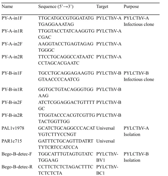
To develop PYLCThV infectious clone, each construct carrying viral genome of 0.7 mer (1.8 kb) and 0.6 mer (1.5 kb) including hairpin region were initially cloned into pUC19b vector and then subcloned to pICH86988 expression vector. (Figure 1). Two amplicons from DNA-A which flanking Sph1, KpaI and KpaI, XmaI restriction enzyme sites are amplified by specific primers based on virus sequence. For DNA-B construction, flanking amplicons containing restriction enzyme sites of SbfI, PstI
Table 1. List of primers used for the construction of an infectious PYLCThV clone
and virus detection.
Name Sequence (5’→3’) Target Purpose
PY-A-in1F TTGCATGCCGTGGATATG TGAGGAAATAG PYLCThV-A PYLCThV-A Infectious clone PY-A-in1R TTGGTACCTATCAAGGTG CGAC PYLCThV-A PY-A-in2F AAGGTACCTGAGTAGAG TGGGC PYLCThV-A PY-A-in2R TTCCTGCAGGCCATAATC CCTAGCACGAATC PYLCThV-A PY-B-in1F TGCCTGCAGGAGAAGTG GTAACCCCAATCG PYLCThV-B PYLCThV-B Infectious clone PY-B-in1R GGTGCTGTACAGGGTGG AAG PYLCThV-B PY-B-in2F ATCTCGGAGGACTGTTTT GC PYLCThV-B PY-B-in2R TTGGTACCCACGTCGTTG TACTGGTTGG PYLCThV-B PAL1v1978 GCATCTGCAGGCCCACAT YGTCTTYCCNGT Universal PYLCThV-A Isolation PAR1c715 GATTTCTGCAGTTDATRT TYTCRTCCATCCA Universal Bego-B-detec-F TGGCATTTGTAGTGTATC TGGAAG PYLCThV-BV1 PYLCThV-B Isolation Bego-B-detec-R CCTTCTCTCTAGACTTTC TCTCTCTA PYLCThV-BC1
Figure 1. Schematic diagram of developing a PYLCThV infectious clone. (A)
pICH86988-PYLCThV-DNA-A and (B) pICH86988-PYLCThV-DNA-B were constructed using recombinant DNA technology.
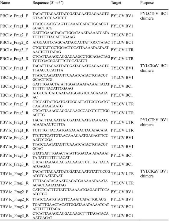
Construction of TYLCKaV/PYLCThV chimeric clone
Previously, the complete TYLCKaV-A and TYLCKaV-B were cloned into the pICH86988 vector by golden-gate cloning method (Engler et al., 2008) in our laboratory (Choi 2018). Based on the virus sequence, total of four YLCKaV/PTLCThV chimeric clones were made by swapping sequences of the TYLCKaV and PYLCThV by Gibson Assembly (GA) method (Gibson et al., 2009). To construct chimeric infectious clones of TYLCKaV-B and PYLCThV-B, empty pICH86988 vector digested with BsaI (Figure 2) and three segments were amplified with a 20-22 nucleotide overlap region between genome segments using GA specific primer pairs (Table 2). Each geminivirus DNA-B genome segments were assembled using Gibson Assembly Master Mix (Transgen Biotech, China) as described in the provided manual. Briefly, 1μl of the linearized vector (40ng/ μl), 1μl of the amplified fragments (33ng/ μl), 1 μl of ultrapure H2O and 5μl of 2X Assembly master mix were mixed and incubated at 50℃ for 15 min. The reaction product were immediately transformed into Trans1-T1 Phage Resistant Competent E.coli Cell (Transgen Biotech, China). Selected colonies were grown at 37℃ for 18 h, and plasmid DNA was extract using the AccuPrep Plasmid Mini Extraction Kit (Bioneer Korea).
Table 2. List of primers used for construction of the PYLCThV/TYLCKaV chimera
clones.
Name Sequence (5’→3’) Target Purpose
PBC1c_Frag1_F TACATTTACAATTATCGATACAATGAGAAGTGGTAACCCCAATCGT PYLCV BV1 PYLCThV BC1 chimera PBC1c_Frag1_R TTATCCAATGTAGTTCAAATCATATTGCACGTGCACTTCG PYLCV BV1
PBC1c_Frag2_F GATTTGAACTACATTGGATAAATAAAATCATATTTTTTTTACATTTGAAG TYLCV BC1 PBC1c_Frag2_R ATGGAGTCCAGCAATAGCAGTATTGCCTATAC TYLCV BC1 PBC1c_Frag3_F CTGCTATTGCTGGACTCCATTAAAATAATAATAACTCTTTATAG PYLCV UTR PBC1c_Frag3_R CTCATTAAAGCAGGACAAGCCTGCAGACTAGTGTCGACGGATTTCTGCATATCT PYLCV UTR
TBC1c_Frag1_F TACATTTACAATTATCGATACAATGAGAAGTGTTAACCCCATTTA TYLCV BV1 TYLCKaV BC1 chimera TBC1c_Frag1_R TTATCCAATATAGTTCAAATCATACTGTACGTGCACTTCG TYLCV BV1
TBC1c_Frag2_F GATTTGAACTATATTGGATAAATAAAATTATATTTTTTTTACATTCGAAG PYLCV BC1 TBC1c_Frag2_R ATGCCATCATCAATAATGGAGTCCAGAAATAAC PYLCV BC1 TBC1c_Frag3_F CTCCATTATTGATGATGGCATTATTGCCGATGTCAATATAATAATG TYLCV UTR TBC1c_Frag3_R CTCATTAAAGCAGGACAAGCCACGTCTTTGGACTTG TYLCV UTR
PBV1c_Frag1_F TACATTTACAATTATCGATACAATGTAAAATAATAATAACTCTTTA PYLCV UTR PYLCThV BV1 chimera PBV1c_Frag1_R TGTTTGTTACAATGAGAGAACTACATACATA PYLCV UTR
PBV1c_Frag2_F TTCTCTCATTGTAACAAACAATGAGAGTTCCAATCCGGA TYLCV BV1 PBV1c_Frag2_R TTATCCAATATAGTTCAAATCATACTGTACGTGCAC TYLCV BV1 PBV1c_Frag3_F GTATGATTTGAACTATATTGGATAA ATAAAATTA TATTTTTTTTACAT PYLCV BC1 PBV1c_Frag3_R CTCATTAAAGCAGGACAAGCTGTTTGTTACAATGAGAG PYLCV BC1
TBV1c_Frag1_F TACATTTACAATTATCGATACAATGTATTGCCGATGTCAATATAAT TYLCV UTR TYLCKaV BV1 chimera TBV1c_Frag1_R TTTTAGATACAAATGAGATGAAAAATAAATAACACCAATATATAG TYLCV UTR
TBV1c_Frag2_F CATCTCATTTGTATCTAAAAATGAGAGTTCCAATCCGG PYLCV BV1 TBV1c_Frag2_R TTATCCAATGTAGTTCAAATCATATTGCACG PYLCV BV1 TBV1c_Frag3_F TGATTTGAACTACATTGGATAAATAAAATCATATTTTTTTTACA TYLCV BC1 TBV1c_Frag3_R CTCATTAAAGCAGGACAAGCTTTTAGATACAAATGAGAT TYLCV BC1
Figure 2. Diagram showing construction of chimeric clones of TYLCKaV-B and PYLCThV-B. (A) pICH86988 vector which is cleaved by Bsa1 is used for
backbone. The BC1 protein, a part of TYLCKaV-B is replaced with PYLCThV-B-BC1 protein by Gibson assembly cloning. (B) pICH86988 vector which is cleaved by Bsa1 is used for backbone. The BC1 protein, a part of PYLCThV-B is replaced with TYLCKaV-B-BC1 protein by Gibson assembly cloning.
Virus inoculation
Agrobacterium-mediated infection of infectious clones was tested using tomato and N.benthamiana. The TYLCKaV, PYLCThV, chimeric constructs were introduced into Agrobacterium strain GV3101 by electroporation. A. tumefaciens harboring begomovirus constructs were grown in LB media containing kanamycin (50 μg/ml) and rifampicin (50 μg/ml) at 30 ℃ shaking incubator. The culture were centrifuged at 4500 rpm, 5min under 4 ℃. Cells were pelleted and resuspended in infiltration medium (10mM MGCl2, 10mM MES, 200μM acetosyringone). A suspension with a final density at 600nm (OD600nm) of 1.0 was incubated at room temperature for 2-3 hours. A. tumefaciens containing pICH86988-TYLCKaV-A and TYLCKAV-B were mixed 1:1, and identically PYLCThV-A with PYLCThV-B, TYLCKaV-A with TYLCKaV-chimera-B, PYLCThV-A with PYLCThV-B in equal proportions. The TYLCV (monopartite) is solely used for positive control. The cell suspension was inoculated into N. benthamiana and tomato using a 1-ml syringe at four-leaf stage. Inoculated plants were grown in a growth chamber with 16 h light/8h dark cycle ay 23 ℃.
Detection of TYLCKaV and PYLCThV accumulation
In order to confirm viral accumulation by PCR, systemically infected leaf samples of tomato and N.benthamiana were collected at 30 days post inoculation (dpi). Total genomic DNA was isolated from plant tissues according to CTAB
method (Choi 2018). For the viral detection, same primer pairs of Gibson assembly construction were used and expected size of fragments were observed only from symptomatic plants.
Infectivity test of infectious clones in pepper
ECW30R were grown to 3-4 weeks old stage in grow chamber and used for inoculation of PYLCThV and PYLCThV/TYLCKaV chimeric clones with two methods. The first, pICH86988-NSs clone was co-suspended in 1:1:1 portion with PYLCThV-A+B, TYLCKaV-A + TYLCKaV chimeric DNA-B and PYLCThV-A + PYLCThV chimeric DNA-B. The suspension containing pICH86988-NSs was infiltrated in same condition as described previously. The second, Sap-inoculum were used with pICH86988-NSs. The 3-4 leaves stage of ECW30R was infiltrated by 1mL of agrobacterium harboring pICH86988-NSs. The infected N. benthamiana by agro-inoculation of PYLCThV-A+B and infected S. lycopersicum by TYLCKaV-A+B, TYLCKaV-A + TYLCKaV chimeric DNA-B were then ground in 0.1M potassium phosphate buffer, pH 7.0, mixed with 400-grit carborundum, and rubbed on the pICH86988-NSs infiltrated leaves of ECW30R. After incubate for 30 minutes in the room temperature, carborundum were washed with distilled water.
Phylogenetic analysis
To infer phylogenetic relationships of PYLCThV among begomoviruses, multiple sequence alignment of BC1 was performed using Molecular Evolutionary Genetics Analysis version 5 (MEGA5). For establishing demarcation of begomoviruses, the International Committee on Taxonomy of Viruses (ICTV) threshold of 89% identified nucleotide sequences were obtained from NCBI data. The final dataset of 274 amino acid positions of begomoviruses were aligned using CLUSTALW and MUSCLE program to optimize the sequence rectangular region. The sequence were then subjected to UPGMA analyses for computing evolutionary distance with 1,000 bootstrap replications (Tumura et al., 2011).
RESULTS
Characterization of PYLCThV
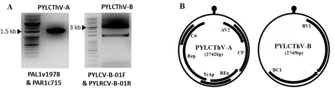
To obtain full-length viral PYLCThV DNA, DNA prepared from yellow leaf curl symptomatic pepper leaf samples and begomovirus universal primers PAL1v1978/PAR1c715 (Rojas et al., 1993) were applied for polymerase chain reaction (PCR). To confirm the identity of begomovirus causing symptoms of leaf samples, an expected size of 1.4 kb amplicon containing geminiviral conserved hairpin sequence (5’-TAATATTAC-3’) in the intergenic-region were obtained (Figure 3A). The amplicon sequence showed 99.5% identity with Pepper yellow leaf
curl Thailand virus (PYLCThV, Genbank Accession No. KX943290.1).
PYLCV-A01F/PYLCV-A01R primers were additionally used for full-length cloning and 2.7 kb size of a DNA-A fragment was obtained. Furthermore, PYLCV-B01F/PYLCVB-01R primers were used to obtain a 2.7 kb DNA-B fragment. Based on the NCBI data, the proteins of PYLCThV was analyzed. A Total six proteins were encoded in DNA-A and two proteins were encoded in DNDNA-A-B (Figure 3B).
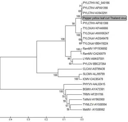
To confirm identity of PYLCThV DNA-B, a phylogenetic tree was constructed using the begomoviral DNA-B BC1 sequences (Figure 4). For establishing demarcation of virus species, the International Committee on Taxonomy of Viruses (ICTV) threshold of 89% identified nucleotide sequences were used (Fauquet et al., 2008). The PYLCThV DNA-B sequence obtained in this study was located in
PYLCThV group and had the highest nucleotide sequence identity with PYLCThV isolate (KX943291, 99%) from Thailand.
Figure 3. Detection and sequence analysis of viral genome. (A) Using the
geminivirus universal primer, ±1.4 kb of PYLCThV DNA-A was detected. PYLCV DNA-B was detected by viral genome specific primer based on NCBI database. (B) Genome organization of PYLCThV containing genimivirus common intergenic region.
Figure 4. Phylogenetic tree based on begomovirus DNA-B BC1 region. The
placement of the PYLCThV isolates in the present study is marked with a box. The sequences of DNA-B BC1 protein were obtained from GenBank and used for UPGMA analysis. The number at the phylogenetic tree represents the bootstrap value (1000 replicates).
Pathogenicity test of the PYLCThV infectious clone in N.
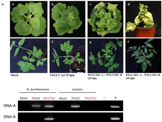
benthamiana and tomato
To confirm the infectivity of the PYLCThV clone, the DNA-A and DNA-B were subcloned into a binary vector pICH86988 and named PYLCThV-A and pICH86988-PYLCTHV-B. The inoculum of A. tumefaciense strain GV3101 containing each clone were mixed at the proportion of 1:1 and infiltrated to true leaves of S.lycopersicum ‘A39’ and N. benthamiana. The infectious clone TYLCV was used as a positive control. At 15 dpi, N. benthamiana and S. lycopersicum inoculated vector only showed no symptoms (Figure 4A-a), whereas plants with the positive control TYLCV started to show typical leaf curling symptoms (Figure 4A-b,f). At 20 dpi, PYLCThV-A + PYLCThV-B infected N. benthamiana started to show leaf curling symptoms and excessive leaf curling with yellow mottling were observed at 25 dpi (figure 4A c). At 45 dpi, acute stunting and chlorosis symptoms were observed (figure 4A d). On the other hand, at 25 dpi, PYLCThV-A + PYLCThV-B inoculated S. lycopersicum showed no symptoms until 45 dpi (Figure 5A g,f)
To confirm that observed symptoms were caused by PTYLCThV, virus genome in systemic leaves was detected by PCR. DNA-A and DNA-B genome segments were amplified in infected N. benthamiana (Figure 5B c). However, PYLCThV inoculated S. lycopersicum showed no PCR detection until 45 dpi (Figure 5B g).
Figure 5. Symptoms of N. benthamiana and S .lycopersicum ‘A39’ infected with PYLCThV. (a)Mock, (b)TYLCV (c) PYLCThV-A+PYLCThV-B (at 25 dpi), (d)
A+B (at 45 dpi). (e)Mock (f) TYLCV (g) PYLCThV-A+PYLCThV-B (at 25 dpi) (h) PYLCThV-PYLCThV-A+PYLCThV-B (at 45 dpi). (B) PYLCThV genome detection by PCR in the systemic leaves of inoculated N.
benthamiana (a) Mock (b) TYLCV (c) PYLCThV-A+PYLCThV and inoculated S. lycopersicum ‘A39’ (e)Mock (f) TYLCV (g) PYLCThV-A+PYLCThV-B. (+)
Table 3. Infectivity test of PYLCThV infectious clone in N. benthamiana and tomato.
Plant Number of plants [infected/inoculated]
Mock TYLCV PYLCThV
N. benthamiana [0/3] [3/3] [6/6]
S. Lycopersicum
Genome swapping analysis of PYLCThV and TYLCKaV
DNA-B
Previously, Choi showed that TYLCKaV had a high infectivity in S.
lycopersicum ‘A39’, which is contrasting to the infectivity of PYLCThV in this study.
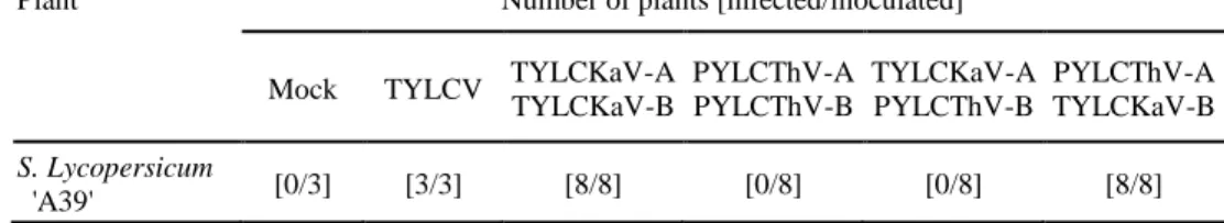
To reveal the genomic region responsible for infectivity in tomato, TYLCKaV and PYLCThV genomes were swapped by mixing subgenomes of each virus: DNA-A + PYLCThV-DNA-B and PYLCThV-DNA-A + TYLCKaV-DNA-B. The symptom development of inoculated tomatoes are monitored until 60 dpi. Fifteen to twenty dpi, TYLCKaV-A+B and swapped combination of PYLCThV-A + TYLCKaV-B started to develop leaf curling symptoms (Figure 6A d,f). However, PYLCThV-A+B and TYLCKaV-A + PYLCThV-B showed no viral symptoms (Figure 6A c,e). Until at 60 dpi, only TYLCKaV-A+B and PYLCThV-A + TYLCKaV-B combination showed leaf curling and retarded plant growth symptoms (Figure 6A d,f). To test virus accumulation, strain specific primer sets were designed and used for PCR detection of each virus. The viral DNA segments were detected in all symptomatic plants (Figure 6B a,d). However, viral DNA was not detected in symptomless plants (Figure 6B b,c). This virus genome swapping experiment result revealed that TYLCKaV-DNA-B genome is a responsible for the infectivity in tomato.
Figure 6. (A) Inoculation of PYLCThV and TYLCKaV DNA-B swapping clones.
(at 60 dpi) (a) Mock, (b) TYLCV, (c) PYLCThV-A+PYLCThV-B, (d) TYLCKaV-A+TYLCKaV-B, (e) TYLCKaV-A+PYLCThV-B, (f) PYLCThV-TYLCKaV-A+TYLCKaV-B,
(B) viral genome detection by PCR of TYLCKaV-A,B and PYLCThV-A,B in systemic leaves from infected S. lycopersicum ‘A39’. (a) TYLCKaV-A+B (b)
PYLCThV-A+B (c) TYLCKaV-A+PYLCThV-B (d) PYLCThV-A + TYLCKaV-B. Geminivirus genome A and B specific detection primer sets were used.
Table 4. Infectivity test of TYLCKaV and PYLCThV DNA-B swapping.
Plant Number of plants [infected/inoculated]
Mock TYLCV TYLCKaV-A TYLCKaV-B PYLCThV-A PYLCThV-B TYLCKaV-A PYLCThV-B PYLCThV-A TYLCKaV-B S. Lycopersicum 'A39' [0/3] [3/3] [8/8] [0/8] [0/8] [8/8]
Mapping the pathogenicity factor of TYLCKaV in tomato.
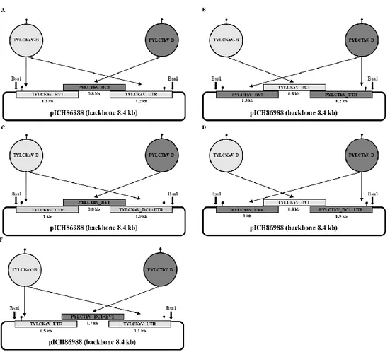
To reveal the pathogenicity factor of TYLCKaV in tomato, the sequence of TYLCKaV and PYLCThV ORFs were compared. TYLCKaV and PYLCThV were consisted of untranslated region (UTR) containing hairpin structure which is important for virus replication in plant cell. Furthermore, two ORFs, BV1 and BC1 which are essential for cell to cell movement and nuclear shuttling were encoded respectively. Sequence analyses showed 80% nucleotide identity and 89% amino acid identity. To delimit the ORFs in DNA-B controlling tomato infection, four chimeric constructs between TYLCKaV and PYLCThV were constructed. Each construct was generated by substitution of BC1 and BV1 proteins of TYLCKaV with PYLCThV (Figure 7). To facilitate the whole protein replacement and to overcome six nucleotides of intergenic region (IR), Gibson Assemby (GA) method was used. Compared to conventional restriction enzyme cloning methods, this cloning method permit us to clone the chimeric clones under limited selectable restriction enzyme condition. The construction of chimeric clone was simultaneously combined up to 20 DNA fragments based on sequence identity with adjacent DNA fragments. A total 4 fragments of 13000 bp were assembled in one 2X master mix (contains exonuclease, DNA polymerase and DNA ligase). These chimeric viruses were mixed with the TYLCKaV DNA-A or PYLCThV DNA-A and inoculated to tomato. At 10-15 dpi, positive control and native TYLCKaV-A+B started to develop symptoms. At the same time, TYLCKaV backbone chimeric constructs replaced with BC1 or BV1
chimeric construct replaced with BC1 or BV1 of TYLCKaV caused no visible symptoms in tomato (Figure 8A g,i) However, unlike the PYLCThV-A+B, The PYLCThV with TYLCKaV backbone showed clear symptoms (Figure 8A e). To confirm virus accumulation, symptomatic leaves and symptomless leaves were collected and virus genome was detected by PCR. The primers of GA were used to verify contamination of original construct infection and as can be seen in figure 8B-c,e,f,h and proper chimeric DNA segment were detected. However no viral DNA were detected in symptomless leaves (Figure 8B d,g,i). Taken together, these results suggest that TYLCKaV UTR region is responsible for pathogenicity in tomato (Figure 8C).
Figure 7. Schematic diagram of PYLCThV/TYLCKaV DNA-B chimera clones.
BV1 and BC1 domains were exchanged by Gibson assembly between TYLCKaV and PYLCThV.
Figure 8. (A) Infectivity test of PYLCThV/TYLCKaV chimeric clones. (B) Viral genome detection by PCR of TYLCKaV-A, PYLCThV-A, and chimeric DNA-B in systemic leaves from infected S. lycopersicum ‘A39’. (C) Schematic summary of mapping of TYLCkaV-B pathogenicity determinant. (a) Mock, (b)
TYLCV (c) TYLCKaV-A+TYLCKaV-B, (d) PYLCThV-A+PYLCThV-B, (e) PYLCThV-A + TYLCKaV backbone with PYLCThV BV1+BC1 chimera, (f) TYLCKaV-A + TYLCKaV-backbone PYLCThV BC1 chimera, (g) PYLCThV-A + PYLCThV-backbone TYLCKaV BC1 chimera, (h) TYLCkaV-A + TYLCKaV-backbone PYLCThV BV1 chimera (i) PYLCThV-A + PYLCThV-TYLCKaV-backbone TYLCKaV BV1 chimera.
Sequence analyses between PYLCThV and TYLCKaV of
intergenic region.
To compare the variation between putative pathogenicity determinant of TYLCKaV and PYLCThV. The sequence of PYLCThV/TYLCKaV intergenic region containing geminivirus conserved stem-loop structure (Figure 9A) are aligned (Figure 9B). The total IR region of 1078nt (PYLCThV) and 1074nt (TYLCKaV) showed 71.92% identity with 292 nt mismatching. The putative loop forming known as viral replication origin of 9 bp 5’-TAATATTAC-3’ was observed in both PYLCThV and TYLCKaV. The GC-rich region that forming stretch of complementary stem region showed 3 SNPs and 1 nucleotide deletion. The minimal DNA binding domain were detected at upstream of hairpin structure. The common ‘GTGTC’ iteron in PYLCThV and TYLCKaV were in different location. Besides, PYLCThV has additional ‘GAATGGGGG’ motif that TYLCKaV is no found in TYLCKaV (Figure 9B).
Figure 9. Comparison of PYLCThV and TYLCKaV intergenic region. (A) The
geminivirus conserved ‘TATAATATTAC’ stem-loop motif is flanked on side by a short stretch of complementary GC-rich motifs. (B) Elements relevant for replication such as TATA-box and iteron are shown in upstream of hairpin structure.
DISCUSSION
In this study, PYLCThV infected pepper leaves were obtained from Thailand and an infectious clones was constructed. The infectivity tests were conducted using the developed PYLCThV infectious clone and TYLCKaV/PYLCThV chimeric clones in tomato, pepper and N. benthamiana. The pathogenicity of PYLCTHV was obvious in N. benthamiana with the rapid deveopment of typical YLCD symptoms. By constrast, tomato and pepper did not show any symptoms and no viral accumulation.
To date, several viral factors that responsible for pathogenicity in Geminivirus have been reported. Zhou et al (2007) verified that nuclear shuttle protein (BV1) determines virulence or avirulence in common bean by using a series of hybrid DNA-B components expressing chimeric Bean dwarf mosaic virus (BDMV) and
Bean golden yellow mosaic virus (BGYMV) NSP. The BDMV with BGYMV NSP
and BDMV with BGYMV N-terminal 1 to 42 amino acids can overcome the BDMV resistance of UI 114. Jupin et al (1995) identified the determinant of DNA replication specificity in monopartite TYLCV. The Rep with 146 nt sequence encompassing the left half of the UTR was strain specifically interacted and replicated.
In our study, contrary to PYLCThV, TYLCKaV showed clear symptoms in both
N. benthamiana and tomato (Choi 2018). The availability of infectious clones
The genome swapping analysis revealed that the B genome of TYLCKaV was responsible for pathogenicity in tomato. Moreover, by mapping analysis, the agro-inoculation harboring five chimeric constructs reveals that the UTR region of TYLCKaV was responsible for tomato infection. In plant cell, once single stranded viral DNA succeeds to invade, the virus starts to form a replication complex by combining DNA with various host factors (Nagar et al., 1995). Then, the Rep protein-host factor complex initiate rolling-cycle replication by nicking the 7th sequence of conserved motif for use as a replication origin (Laufs et al., 1995). By comparing hairpin structures of TYLCKaV and PYLCThV, we found variations including one deletion and three SNPs in GC-rich region. Furthermore common geminiviral iteron of ‘GTGTC’ was positioned in different location, and PYLCThV has additional ‘GAATGGGGG’ motif that is no found in TYLCKaV. Taken together, we speculated that the difference of pathogenicity between TYLCKaV and PYLCThV may be derived from the variation of these motifs.
Even though the putative pathogenicity factor of TYLCkaV for tomato infection was is identified in this study, the successful infection of pepper by infectious clone is still elusive. A recent study showed that the pepper infection can be caused by acquisition of satellite DNA molecule that resulted in more aggressive crop-adaptation (Patil et al., 2018). Additionally, the beta satellite usually related with transcriptional and post-transcriptional gene silencing (Yang et al., 2011). Based on these observation, we used TSWV-NSs protein as RNAi suppressor (Schnettler et al., 2010) when sap-inoculation. Finally, we could infect C. annuum ‘ECW30R’ but
unfortunately only one out of fifty plants was showed clear symptoms in the plant inoculated with TLYCKaV-A + TYLCKaV with PYLCThV BC1 (Figure 10). Although this method could not be repeated, suppression of the nene silencing may be helpful for successful Agrobacterium-mediated infection. Additionally previous reports showed that co-infection of helper component protein (HCpro) of Plum pox
virus (PPV) with Cucumber vein yellowing ipomovirus (CVYV) can suppress the
resistance conferred by RNAi defense mechanism (Mbanzibwa et al., 2009; Hanley-Bowdoin et al., 2013, Valli et al., 2006). For the further study, we are testing transmission using Korean Q type of B. tabaci. The Figure 11 shows leaf curling symptoms observed at 15 dpi in EXW30R inoculated with a whitefly-mediated method.
In South Korea, there has been no report on the incidence of TYLCD in pepper. TYLCV incidence in tomato was first reported in 2008 (Ko et al., 2014; Kwak et al., 2008). Owing to global warming the average temperature of Korea is continuously rising and the whitefly population could be rapidly increase. This study provides molecular basis of understanding TYLCV and PYLCV infectivity and the interaction between virus and plants.
Figure 10. (A) C. annuum ‘ECW30R’ infected by TYLCKaV + TYLCKaV backbone chimeric constructs with PYLCThV BC1. (a) mock, (b) leaf curling
symptoms at 14 dpi (c) severe leaf curling with yellow vein mosaic and shortened internodes symptom at 60 dpi. (B) PCR detection by virus genome specific
Figure 11 Confirmation of C. annuum ‘ECW30R’ as a natural host using insect vectors. (A): TYLCKaV-viruliferous Bemisia tabaci were released in the insect cage.
REFERENCES
Ali Z, Ali S, Tashkandi M, Zaidi SS, Mahfouz MM. (2016)
CRIPR/Cas9-mediated immunity to geminiviruses: defferential interference and evasion
Sci Rep 6:30223
Ashraf MA, Shahid AA, Rao AQ, Bajwa KS, Husnain T. (2014) Functional
characterization of a bidirectional plant promoter from cotton leaf curl
Burewala virus using an Agrobacterium-mediated transient assay. Viruses
6(1):223-42
Chiemsombat P, Srikamphung B, Yule S. (2018) Begomoviruses associated to
pepper yellow leaf curl disease in Thailand J Agri Res 3(7): 000183.
Chatterji A, Beachy RN, Fauquet CM. (2001) Expression of the oligomerization
domain of the replication-associated protein (Rep) of Tomato leaf curl New
Delhi virus interferes with DNA accumulation of heterologous geminiviruses J Biol Chem 276(27):25631-8.
Choi S. (2018) Identification and inheritance of new sources of resistance against Cucumber mosaic virus Isolate P1 in Capsicum and Development of a Tomato yellow leaf curl Kanchanaburi virus infectious clone. (Unpublished masters’
thesis). Seoul national university, Seoul, Korea.
Choi S, Lee JH, Kang WH, Kim J, Huy HN, Park SW, Son EH, Kwon JK, Kang BC. (2018) Identification of Cucumber mosaic resistnace 2 (cmr2) that confers
resistance to a new Cucumber mosaic virus isolate P1 (CMV-P1) in pepper (Capsicum spp.) Front Plant Sci 9:1106
Csorba T, Pantaleo V, Burgyan J. (2009) RNA silencing: an antiviral mechanism Adv Virus Res 75:35-71
De Barro PJ, Liu SS, Boykin LM, Dinsdale AB. (2011) Bemisia tabaci: a
statement of species status Annu Rev Entomol 56:1-19
of plant-pathogen interactions Nat Rev Genet 11(8):539-48
Diaz-Pendon JA, Truniger V, Nieto C, Garcia-Mas J, Bendahmane A, Aranda MA. (2004) Advances in understanding recessive resistance to plant viruses Mol Plant Pathol 223-333
Fauquet CM, Briddon RW, Brown JK, Moriones E, Stanley J, Zerbini M, Zhou X. (2008) Geminivirus strain demarcation and nomenclature Arch Virol
153(4):783-821
Fondong VB. (2013) Geminivirus protein structure and function Mol Plant Pathol
6:635-49
Gafni Y and Epel BL. (2002) The role of host and viral proteins in intra- and
inter-cellular trafficking of geminiviruses physiological and molecular plant
pathology 60:231-241
Green, SK, Tasi WS, Shih SL, Rezaian MA, Duangson U. (2003) Molecular
characterization of a new begomovirus associated with tomato yellow leaf curl and eggplant yellow mosaic diseases in Thailand. Plant Disease 87,446-446
Hanley-Bowdoin L, Bejarano ER, Robvertson D, Manssor S. (2013)
Geminiviruses: masters at redirecting and reprogramming plant processes Nat
rev microbial 11:777-88
Hanley-Bowdoin L, Settlage SB, Orozco BM, Nagar S, Robertson D. (2000)
Geminiviruses: models for plant DNA replication, transcription, and cell cycle regulation Crit Rev Biochem Mol Biol 2:105-40
Harrison BD. (1985) Advances in geminivirus research Annu. Rev. Phytopathol
23:83-96
Jones JD and Dangl JL. (2006) The plant immune system Nature
444(7117):323-9
Jupin I, Hericourt F, Benz B, Gronenborn B. (1995) DNA replication specificity
of TYLCV geminivirus is mediated by the amino-terminal 116 amino acids of the Rep protein FEBS Lett 365(2):166-20
Kenyon L, Kumar S, Tasi WS, Hughes Jd. (2014) Virus diseases of peppers
(Capsicum spp.) and their control Adv Virus Res 90:297-354
Koeda S, Kesumawati E. Tanaka Y, Hosokawa M. Doi M, Kitajima A. (2016)
Mixed infection of begomoviruses on pepper plants at northern Sumatra, Indonesia (2016) Trop Agr Develop 60(2):59-64
Koeda S, Homma K, Tanaka Y, Kesumawati E, Zakaria S, Kanzaki S. (2017)
Highly efficient agroinoculation method for tomato plants with Tomato yellow
leaf curl Kanchanaburi virus Hort J 4:479-486
Ko SJ, Choi DS, Ma KC, Kim DI, Kim HW, Kim MK, Choi HS. (2014) Pattern
of the occurrence of Tomato yellow leaf curl virus on cultivation Res in Plant 20,303-306
Kourelis J, van der Hoorn RAL. (2018) Defended to the Nines: 25 years of
resistance gene cloning identifies nine mechanisms for R protein function Plant
Cell 30:285-299
Kwak HR, Kim MK, LEE GS, Kim CS, Kim MJ, Kim JD, Choi HS. (2002)
Molecular characterization of Tomato yellow leaf curl virus (TYLCV) isolated firstly in Korea Research in Plant Disease 24,238
Laufs J, Traut W, Heyraud F, Matzeit V, Rogers SG, Schell J, Gronenborn B.
(1995) In vitro cleavage and joining at the viral origin of replication by the replication initiator protein of tomato yellow leaf curl virus Proc Natl Acad Sci
U S A 92(9):3879-83
Lapidot M, Karniel U, Gelbart D, Fogel D, Evenor D, Kutsher Y, Makhbash Z, Nahon S, Shlomo H, Chen L, Reuveni M, Levin I. (2015) A novel route
controlling begomovirus resistance by the messenger RNA surveillance factor pelota PloS Genet 11(10):e1005538
Lapidot M, Friedmann M. (2002) Breeding for resistnace to whitefly-transmitted
geminiviruses Ann Appl Biol 140:109-127
Li F, Pignatta D, Bendix C, Brunkard JO, Cohn MM, Tung J, Sun HY, Kumar P, Baker B. (2012) MicroRNA regulation of plant innate immune receptors
Proc Natl Acad Sci USA 109:1790-1795
Mbanzibwa DR Tian Y, Mukasa SB, Valkonen JP. (2005) Cassava brown steak virus (potyviridae) encodes a putative Maf/HAM1 pyrophosphatase implication
in reduction of mutations and P1 proteinase that suppresses RNA silencing but contains no HC-Pro J Virol 83:6934-6940
Nagar S, Pedersen TJ, Carrick KM, Hanley-Bowdoin L, Robertson D. (1995) A
geminivirus induces expression of a host DNA synthesis protein in terminally differentiated plant cells Plant Cell 6:705-19
Nash, TE, Dallas MB, Reyes MI, Buhrman GK, Ascencio-Ibañez JT, Hanley-Bowdoin L. (2011) Functional analysis of a novel motif conserved across
geminivirus Rep proteins J Virol 3:1182-92
Kanakala S, Ghanim, M. (2018) Whitefly-transmitted begomovoruses and
advances in the control of their vectors In Genes, and transgenics for virus
resistance in plants Patil BL (pp.201-220). New delhi, Caister Academic press Pradhan B, Van Tien V, Dey N, and Mukhgerjee SK. (2017) Molecular biology
of geminivirus DNA replication Avid Science 2-31
Pumplin N, Vionnet O. (2013) RNA silencing suppression by plant pathogens:
defence, conter-defence and conter-conter-defence Nat Rev Microbiol 11:745-760
Rojas MR, Hagen C, Lucas WJ, Gilbertson RL. (2005) Exploiting Chinks in the
plant’s armor: evolution and emergence of geminiviruses Annu Rev Phytopathol 43:361-94
Ronde D. Butterbnach P, Kormelink R. (2014) Dominant resistance against plant
viruses Front in Plant Sci 5, 307
Schnettler E, Hemmes H, Huismann R, Goldbach R, Prins M, Kormelink R.
(2010) Diverging affinity of tospovirus RNA silencing suppressor proteins, NSs, for various RNA duplex molecules J Virol 84:11542-54
Scholthof KB, Adkins S, Czosnek H, Palukaitis P, Jacquot E, Hohn T, Hohn B, Saunders K, Candresse T, Ahlquist P, Hemenway C, Foster GD. (2011)
Top 10 plant viruses in molecular plant pathology Mol Plant Pathol 9:938-54
Shih SL, Tsai WS, Lee LM, Wang JT, Green SK, Kenyon L. (2010) First report
of tomoato yellow leaf curl Thailand virus associated with pepper leaf curl disease in Taiwan Plant Dis 94(5):637
Soosaar JL, Burch-Smith TM, Dinesh-kumar SP. (2005) Mechanisms of plant
resistance to viruses Nat Rev Microbiol, 3:789-98
Tamura K, Peterson D, Peterson N, Stecher G, Nei M, Kumar S. (2011) MEGA5:
Molecular Evolutionary Genetics Analysis using Maximum Likelihood, Evolutionary Distance, and Maximum Parsimony Methods Molecular Biology
and Evolution
28: 2731-2739.
Ting JPand Davis BK. (2005) CATERPILLER: a novel gene family important in
immunity, cell death, and deseases Annu Rev Immunol 23:387-414
Truniger V, Aranda Ma. (2009) Recessive resisance to plant viruses Adv Virus Res
75:119
Valli A, Martin-Hernandez AM, Lopez-Moya JJ, Gracia JA. RNA silencing
suppression by a second copy of the p1 serine protease of Cucumber vein
yellowing ipomovirus, a member of the family potyviridae that lacks the
cysteine protease HCPro (2006) J virol 20:10055-63
Voinnet O, Pinto YU, Baulcombe DC. (1999) Suppression of gene silencing: a
general stategy used by diverse DNA and RNA viruses of plants Proc Natl Acad
Sci USA 96:14147-14152
Yang X, Xie Y, Raja P, Li S, Wolf JN, Shen Q, Bisaro DM, Zhou X. (2011)
Suppression of methylation-mediated transcriptional gene silencing by βC1-SAHH protein interaction during geminivirus-betasatellite infection PLoS
Pathog 7(10):e1002329
Zerbini FM, Briddon RW, Idris A, Martin DP, Moriones E, Roumagnac P. (2017)
ICTV Virus Taxonomy Profile: Geminiviridae J Gen Virol 98(2):131-133
Zhou YC, Garrido-Ramiez ER, Sudarshana MR, Yendluri S, Gilvertson RL.
(2007) The N-terminous of begomocirus nuclear shuttle protein (BV1) determines virulence of avirulence in Phaseolus vulgaris Mol Plant Microbe
ABSTRACT IN KOREAN
황화잎말림바이러스 (Yellow leaf curl virus)는 Geminivirus 계통의 Begomovirus 그룹에 속하는 원형의 단일가닥 DNA 바이러스로 열대 및 아열대 국 특히 토마토와 고추에서 심각한 손실을 일으킨다. 본 실험에서는 고추에 감염된 PYLCThV를 분리하여 유전체를 확인 및 분석했다. PYLCThV 유전체 정보를 바탕으로 recombinant DNA technology를 이용하여 바이러스 감염 클론을 개발했다. PYLCThV 감염클론은 agroin filtration법으로 담배, 토마토, 고추에 접종했다. 접종된 담배는 바이러스 증상이 확인된 반면, 토마토와 고추에서는 PYLCThV의 감염 증상이 나타나지 않았다. 바이러스의 감염 여부는 중합효소 연쇄반응(PCR법을 이용하여 재 확인 하였다. 이 결과를 바탕으로, 실험실에 보유하고 있던 병원성이 높은 TYLCKaV 감염클론을 사용하여 게놈 교환 실험을 수행했다. 그 결과 TYLCKaV-B 게놈이 토마토의 병원성을 일으킨다는 것을 밝혔다. 또한 TYLCKaV-B 게놈의 병원성 인자를 매핑하기 위하여 5개의 키메라 클론을 제작하였다. 그 결과 TYLCKaV의 유전자간부위 (intergenic region)가 토마토의 병원성을 결정하는데 핵심적인 역할을 한다는 것이 밝혀 졌다. PYLCThV와 TYLCKaV간의 병원성인자인 intergenic region간 변이를 확인해 본 결과 바이러스 유전자 복제 개시와 관련된 조절 유전자 (regulatory gene)들의 변이를 발견할 수 있었다. 본 연구에서 개발된 PYLCThV 감염 클론은 앞으로 병저항성
연구에 유용한 도구로 이용될 것이다. 또한 토마토의 병원성을 결정하는 인자는 앞으로 Begomovirus와 식물간의 상호작용을 이해하는데 분자적 기초를 제공할 것이다.
주요어: Pepper yellow leaf curl Thailand virus (PYLCTV), Tomato yellow leaf curl Kanchanaburi virus TYLCKaV), Pathogenicity factor, Intergenic region (IR)