Tumor suppressor p53 pathway mediates
glucolipotoxicity-induced apoptosis of the
rat cardiomyoblast cell
Hye Jin Wang
Department of Medical Science
The Graduate School, Yonsei University
Tumor suppressor p53 pathway mediates
glucolipotoxicity-induced apoptosis of the
rat cardiomyoblast cell
Hye Jin Wang
Department of Medical Science
The Graduate School, Yonsei University
Tumor suppressor p53 pathway mediates
glucolipotoxicity-induced apoptosis of the
rat cardiomyoblast cell
Directed by Professor Hyun Chul Lee
The Master’s Thesis submitted to the Department of
Medical science, the Graduate School of Yonsei
University in partial fulfillment of the requirements for the
degree of Master of Medical Science.
Hye Jin Wang
December 2009
This certifies that the master’s thesis of
Hye Jin Wang is approved.
___________________________
Thesis Supervisor: Hyun Chul Lee
___________________________
Thesis Committee Member: Kyung Sup Kim
___________________________
Thesis Committee Member: Byung Wan Lee
The Graduate School
Yonsei University
<
Table of contents>
ABSTRACT--- 1
I. INTRODUCTION--- 3
II. MATERIALS AND METHODS
1. Cell culture--- 6
2. Treatment H9c2 cells with palmitic acid --- 6
3. Preparation of nuclear extracts for Nrf2--- 7
4. Western blot analysis--- 7
5. DNA fragmentation--- 8
6. Detection of ROS--- 9
7. Cell survival assays--- 9
8. Chromatin immunoprecipitation assay--- 10
9. Annexin V-PI staining--- 11
III. RESULTS
1.
Combined treatment with high glucose and palmitate induces
apoptosis in H9c2 cells--- 13
2. Combined treatment with high glucose and palmitate generates
intracellular ROS--- 15
3. Glucolipotoxicity upregulates p53, Nrf2 and cleaved caspase-3,
and downregulates NQO1 expression--- 17
4. p53 interacts with the ARE region of the NQO1 gene promoter
--- 19
5. The p53 inhibitor, pifithrin-α, protects H9c2 cells from
glucolipotoxicity-induced apoptosis--- 21
6. PFT-α inhibits glucolipotoxicity-induced intracellullar ROS
generation--- 23
7. Scavenging of ROS by N-acetyl-L-cysteine decreases
glucolipotoxicity-induced p53 expression and normalizes NQO1
levels--- 25
IV. DISCUSSION--- 28
V. CONCLUSION--- 32
REFERENCES--- 34
LIST OF FIGURES
Figure 1. Combined treatment with high glucose and palmitate
(glucolipotoxicity) induces apoptosis in cardiomyocytes--- 14
Figure 2. Glucolipotoxicity increases ROS content in
cardiomyocytes---
16
Figure 3. Glucolipotoxicity differentially affects expression of p53,
Nrf2, and NQO1 proteins, and cleaved caspase-3 levels--- 18
Figure 4. p53 interacts with the NQO gene promoter--- 20
Figure 5. Inhibition of p53 with PFT-α protects H9c2 cells from
glucolipotoxicity-induced apoptosis---
22
Figure 6. Inhibition of p53 with PFT-α attenuates
glucolipotoxicity-induced intracellular ROS generation--- 24
Figure 7. NAC decreases glucolipotoxicity-induced p53 expression
and normalizes NQO1 levels--- 26
Figure 8. Model of p53-dependent apoptosis in H9c2 cells via
inhibition of the anti-apoptotic Nrf2-NQO1 pathway--- 32
<ABSTRACT>
Tumor suppressor p53 pathway mediates glucolipotoxicity-induced apoptosis of the rat cardiomyoblast cell
Hye Jin Wang
Department of Medical Science The Graduate School, Yonsei University
(Directed by Professor Hyun Chul Lee)
Reactive oxygen species (ROS) generated by exposure to excessive
levels of glucose and free fatty acids (glucolipotoxicity) have been reported to
induce apoptosis. Here, we investigated the glucolipotoxic effects of high
glucose and palmitic acid (C16:0) on the rat cardiomyoblastic cell line, H9c2,
focusing on the role of the tumor suppressor, p53. Combined exposure to
high levels of glucose (30 mM) and 250 µM palmitate resulted in a 2.2-fold
increase in ROS generation compared to 30 mM glucose only. This increase
caspase-3, as well as increased expression of nuclear factor erythroid 2-related
factor 2 (Nrf2). Somewhat unexpectedly, the Nrf2 target, NAD(P)H:quinine
oxidoreductase-1 (NQO1), a detoxifying enzyme, was downregulated.
Pifithrin-α, an inhibitor of p53, attenuated glucolipotoxicity-induced ROS
generation and p53 expression, restoring p53 to normal levels. Chromatin
immunoprecipitation analysis revealed that upregulated p53 interacted with
the Nrf2-binding ARE region of the NQO1 promoter, and blocked Nrf2–
mediated NQO1 expression, causing greater myoblast apoptosis.
Pretreatment with the ROS scavenger, N-acetyl-L-cysteine decreased
glucolipotoxicity-induced p53 expression and normalized NQO1 levels.
These results indicate that glucolipotoxicity-induced p53 elevation mediates
apoptosis of rat cardiomyoblast cells through a dual mechanism: stimulation of
a pro-apoptotic caspase signaling pathway and inhibition of the anti-apoptotic
Nrf2-NQO1 signaling pathway.
Key work: p53, NQO1, Glucolipotoxicity, ARE region, Reactive oxygen species(ROS), palmitate, hyperglycemia, apoptosis
Tumor suppressor p53 pathway mediates glucolipotoxicity-induced apoptosis of the rat cardiomyoblast cell
Hye Jin Wang
Department of Medical Science The Graduate School, Yonsei University
(Directed by Professor Hyun Chul Lee)
I. INTRODUCTION
The incidence of diabetes and its complications has reached epidemic
proportions and is increasing steadily. Plasma levels of glucose and free
fatty acids in diabetic patients are pathologically elevated. A number of
studies have confirmed that hyperglycemia and hyperlipidemia can induce
intracellular toxicity1-3. The saturated long chain fatty acid palmitate, in
particular, induces cell death in many cell types4-7. Although glucotoxicity8
or lipotoxicity9 alone can induce apoptosis, when combined they synergize,
Glucolipotoxicity-induced oxidative stress is reported to play a key role in
apoptosis in cardiac myocytes, but the underlying molecular mechanisms
remain unclear.
Increased levels of reactive oxygen species (ROS) associated with oxidative
stress induce an increase in nuclear factor erythroid-related factor 2 (Nrf2).
Nrf2 binds to antioxidant response elements (AREs) in the promoters of ROS
scavenging and detoxification enzymes, including NAD(P)H:quinine
oxidoreductase-1 (NQO1) and glutathione S-transferases, increasing their
expression14. The resulting Nrf2-induced upregulation of antioxident
enzymes is reported to protect cells against ROS15. The tumor suppressor,
p53, which is upregulated in response to genotoxic stress and causes cell-cycle
arrest and cell death, is also upregulated by ROS11. Like Nrf2, p53 is also
known to bind to the AREs of ROS scavenging and detoxification enzymes
that are induced by Nrf213. Thus, under ROS stress, p53 can inhibit
Nrf2-induced transcription of antioxidant enzymes through competitive binding to
AREs13.
Because p53 is a key apoptotic regulator and responds to ROS, we
examined whether p53 is involved in glucolipotoxicity-induced cell death in
H9c2 cells, a rat cardiomyoblast cell line. Specifically, we hypothesized that
the p53-Nrf2/ARE pathway might be an additional mechanism that contributes
that glucolipotoxicity-induced apoptosis of H9c2 cells is mediated through
dual p53-dependent mechanisms: stimulation of a pro-apoptotic caspase
signaling pathway by elevated ROS, and inhibition of the anti-apoptotic
Nrf2-NQO1 signaling pathway through binding of p53 to cis regulatory ARE sites
II. MATERIALS AND METHODS
1. Cell culcure
H9c2 rat cardiomyoblasts were maintained at 37°C, 5% CO2 in
DMEM/F-12 (WelGENE, Daegue, South Korea) supplemented with 10% bovine calf
serum (WelGENE, Daegue, South Korea), penicillin (100 U/ml), and
streptomycin (100 µg/ml).
2. Treatment H9c2 cells with palmitic acid.
H9c2 cells were incubated for 18 h in DMEM/F12 medium containing
normal (5 mM) or high (30 mM) glucose with 0.25% (wt/vol) BSA alone
(control) or 0.25 mM palmitate (C16:0) complexed to 0.25% BSA. The
0.25 mM palmitic acid medium was prepared as previously described16.
Briefly, a 20 mM solution of palmitic acid in 0.01 mol/l NaOH was incubated
at 70°C for 30 min. Then, 900 µl of 5% BSA and 300 µl of the palmitic
acid/NaOH solution was mixed together. It has been shown that the addition
3. Preparation of Nuclear extracts for Nrf2
To prepare nuclear extracts, 10cm culture dish cell rinsed 2 times using
PBS and harvested cell pellets. The pellets were suspended in 1ml buffer A
(10mM Hepes pH 7.9, 10mM KCl, 0.1mM EDTA, 0.1mM EGTA, and 1mM
dithiothreitol) and incubated on ice for 15min. After incubation, added 15µl
10% NP-40 and centrifugation at 12,000g for 5min. The supernatant was
removed and the pellet was resuspended in 200µl buffer C [20mM Hepes pH
7.9, 0.4M NaCl, 1mM EDTA, 1mM EGTA, 1mM ditiothreitol, and protease
inhibitor cocktail (Roche applied science, Indianapolis, USA)] by vortexing
for 5s, and then shaking incubated 4℃ for 30min. After centrifugation at
12,000g for 10min on 4℃, the supernatant was collected and quantified
protein concentrations with the Bradford’s reagent (Sigma-Aldrich, St. Louis,
USA)
4. Western blot analysis
For western blot analysis, H9c2 cells were rinsed with ice-cold PBS and
scraped on ice into lysis buffer that contained 50mM Tris-Cl (pH8), 120mM
NaCl, 1mM EGTA, 1mM EDTA, 0.5% NP-40, protease inhibitor cocktail
(Roche applied science). The cell lysates were then shaking for 30 min in 4℃.
Cell debris were removed by centrifugation (12,000g for 15 min in 4℃), the
Bradford’s reagent (Sigma-Aldrich). Protein samples were boiled for 5min
in Laemmli sample buffer, fractionated by SDS-polyacrylamide gel in a 12%
polyacrylamide gel and electrotransferred onto PVDF membrane (Millipore
Corporation, Bedford, USA). Membrane were incubated 2h at 4℃ in a
blocking solution of 5% non-fat powdered milk in T-phosphate-buffered saline
(phosphate-buffered saline, pH 7.5, containing 0.1% Tween 20). p53, NQO1,
Nrf2 (Santa-Cruz biotechnology, CA, USA), cleaved caspase3 (Cell signaling)
and β-actin (Sigma-aldrich) antibodies were used as primary antibodies,
diluted 1:1000. Horseradish peroxidase-conjugated goat anti-mouse IgG,
anti-rabbit IgG and anti-goat IgG (Santa-Cruz biotechnology, CA, USA) were
used as secondary antibodies (at dilution of 1:5000). Development of the
western blot was performed using an enhanced chemioluminisence western
blotting analysis system (Animal Genetics, Inc., South Korea). To confirm p53
involvements, we used pifithrin-α (PFT-α) (Chemicon, MA, USA), as a p53
inhibitor at the same time with palmitic acid.
5. DNA fragmentation
After treating with glucose (5mM or 30mM) with or without 250µM
palmitic acid for 18 h, H9c2 cells were harvested into ice-cold PBS, and an
equal volume of 2X lysis buffer (200 mM HEPES [pH 7.5], 2% Triton X-100,
incubated on ice for 30 min and microcentrifuged at maximum rpm for 15 min
at 4°C. After removing the supernatant, the cells were treated with 0.1
mg/ml RNase A for 1 h at 37ºC and then with 0.2 mg/ml proteinase K for 3 h
at 56ºC. Genomic DNA was then extracted with phenol/chloroform and
dissolved in TE buffer (10 mM Tris–Cl, 1 mM EDTA, pH 8.0). DNA
fragmentation was analyzed by electrophoresis in 1.8% agarose gels.
6. Detection of ROS
Accumulation of ROS in H9c2 cells treated with normal glucose, high
glucose and palmitic acid (for 3hrs) was monitored using the
fluorescence-generating probe H2DCFDA (Invitrogen, Oregon, USA). Cells (2X10⁵
cells/60mm dish) were treated with 5µM H2DCFDA. After 30-min
incubation at 37°C, cells were examined under a fluorescence microscope set
at 488 nm for excitation and 530 nm for emission, and sorted by flow
cytometry.
7. Cell survival assays.
H9c2 cells were dispensed in 24-well plates at a density of 5X10⁴
cells/well. MTT [3-(4,5-dimethylthiazol-2-yl)-2,5-diphenyl-tetrazolium
bromide] assay was used to determine cell viability following the
manufacturer’s protocols. The MTT labeling reagent (5 mg/ml MTT in PBS)
was incubated for 4 hrs. After discard of media 250µl DMSO was added to
stained cells to solubilize the purple crystals at a 1:1 ratio, and 200µl samples
moved to 96-well microplate. Absorbance of the samples was read using a
microplate reader at 490 nm.
8. Chromatin immunoprecipitation assay
H9c2 cells were grown in 150-mm dishes and treated normal glucose, high
glucose and palmitate (for 18hrs). The cells were fixed using 1%
formaldehyde in medium for 10 min at room temperature. The cells were
treated with 0.125M glycine for 5 min to stop fixation and washed twice with
ice-cold PBS. The cells were scraped in PBS, collected by centrifugation, and
lysed in lysis buffer (5 mM HEPES, pH 8.0, 85 mM KCl, 0.5% Nonidet P-40,
1 mM PMSF supplemented with protease inhibitors). After centrifugation,
nuclear pellet was resuspended in nuclear lysis buffer (50 mM Tris-HCl, pH
8.0, 10 mM EDTA, 0.8% SDS, 1 mM PMSF supplemented with protease
inhibitors) and sonicated for four cycles (30-s pulse and 30-s rest on ice). The
sonication conditions were optimized to determine generation of DNA
fragments between 500 and 1000 base pairs in length. Sheared chromatins
were immunocleared with protein A/G-agarose slurry (Santa-Cruz
biotechnology, CA, USA) for 1 h at 4 °C. A portion of the precleared
was immunoprecipitated with IgG (control), p53 antibody. The
immunoprecipitates were washed sequentially with wash low salt buffer; with
high salt buffer; with LiCl buffer; and TE buffer for 5min each. Protein-DNA
complexes were resuspended in TE buffer for reversal of cross-links by
heating at 65 °C overnight. Then, DNA was purified using proteinase K
treatment at 50 °C for 3 h followed by phenol extraction and ethanol
precipitation. PCR was performed using 1:100-diluted inputDNA and 3µl of
immunoprecipitated DNA from a 30µl sample. The association of p53 with
endogenous NQO1 promoter ARE region in H9c2 cells was measured by PCR
on immunoprecipitated chromatin using the following primers spanning the
NQO1 promoter containing AREs:
F;5’-GCAGTTTCTAAGAGCAGAATC-3’ R; 5’-TTAGTCCTTGGTCAGATGTGG-F;5’-GCAGTTTCTAAGAGCAGAATC-3’. The PCR products were run on
2% agarose gels and visualized by ethidium bromide staining.
9. Annexin V-PI staining
Early apoptosis and necrosis were identified by means of double
fluorescence staining with Annexin V-FITC and propidium iodide(PI)
stainings. Apoptotic cells translocate phosphatidylserine from the inner site of
the plasma membrane to the outer surface while the membrane remains
physically intact. Apoptotic cells were therefore stained with Annexin V-FITC,
fluorescence when excited at 450– 480 nm, and exclude PI, a DNA dye
unable to cross the plasma membrane. Necrotic cells lose the physical integrity
of their plasma membrane and therefore stained with both annexin V-FITC
and PI. Cells that were neither apoptotic nor necrotic were not stained with
either dye. Samples (5×10⁵ cells per sample) were double stained according to the manufacturer’s instructions, and immediately analyzed on a FACScan
III. RESULTS
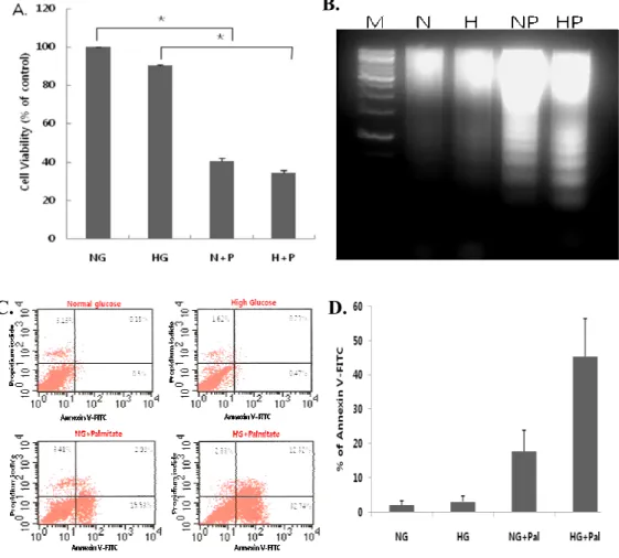
1. Combined treatment with high glucose and palmitate induces apoptosis in H9c2 cells.
H9c2 cells were treated with 5 or 30 mM glucose alone or together with 250 µM palmitic acid for 18 h. High glucose treatment alone did not
decrease cell viability (Fig. 1A) or induce apoptosis (Fig. 1B, C, D).
However, palmitate in combination with either normal or high glucose reduced
cell viability, as assessed by MTT (Fig. 1A), and increased apoptosis, as
measured by both DNA fragmentation (Fig. 1B) and annexin V-propidium
iodide (PI) staining (Fig. 1C and D). The combination of high glucose and
palminate was particularly effective, significantly reducing viability
B.
C. D.
Fig 1. Combined treatment with high glucose and palmitate (glucolipotoxicity) induces apoptosis in cardiomyocytes. H9c2 cells were treated with 5 or 30 mM
glucose alone or in combination with 250 µM palmitate for 18 h. High glucose (HG or H) combined with palmitate (A) reduced cell viability, as measured by MTT assays (***P < 0.001), and (B-D) increased apoptosis, as measured by DNA fragmentation (B) and annexin V-PI staining (C, D). High glucose alone did not affect viability or apoptosis. The data are presented as means ± SEs of three independent experiments.
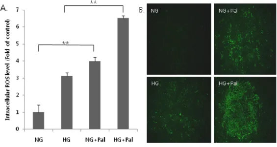
2. Combined treatment with high glucose and palmitate generates intracellular ROS.
To investigate the generation of intracellular ROS, we incubated H9c2 cells for 3 h in medium containing normal glucose (5 mM), high glucose (30
mM), or 250 µM palmitic acid alone, or with a combination of glucose (low
and high) with 250 µM palmitic acid. After 3 h, cells were incubated with
the fluorescent ROS probe, H2DCFDA, for 30 min, and fluorescence was
detected by flow cytometry (Fig. 2A) and fluorescence microscopy (Fig. 2B).
As shown in Figure 2A, 250 µM plamitate alone significantly increased ROS
levels in H9c2 cells compared with 5 mM glucose, whereas 30 mM glucose
alone did not. Under glucolipotoxic conditions (30 mM glucose and 250 µM
palmitate), ROS generation was increased 2.2-fold compared with normal
glucose (P < 0.05). Thus, of all incubation conditions used, the combination
of high glucose and palmitate induced the greatest increase in ROS production.
Fig 2. Glucolipotoxicity increases ROS content in cardiomyocytes. Total ROS
content in H9c2 cells was assayed using the fluorescent ROS probe, DCF-DA, after a 3-h exposure to palmitate and glucose. ROS fluorescence was measured by (A) flow cytometry and (B) fluorescence microscopy. Note that palmitate (Pal) with high glucose (HG) led to a significant increase in ROS (*P < 0.05). The data are presented as the mean (±S.E) of three independent experiments.
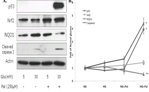
3. Glucolipotoxicity upregulates p53, Nrf2 and cleaved caspase-3, and downregulates NQO1 expression.
Whole-cell lysates were used for detection of p53, NQO1 and cleaved
caspase-3 by western blotting, and nuclear extracts were used for Nrf2
detection. As shown in Figure 3, p53 levels were increased approximately
4-fold in cells incubated with palmitate and high glucose compared to cells in
normal glucose (P < 0.001), and cleaved caspase-3 levels were increased about
4.5-fold (P < 0.001). Nrf2 expression was also increased in the presence of
high glucose plus palmitate; but interestingly, expression of the Nrf2 target,
NQO1, was decreased (P < 0.05). These results demonstrate that
glucolipotoxicity increased p53 and Nrf2 expression but reduced expression of
A. B.
Fig 3. Glucolipotoxicity differentially affects expression of p53, Nrf2, and NQO1 proteins, and cleaved caspase-3 levels. H9c2 cells were exposed to glucose and
palmitate for 18 h, and p53, Nrf2, NQO1 and cleaved caspase-3 levels were determined by western blotting. p53, cleaved caspase-3, and Nrf2 protein levels were increased only under high glucose (30 mM) plus palmitate (250 µM) conditions. Although Nrf2 levels were increased, the levels of its target, NQO1, were decreased. The data are presented as means ± SEs of three independent experiments.
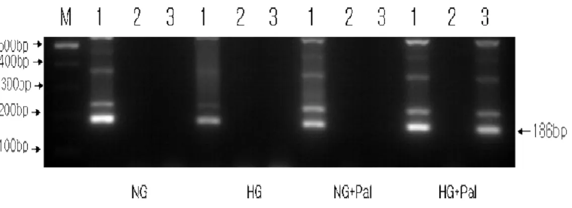
4. p53 interacts with the ARE region of the NQO1 gene promoter.
It was recently reported that p53 binds to the ARE region of the promoters of several antioxidant enzymes in mouse hepatocarcinoma cells13. To
determine whether p53 interacts with endogenous ARE-containing regions of
the NQO1 promoter in H9c2 cells under glucolipotoxic conditions, we used
ChIP assays. As shown in Figure 4, anti-p53 antibodies immunoprecipitated
the NQO1 promoter only from chromatin prepared from H9c2 cells incubated
with high glucose and palmitate, indicating that p53 interacts with this region
under glucolipotoxic conditions. Because Nrf2 also binds to ARE regions of
antioxidant enzymes, including NQO1, these results imply that p53
Fig 4. p53 interacts with the NQO1 gene promoter. p53 binding to the
ARE-containing region of the endogenous NQO1 promoter was determined using ChIP assays. Chromatin prepared from H9c2 cells treated with high glucose and palmitate was immunoprecipitated with mouse IgG or p53 antibodies, as indicated. Immunoprecipitated chromatin was amplified by PCRs using a primer pair bracketing the ARE-containing proximal promoter region (-379 to -533) of the rat NQO1 gene (NQO1[ARE]).
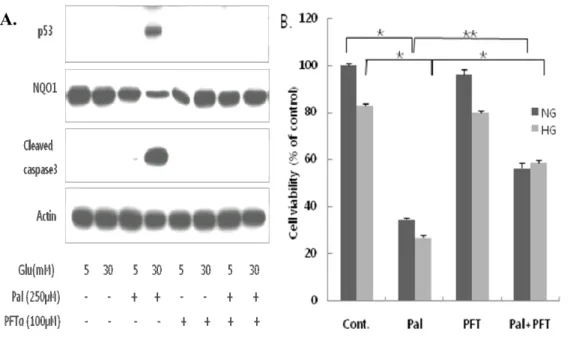
5. The p53 inhibitor, pifithrin-α, protects H9c2 cells from glucolipotoxicity-induced apoptosis.
To investigate the involvement of p53 in this apoptosis pathway, we examined the effects of pifithrin-α (PFT-α), a p53 inhibitor. Whole-cell
lysates were prepared from H9c2 cells co-treated for 18 h with 100 µM PFT-α,
30 mM glucose, and 250 µM palmitate. Apoptosis and viability were
analyzed by western blotting (Fig. 5A) and MTT assays (Fig. 5B), respectively.
The increases in p53 expression and cleaved caspase-3 levels induced by
combined exposure to palmitate and high glucose were prevented by
co-treatment with PFT-α (Fig. 5A). PFT-α also partially, but significantly,
reduced the level of apoptosis induced by glucolipotoxic conditions (Fig. 5B).
Note that PFT-α did not affect cell viability when added to cultures containing
normal or high glucose alone. Interestingly, PFT-α markedly increased NQO1
expression compared to palmitate-treated cells (Fig. 5A), suggesting that
endogenous expression of NQO1 is highly sensitive to p53. These results
further suggest that p53 acts through inhibition of NQO1 to play a key role in
A.
Fig 5. Inhibition of p53 with PFT-α protects H9c2 cells from glucolipotoxicity-induced apoptosis. H9c2 cells were incubated with high glucose (30 mM) and
palmitate (250 µM) in the absence and presence of PFT-α (100 µM) for 18 h. (A) Cells were harvested for western blot analysis of p53, NQO1, and cleaved caspase-3. (B) Cell viability was assessed by MTT assays. PFT-α increased survival in H9c2 cells incubated with both high glucose and palmitate (***P < 0.001, *P < 0.05). Cells treated with normal or high glucose only were unaffected by PFT-α.
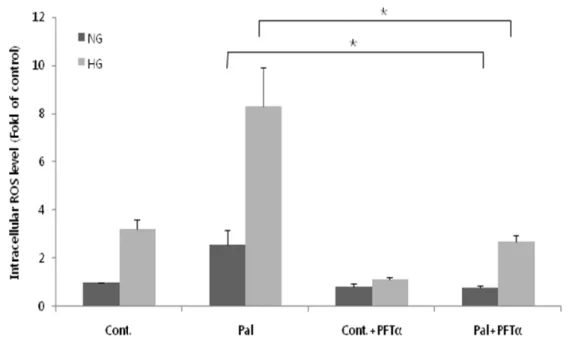
6. PFT-α inhibits glucolipotoxicity-induced intracellullar ROS generation. To investigate the effect of PFT-α on glucolipotoxicity-induced
intracellular ROS generation, we incubated H9c2 cells for 3 h with glucose alone or in combination with palmitate in the absence and presence of 100 µM PFT-α. The increased levels of ROS induced by high glucose (P < 0.05, high vs. low glucose) or high glucose and palmitate (P < 0.001, Pal vs. Cont), which were similar to those shown in Figure 1, were significantly reduced by PFT-α (Fig. 6). These results suggest that p53 plays a role in mediating the glucolipotoxicity-induced elevation of intracellular ROS levels.
Fig 6. Inhibition of p53 with PFT-α attenuates glucolipotoxicity-induced intracellular ROS generation. Total ROS content in H9c2 cells was assayed after
a 3-h exposure to glucose alone or glucose together with palmitate in the absence and presence of PFT-α using the fluorescent ROS probe, DCF-DA. Fluorescence due to intracellular ROS was measured by flow cytometry. The increase in ROS induced by palmitate (Pal) and high glucose (HG) was significantly inhibited by PFT-α (***P < 0.001). The data are presented as means ±SEs of three independent experiments.
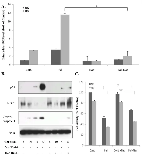
7. Scavenging of ROS by N-acetyl-L-cysteine decreases glucolipotoxicity-induced p53 expression and normalizes NQO1 levels.
To investigate the relationship between p53 expression and ROS, we repeated the experiments shown in Figure 4 in the presence of the ROS scavenger, N-acetyl-L-cysteine (NAC), preincubating with NAC for 1 h and incubating with normal/high glucose with or without palmitate for 3 h. As shown in Figure 7A, NAC markedly attenuated the increase in ROS levels induced by high glucose and palmitate. As expected, glucolipotoxic conditions increased p53 expression and cleaved caspase-3 levels, and decreased NQO1 expression. Co-treatment with NAC under these conditions significantly decreased p53 expression and cleaved caspase-3 levels; it also completely restored NQO1 expression (Fig. 7B), which had been reduced to undetectable levels in the absence of NAC. Similar effects of NAC on caspase-3 levels and NQO1 expression were observed in cells cultured with the combination of low glucose and palmitate (Fig. 7B), whereas only minimal and variable effects on p53 expression were observed under these conditions. NAC also improved cell viability in the presence of palmitate, but did not affect the viability of cells treated with normal or high glucose alone (Fig. 7C). Taken together, these results suggest that increased p53 expression might interfere with Nrf2-mediated induction of ARE-dependent NQO1 expression.
A.
B. C.
Fig 7. NAC decreases glucolipotoxicity-induced p53 expression and normalizes NQO1 levels. H9c2 cells were preincubated with 1 mM NAC for 1 h and then
incubated with normal (5 mM) or high (30 mM) glucose alone or together with 250 µM palmitate for 18 h, and then assayed by western blotting and MTT assay. For ROS experiments, cells were incubated as described, except that the final incubation was 3 h instead of 18 h on the same conditions above. (A) NAC significantly attenuated elevated ROS levels induced by high glucose and palmitate. (B) NAC
cleaved caspase-3 levels, and increased NQO1 expression. (C) NAC increased the viability of cells incubated with low or high glucose in the presence of palmitate (***P < 0.001, *P < 0.05). The data are presented as means ± SEs of three independent experiments.
IV. DISCUSSION
Chronic hyperglycemia and hyperlipidemia in patients with diabetes are
known to generate ROS, leading to apoptosis of many cell types, including
pancreatic β-cells17 and cardiomocytes18. In the present study, we focused on
glucolipotoxicity-induced apoptosis in cardiomyocyte and the underlying
apoptotic pathways. Our results demonstrated that glucolipotoxicity-induced
apoptosis of H9c2 cells was mediated by stimulation of a pro-apoptotic
pathway involving both increased generation of ROS and upregulation of p53
and caspase-3, as well as by suppression of an anti-apoptotic pathway
involving competitive binding of p53 to ARE-containing promoter regions
required for Nrf2-mediated induction of NQO1. Three aspects of our results
deserve highlighting.
First, our finding that glucotoxicity or lipotoxicity alone are partially
capable of inducing apoptosis in H9c2 cardiomyoblast cells (Figs. 1, 2, 3 and
7) is consistent with some previous reports however, there is some controversy
about whether glucotoxicity alone is capable of inducing apoptosis, especially
in cardiomyocytes1, 18-19. On the other hand, palmitate-induced lipotoxicity
has been found to induce apoptosis in cardiomyocytes in all reports1, 19-23.
One of these studies reported that glucotoxicity and lipotoxicity synergized in
inducing apoposis19, but another did not1. In other systems, notably
glucose combined with palmitic acid consistently caused apoptosis (Figs. 1, 3,
5 and 7), and clarifying the concept of synergistic glucolipotoxicity in H9c2
cardiomyoblasts. Our observations that high glucose in the presence of
palmitic acid induced apoptotic cell death and increased intracellular ROS
levels in H9c2 cells, taken together with the finding that NAC reduced ROS
levels and attenuated apoptosis (Fig. 7A and C), suggest that
glucolipotoxicity-induced apoptosis was triggered by intracellular ROS
generation. By extension, our results imply that there is a threshold of
intracellular ROS level which is a starting point of apoptosis.
Second, in concordance with previous reports, we found that
glucolipotoxicity induced intracellular ROS generation, resulting in increased
expression of p53 and higher levels of cleaved caspase-3 in H9c2 cells. The
tumor suppresser protein, p53, is a transcription factor that is well known to
promote either cell-cycle arrest or apoptosis25-26. In addition, a number of
studies have shown that elevated levels of ROS are capable of inducing p53
expression by producing DNA damage20-21. Interestingly, p53 expression
was not affected by glucotoxicity or lipotoxicity alone, but was markedly
increased by glucolipotoxicity (Figs. 3 and 5), suggesting that high levels of
ROS may be required to induce p53 expression. The finding that treatment
with NAC concomitantly abrogated both p53 expression and intracellular ROS
levels hints at an important pro-apoptotic pathway linking p53 and ROS in this
(i.e., p53 affects on ROS) is provided by the observation that inhibition of p53
activity by PFT-α markedly lowered intracellular levels of ROS (Fig. 6).
This mechanism is consistent with previous reports that p53 transcriptionally
activates a specific subset of genes encoding ROS-generating enzymes27-28
and/or inhibits antioxidant enzymes such as NQO1. These reports and our
results together suggest the existence of a positive feedback loop in which
glucolipotoxicity-induced intracellular ROS generation stimulates p53
expression, which in turn induces an accumulation of ROS. Collectively, the
effects of NAC and PFT-α on ROS levels, p53 expression, and cleaved
caspase-3 levels described here (Fig. 6 and 7) indicate that p53-dependent
mechanisms may play an important role in the development of apoptosis in
cardiomyocyte on the glucolipotoxicity condition.
Third, we investigated the role of inhibition of the Nrf2-NQO1 signaling
pathway, which is an important defense mechanism against oxidative stress29,
in glucolipotoxicity-induced, p53-mediated apoptosis in rat cardiomyoblasts.
Once dissociated from its negative regulator Keap1, Nrf2 activates antioxidant
genes, such as those for NQO1, glutathione S-transferase and
r-glutamylcysteine synthase, by binding to ARE regions in the promoters of the
corresponding genes13. In this study, Nrf2-dependent activation of the
antioxidant gene, NQO1, was essentially abolished by upregulation of p53
our ChiP assays confirm that p53 binds to ARE regions of the NQO1 promoter
(Fig. 4), as previously reported13. Thus, despite upregulation of Nrf2 by
glucolipotoxicity, p53 apparently represses Nrf2 transcriptional activity by
displacing NRf2 bound to the NQO1 promoter, resulting in the
near-abrogation of NQO1 expression, diminished antioxidant activity, and further
/p53-dependent apoptosis. However, additional p53- and
ROS-independent apoptotic pathways also contribute to the observed cardiomyocyte
cell death because NAC and PFT-α did not fully abolish
glucolipotoxicity-induced apoptosis.
In summary, high glucose (30 mM) in the presence of palmitic acid
(250 µM) induced apoptotic cell death in cardiomyocytes. This
glucolipotoxicity-induced apoptosis was p53-dependent and was mediated
through a dual mechanism: ROS-dependent stimulation of pro-apoptotic
signaling through the caspase cascade, and inhibition of the anti-apoptotic
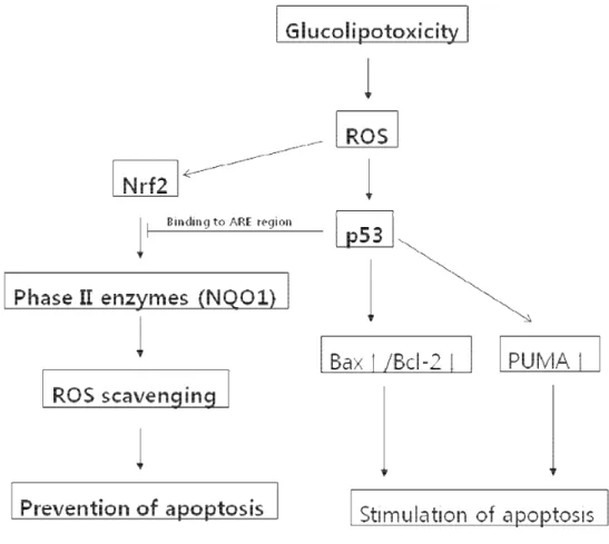
V. CONCLUSION
Fig 8. Model of p53-dependent apoptosis in H9c2 cells via inhibition of the anti-apoptotic Nrf2-NQO1 pathway. The glucolipotoxicity caused by combined
exposure to high glucose (30 mM) and palmitic acid (250 µM) generates ROS, which increase p53 and Nrf2 levels. Induction of Nrf2, a positive transcriptional regulator of ROS-scavenging phase II enzymes, leads to a reduction in ROS levels and thereby prevents apoptosis. However, p53 interrupts Nrf2 transcriptional activity by binding to ARE regions. Hence, ROS accumulate and then lead to stimulation of apoptosis by p53.
To identify the apoptosis pathway by palmitic acid and high concentrated glucose induced glucolipotoxicity, we performed several experiments, and we found p53 involved pathway.
We obtain the following results.
1. ROS generation by palmitate and high concentrated glucose increases p53
expression, and Nrf2 was interrupted transcriptional activity by p53, therefore
NQO1 which is a representative of phase II enzymes was decreased.
2. We could reduce apoptosis of H9c2 cells by treatment with pifithrin-α as a
p53 inhibitor on glucolipotoxicity conditions.
3. We demonstrated that p53 was increased by ROS. When ROS was
scavenged by N-acetyl-L-cystein (NAC), p53 was decreased and cell viability
REFERENCE
1. Dyntar D, Eppenberger-Eberhardt M, Maedler K, Pruschy M, Eppenberger
HM, Spinas GA, et al. Glucose and palmitic acid induce degeneration of myofibrils and modulate apoptosis in rat adult cardiomyocytes. Diabetes 2001;50:2105-13.
2. Listenberger LL, Ory DS, Schaffer JE. Palmitate-induced apoptosis can occur
through a ceramide-independent pathway. J Biol Chem 2001;276:14890-5.
3. Fiordaliso F, Leri A, Cesselli D, Limana F, Safai B, Nadal-Ginard B, et al.
Hyperglycemia activates p53 and p53-regulated genes leading to myocyte cell death. Diabetes 2001;50:2363-75.
4. Blazquez C, Galve-Roperh I, Guzman M. De novo-synthesized ceramide
signals apoptosis in astrocytes via extracellular signal-regulated kinase. FASEB J 2000;14:2315-22.
5. de Vries JE, Vork MM, Roemen TH, de Jong YF, Cleutjens JP, van der
Vusse GJ, et al. Saturated but not mono-unsaturated fatty acids induce apoptotic cell death in neonatal rat ventricular myocytes. J Lipid Res 1997;38:1384-94.
6. Paumen MB, Ishida Y, Muramatsu M, Yamamoto M, Honjo T. Inhibition of
carnitine palmitoyltransferase I augments sphingolipid synthesis and palmitate-induced apoptosis. J Biol Chem 1997;272:3324-9.
7. Shimabukuro M, Zhou YT, Levi M, Unger RH. Fatty acid-induced beta cell
apoptosis: a link between obesity and diabetes. Proc Natl Acad Sci U S A 1998;95:2498-502.
8. Grill V, Bjorklund A. Dysfunctional insulin secretion in type 2 diabetes: role
9. Unger RH. Lipotoxicity in the pathogenesis of obesity-dependent NIDDM. Genetic and clinical implications. Diabetes 1995;44:863-70.
10. Roduit R, Morin J, Masse F, Segall L, Roche E, Newgard CB, et al. Glucose
down-regulates the expression of the peroxisome proliferator-activated receptor-alpha gene in the pancreatic beta -cell. J Biol Chem 2000;275:35799-806.
11. Wang S Fau - Leonard SS, Leonard Ss Fau - Ye J, Ye J Fau - Ding M, Ding
M Fau - Shi X, Shi X. The role of hydroxyl radical as a messenger in Cr(VI)-induced p53 activation. Am J Physiol Cell Physiol 2000;279:C868-75.
12. Qiu X, Ting Z, Gu Y, Zhang X, Gan X, Fang Y, et al. Linalool preferentially
induces robust apoptosis of a variety of leukemia cells via upregulating p53 and cyclin-dependent kinase inhibitors. Toxicology 2009.
13. Faraonio R, Vergara P, Di Marzo D, Pierantoni MG, Napolitano M, Russo T,
et al. p53 suppresses the Nrf2-dependent transcription of antioxidant response genes. J Biol Chem 2006;281:39776-84.
14. Johnson JA, Johnson DA, Kraft AD, Calkins MJ, Jakel RJ, Vargas MR, et al.
The Nrf2-ARE pathway: an indicator and modulator of oxidative stress in neurodegeneration. Ann N Y Acad Sci 2008;1147:61-9.
15. Villeneuve Nf Fau - Sun Z, Sun Z Fau - Chen W, Chen W Fau - Zhang DD,
Zhang DD. Nrf2 and p21 regulate the fine balance between life and death by controlling ROS levels. Cell Cycle 2009;8:3255-6.
16. Spector AA. Structure and lipid binding properties of serum albumin.
Methods Enzymol 1986;128:320-39.
17. Kim WH, Lee JW, Suh YH, Lee HJ, Lee SH, Oh YK, et al. AICAR
potentiates ROS production induced by chronic high glucose: roles of AMPK in pancreatic beta-cell apoptosis. Cell Signal 2007;19:791-805.
18. Cai L, Li W, Wang G, Guo L, Jiang Y, Kang YJ. Hyperglycemia-induced apoptosis in mouse myocardium: mitochondrial cytochrome C-mediated caspase-3 activation pathway. Diabetes 2002;51:1938-48.
19. Ghosh S, An D, Pulinilkunnil T, Qi D, Lau HC, Abrahani A, et al. Role of
dietary fatty acids and acute hyperglycemia in modulating cardiac cell death. Nutrition 2004;20:916-23.
20. Kong Jy Fau - Rabkin SW, Rabkin SW. Lovastatin does not accentuate but is
rather additive to palmitate-induced apoptosis in cardiomyocytes. Prostaglandins Leukot Essent Fatty Acids 2002;67.
21. Miller TA, LeBrasseur NK, Cote GM, Trucillo MP, Pimentel DR, Ido Y, et al.
Oleate prevents palmitate-induced cytotoxic stress in cardiac myocytes. Biochem Biophys Res Commun 2005;336:309-15.
22. Miller TA, Icli B, Cote GM, Lebrasseur NK, Borkan SC, Pimentel DR, et al.
Palmitate alters neuregulin signaling and biology in cardiac myocytes. Biochem Biophys Res Commun 2009;379:32-7.
23. Hickson-Bick Dl Fau - Sparagna GC, Sparagna Gc Fau - Buja LM, Buja Lm
Fau - McMillin JB, McMillin JB. Palmitate-induced apoptosis in neonatal cardiomyocytes is not dependent on the generation of ROS. Am J Physiol Heart Circ Physiol 2002;282.
24. Poitout V, Robertson RP. Glucolipotoxicity: fuel excess and beta-cell
dysfunction. Endocr Rev 2008;29:351-66.
25. Waldman T Fau - Kinzler KW, Kinzler Kw Fau - Vogelstein B, Vogelstein B.
p21 is necessary for the p53-mediated G1 arrest in human cancer cells. Cancer Res 1995;55:5187-90.
26. Villunger A, Michalak EM, Coultas L, Mullauer F, Bock G, Ausserlechner
27. Polyak K, Xia Y, Zweier JL, Kinzler KW, Vogelstein B. A model for p53-induced apoptosis. Nature 1997;389:300-5.
28. Matoba S, Kang JG, Patino WD, Wragg A, Boehm M, Gavrilova O, et al. p53
regulates mitochondrial respiration. Science 2006;312:1650-3.
29. Motohashi H, Yamamoto M. Nrf2-Keap1 defines a physiologically important
stress response mechanism. Trends Mol Med 2004;10:549-57.
<ABSTRACT (IN KOREAN)> 종양 억제 p53 경로를 통한 rat 심근배아세포의 당지방독성에 의한 세포자살 왕혜진 연세대학교 대학원 의과학과 (지도교수 이현철) 고혈당은 자유지방산과의 상승작용으로 활성산소(ROS)를 생성하고 세포자살을 일으킨다. 이번 실험에서 우리는 palmitic acid(C16:0)와 높은 당의 당지방독성 효과를 rat의 심근세포주인 H9c2 를 이용하여 실험하였고 종양 억제 p53 과 관련된 세포자살 경로에 초점을 맞추었다.
H9c2 세포에 5mM normal glucose, 30mM high glucose와 250 µM palmitic acid를 각각 처리하거나 섞어서 처리하였다. 세포의 생존과 자살은 각각 MTT assay와 western blot을 이용한 cleaved caspase 3 를 통해 확인하였고
30mM high glucose와 250µM의 palmitat를 함께 처리 했을 때에만 p53 과 cleaved caspase 3 의 발현이 증가하는 것을 확인 할 수 있었고 5mM glucose condition과 비교 했을 때 ROS 생성이 2.5 배 증가하는 것을 확인 할 수 있었다. p53 억제제인 pifithrin-α를 처리 한 경우 당지방독성에 의해 증가된 ROS와 p53 발현이 모두 감소하는 것을 확인 할 수 있었다. 250µM palmitic acid가 처리된 경우, Nrf2 의 발현이 증가되는 반면 NQO1 의 발현은 감소하는 것을 확인하였고 pifithrin-α를 처리 하였을 때 NQO1 의 발현이 다시 증가하는 것을 볼 수 있었다. 염색체 면역 침강법을 이용하여 p53 이 NQO1 의 ARE 지역과 결합하는 것을 확인하였다. Rat 심근세포주에서 당지방독성에 의해 ROS가 증가하고 증가된 ROS에 의해 p53 의 발현이 증가하였다. 증가된 p53 은 Nrf2 의 작용을 방해함으로써 NQO1 의 발현을 막아 다시 ROS를 생성하게 되면서 결과적으로 세포자살 억제 경로를 막게 된다. 이번 실험에서 우리는 증가된 p53 이 원래 자신의 세포자살 활성 능력과 함께 Nrf2 의 세포자살 억제경로를 방해하는 작용도 같이 함을 보였다.
핵심 단어: p53, NQO1, 당지방독성, ARE region, 활성산소종 (ROS),