Introduction
Introduction
대장암의 치료는 오랜 기간 동안 fluoropyrimidine 기반 화학 요법으 로 구성되어 왔다. 최근에는 전이성 대장암 치료를 위한 치료 기준이 바 뀌어 irinotecan과 oxaliplatin을 포함하는 병용 화학 요법으로 처치되 고 있다. 전통적인 화학 요법의 상당한 개선에도 불구하고 이 치료에는 한계가 있다고 알려져 있다. 따라서 항암 효능을 높이기 위한 다양한 화 학 요법들과 함께 여러 가지 새로운 표적 및 약제 발굴이 계속되어 오고 있다[1]. Vinpocetine은 대수리 식물에서 추출한 알칼로이드 성분인 vin-camine의 반합성 유도체이다[2]. Vinpocetine은 뇌혈관 질환의 치료 에 널리 사용되어 왔으며 그 치료 효능은 대뇌 혈류를 개선하고 신경 손 상을 약화시킨다고 한다[3-5]. 또한 vinpocetine은 대뇌 평활근 세포 를 이완시켜 세포 내 cGMP를 분해하는 효소인 phosphodiesterase type-1 (PDE-1)을 선택적으로 억제하여 대뇌 혈류를 향상시킨다고 알려져 있다[6,7]. 전압 의존적 Na+ 채널과 Ca2+ 채널도 vinpocetine 에 의해 억제된다고 한다[8,9]. 심혈관계에서 vinpocetine이 검증되기 도 하였는데 다양한 종의 말초 혈관에서 vinpocetine의 혈관 이완 효능 은 PDE-1을 억제함으로써 매개되어 나타났다[10]. Wu 등[11]의 연구 에서는 in vitro 및 in vivo 실험연구에서 심장 비대, 섬유증 및 병리학 적인 심장 리모델링에서 vinpocetine의 역할이 규명되기도 하였다. 한 편 vinpocetine은 PDE-1 독립적인 nuclear factor-κB (NF-κB) 신 호전달경로의 억제와 interleukin-1β 및 tumor necrosis factor-α 와 같은 염증성 사이토카인들의 방출 저해를 통해 여러 가지 염증성 질 환들에서 항염증 효과를 유도하는 것으로 밝혀졌다[12,13]. 최근에는 vinpocetine은 세포주기를 억제함으로써 여러 인간 유방암 세포들의 증식을 강력하게 억제하고, 이종 이식 동물의 유방암 형성을 저해한다 고 보고되었다[14]. 본 연구에서는 vinpocetine이 대장암 세포의 증식에 미치는 영향을 조사하고 그 억제 기전을 조사하였다. Vinpocetine은 세포주기 조절 및 Int J Oral Biol 46:7-14, 2021pISSN: 1226-7155 • eISSN: 2287-6618 https://doi.org/10.11620/IJOB.2021.46.1.7
Vinpocetine inhibits the proliferation and induces
apoptosis in human colon cancer cells
Mi-Kyoung Kim
1, Hyun-Joo Park
1, Yeon Kim
1, Yong-Il Kim
2, Soo-Kyung Bae
3, Hyung Joon Kim
1*,
and Moon-Kyoung Bae
1*
1Department of Oral Physiology, Pusan National University School of Dentistry, Yangsan 50612, Republic of Korea 2Department of Orthodontics, Pusan National University School of Dentistry, Yangsan 50612, Republic of Korea 3Department of Dental Pharmacology, Pusan National University School of Dentistry, Yangsan 50612, Republic of Korea
Vinpocetine induces anti-inflammatory effects in various inflammatory diseases via the inhibition of phosphodiesterase
type-1-independent nuclear factor-κB signaling pathway and the release of inflammatory cytokines. In this study,
we investigated the effect of vinpocetine on the proliferation of colon cancer cells and its underlying molecular
mechanisms. Our data showed that vinpocetine inhibits the viability and proliferation of colon cancer cells.
Vinpocetine treatment induced cell death in HCT116 cells, which the percentages of sub-G1 phase were significantly
increased, and the apoptosis-related genes were regulated after HCT116 cells were treated with vinpocetine. In sum,
our findings indicated that vinpocetine could be a therapeutically useful candidate in the treatment of colon cancer.
Keywords:
Vinpocetine, Colon cancer cells, Apoptosis, Proliferation
Received December 30, 2020; Accepted January 6, 2021
*Correspondence to: Moon-Kyoung Bae, E-mail: [email protected] https://orcid.org/0000-0003-3948-4922 *Correspondence to: Hyung Joon Kim, E-mail: [email protected] https://orcid.org/0000-0002-2962-9553
Copyright © The Korean Academy of Oral Biology
CC This is an open-access article distributed under the terms of the Creative Commons Attribution Non-Commercial License
(http://creativecommons.org/licenses/by-nc/4.0/), which permits unrestricted non-commercial use, distribution, and reproduction in any medium, provided the original work is properly cited.
세포자멸사를 통해 대장암 세포의 증식을 억제시킨다는 다양한 실험법 을 통하여 증명하였다.
Materials and Methods
Materials and Methods
1. 실험재료
Vinpocetine은 Tocris Bioscience (Bristol, UK)에서 구입하였다. 3-[4-Dimethylthiazol-2-yl]-2,5-diphenyltetrazolium bromide (MTT), agarose 및 propidium iodide (PI) 시약은 모두 Sigma-Aldrich (St. Louis, MO, USA) 제품을 사용하였다. Bcl-2 항체와 β-actin 항체는 각각 Cell Signaling (Boston, MA, USA)과 Bio-world Technology (St. Louis Park, MN, USA)에서 구입하였다. X-linked inhibitor of apoptosis protein (XIAP), poly (ADP-ribose) polymerase (PARP) 항체는 Santa Cruz Biotechnology (Dallas, TX, USA)에서 구매하였다. Annexin V-fluorescein isothiocyanate (FITC) apoptosis detection kit는 BD Bioscience (San Diego, CA, USA)로부터 구입하였다.
2. 세포 배양
실험에 사용된 대장암 세포주인 HCT116, SW480, SW620은 37℃, 95% 습도, 5% CO2 배양기에서 배양하였으며, 2–3일에 한 번
씩 계대 배양하였다. 배양 배지는 10% fetal bovine serum과 1% penicillin streptomycin 항생제를 포함한 Dulbecco’s modified Eagle’s medium 배지를 사용하였으며, 이들 제품은 모두 Gibco Life Technologies (Grand Island, NY, USA)에서 구매하였다.
3. MTT assay
24-well plate의 well당 4 × 10⁴개의 대장암 세포를 500 µL의
배양 배지에 넣어주었다. 대조군으로 용매제인 dimethyl sulfoxide (DMSO)를 처리하였고, 1–100 µM의 농도로 vinpocetine을 처리하 였다. 각 24시간, 48시간, 72시간마다 MTT (5 mg/mL) 용액을 50 µL 넣고 3시간 동안 반응시킨다. 배지는 제거하고, 생성된 formazan 결정들은 DMSO로 용출하여 540 nm에서 흡광도를 측정하였다.
4. Colony formation assay
Base layer를 만들기 위해 24-well plate에 0.9% soft agar를 포함 한 배양 배지 500 µL를 넣어준다. 4℃에서 30분 정도 굳혀준 후, 37℃ 배양기에 넣는다. 그 위로 HCT116 세포 1 × 10⁴개와 0.4% soft agar가 들어간 배양 배지 500 µL를 얹어준 후, 약 10분 동안 4℃에서 굳혀준다. 배양 배지 250 µL에 원하는 농도의 vinpocetine을 잘 섞어 서 세포층 위에 처리해주었다. 14일 정도 관찰하면서 암세포 집락 형성 정도를 광학 사진으로 촬영하였다.
5. 유세포 분석(flow cytometry analysis)
FITC 형광물질이 표지된 Annexin V와 PI 염색을 통해 세포자멸사 를 확인하였다. HCT116 세포에 원하는 시간 동안 vinpocetine을 처 리한 후, 1 mL당 1 x 10⁶이 되도록 수집한다. Phosphate-buffered
saline (PBS)로 두 번 세척 후, binding buffer에 세포를 모아준다. 1 x 10⁵ 세포가 들어있는 100 µL에 Annexin V-FITC와 PI를 잘 섞어준 후
빛을 차단한 상태로 상온에서 15분간 반응시킨다. 400 µL의 binding buffer를 추가해주고, 그중 10,000개의 세포를 유세포 측정 분석기 (BD Bioscience)로 분석하였다.
6. TUNEL assay
세포자멸사가 유도되었는지 DeadEndTM Fluorometric TUNEL
System (Promega, Madison, WI, USA)을 이용하여 확인하였다. 간단히 설명하면, vinpocetine을 처리한 HCT116 세포를 4% form-aldehyde 용액으로 4℃에서 25분 동안 고정시키고, 0.2% Triton X-100 용액으로 상온에서 5분 동안 반응시켜 세포투과성을 높여준 다. 100 µL의 equilibration buffer와 반응 후, 세포자멸사에 의해 잘 려진 DNA의 3’ 말단 부위에 결합할 수 있는 TdT-mediated dUTP nick end labeling (TUNEL) 혼합액을 37℃에서 1시간 동안 처리하 였다. 4′,6-diamidino-2-phenylindole (DAPI)가 포함된 고정 용액 으로 대조 염색을 해준다. 표지된 DNA 단편들을 형광현미경(Nikon, Shizuoka, Japan)으로 관찰하였다.
7. 역전사-중합효소연쇄반응(reverse transcriptase
polymerase chain reaction, RT-PCR)
HCT116 세포에서 전체 RNA를 얻기 위해 TRIzol reagent kit (Invitrogen, Gaithersburg, MD, USA)를 이용하였다. Reverse transcription kit (Promega)를 이용하여 RNA로부터 cDNA를 합 성하였다. cDNA는 β-actin (sense: 5’-GACTACCTCATGAA-GATC-3’, antisense: 5’-GATCCACATCTGCTGGAA-3’)과 cyclin-dependent kinase 6 (CDK6) (sense: 5’-CGCTGACCAGCAG-TACGAAT-3’, antisense: 5’-TGGACTGGAGCAAGACTTCG-3’), cyclin D1 (sense: 5’-TGCTCCTGGTGAACAAGCTC-3’, anti-sense: 5’-GGCAGTCTGGGTCACACTTG-3’), cyclin E2 (anti-sense: 5’-GGTGCTTGCAGTGAAGAGGA-3’, antisense: 5’-TCGGTG-GTGTCATAATGCCT-3’), cyclin B1 (sense: 5’-AGCCAGAACCT-GAGCCTGTT-3’, antisense: 5’-ACATGGCAGTGACACCAACC-3’) primer를 이용하여 중합효소연쇄반응(PCR)을 시행하였다.
8. Western blot analysis
Vinpocetine을 처리한 세포들은 PBS로 세척 후 세포용해액(40 mM Tris–Cl, 10 mM EDTA, 120 mM NaCl, 0.1% NP-40, protease
inhibitor cocktail)으로 용해시켜서 단백질을 얻는다. 30 µg의 단백질 을 sodium dodecyl sulphate–polyacrylamide gel electrophoresis (SDS-PAGE)로 크기별로 분리하였고, 이를 nitrocellulose mem-brane (GE Healthcare, Marlborough, MA, USA)으로 전이시켰다. Membrane은 blocking buffer (1×TBS, 0.1% Tween-20, 5% skim milk)로 1시간 동안 상온에서 처리하였다. 원하는 단백질의 1차 항체를 4℃에서 16시간 반응시킨 후, horseradish peroxidase (HRP) 가 결합된 2차 항체를 상온에서 2시간 동안 반응시켰다. Enhanced chemiluminescence (ECL, GE Healthcare) 용액을 이용하여 발현 정도를 확인하였다.
9. 통계처리
본 실험 결과는 평균 세 번 이상의 실험으로부터 얻은 값으로 평균값 을 구하였다. 대조군과 실험군 간의 통계적 차이는 GraphPad Prism (version 5.0; GraphPad Software Inc., San Diego, CA, USA) 프 로그램을 이용하여 unpaired 2-tailed Student’s t-test 분석을 실시 하여 측정하였다. p값은 0.05 미만일 때 통계적 유의성이 있다고 판단 하였다.
Results
Results
1. Vinpocetine에 의한 대장암 세포주의 생존율과 증식 억제
효과
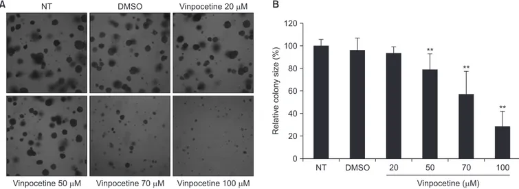
Vinpocetine이 대장암 세포주의 생존율을 조절할 수 있는지 확인하 기 위해 MTT assay를 진행하였다. 대장암 세포주인 HCT116에 다양 한 농도의 vinpocetine을 처리하여 세포의 생존 정도를 확인한 결과, Fig. 1A와 같이 vinpocetine의 농도가 증가할수록 그리고 반응 시간이 길어질수록 세포 생존율이 감소되었다. Vinpocetine을 70 µM의 농도 로 48시간 이상 배양 시, 50% 이상의 세포 생존 감소 효과를 보였다. HCT116뿐만 아니라 다른 대장암 세포주인 SW480과 SW620 세포주 에서 생존 억제 효과를 확인해보았다. Fig. 1B에서 보여주듯이 SW480 과 SW620 세포에서도 vinpocetine이 농도 의존적으로 세포 생존율을 억제함을 알 수 있었다. 다음으로 colony formation assay를 수행하여 vinpocetine이 HCT116 세포의 anchorage-independent growth 에 미치는 영향을 확인해보았다. Vinpocetine을 Fig. 2A에 나타낸 농 도로 처리하여 집락 형성 정도를 관찰하였다. 2주 동안 관찰한 결과, vinpocetine이 농도 의존적으로 HCT116 세포의 증식을 억제하여 대 장암 세포주의 집락 형성을 저해하였다. 대조군에 비해 vinpocetine을 70 µM, 100 µM 처리한 군에서 집락 크기가 각각 50%, 30%까지 감 DMSO 150 100 50 Cell viability (%) Vinpocetine ( M) 0A
B
C
10 24 hr 48 hr 72 hr 1 20 50 60 70 80 90 100 * ** ** ** **** ** ** ** ** ** ** ** * DMSO 150 100 50 Cell viability (%) Vinpocetine ( M) 0 48 hr 72 hr 20 50 70 100 SW480 SW620 *** ** ** ** ** **** DMSO 150 100 50 Cell viability (%) Vinpocetine ( M) 0 48 hr 72 hr 20 50 70 100 **** ** ** ** ** ** **Fig. 1. Effect of vinpocetine on cell viability in human colon cancer cells. HCT116 (A), SW480 and SW620 (B) were cultured with varying concentrations of vinpocetine or ve-hicle (dimethyl sulfoxide, DMSO) for indicated times. Cell viability was determined with 3-[4-Dimethylthiazol-2-yl]-2,5-diphenyltetrazo-lium bromide (MTT) assay. Data represents the mean of at least three experiments. *p < 0.05 vs. DMSO; **p < 0.01 vs. DMSO.
소하였다(Fig. 2B). 이로써 vinpocetine은 대장암 세포주의 생존율 감 소 및 세포 증식 억제 효과를 가지고 있음을 확인하였다.
2. Vinpocetine이 대장암 세포주의 세포주기 조절에 미치는
영향
Vinpocetine의 대장암 세포 증식 억제 효과가 세포주기 조절과 관련 되었는지 여부를 조사하기 위하여 DNA 염색을 할 수 있는 PI 염색액을 이용하여 세포주기를 분석하였다. HCT116 세포에 vinpocetine을 50 µM, 70 µM 처리하여 72시간 동안 배양하였다. 이 세포들을 회수하여 유세포 분석을 한 결과, Fig. 3에서 보여주듯이 vinpocetine 처리군에 서 M기의 세포는 감소하여 세포의 증식이 억제되었고, 세포자멸사로 인한 DNA 단편 및 핵의 응축이 일어나고 있는 sub-G1기의 세포가 눈 에 띄게 증가하였다.3. Vinpocetine이 대장암 세포주의 세포자멸사에 미치는 영향
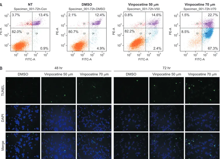
앞서 살펴본 바와 같이 vinpocetin이 HCT116 세포의 생존율과 증식 을 억제하였고, 세포주기를 조절하여 sub-G1기의 세포를 증가시켰다. Vinpocetine의 이러한 효과가 세포자멸사와 연관성이 있는지 추가적 으로 조사해보았다. 먼저 FITC가 표지된 Annexin V를 이용한 유세포 분석을 실시하였다. HCT116 세포에 72시간 동안 70 µM의 vinpo-cetine을 처리한 결과를 살펴보면 초기 세포자멸사 분획(Q4)이 67% 까지 증가하였고, 후기 자멸사 분획(Q3)도 22%까지 증가하였다(Fig. NT DMSO Vinpocetine 20 MVinpocetine 50 M Vinpocetine 70 M Vinpocetine 100 M
A
B
NT DMSO 120 100 80 60 40 20 Relative colony size (%) Vinpocetine ( M) 0 ** 20 50 70 100 ** **Fig. 2. Effect of vinpocetine on cell proliferation in human colon cancer cells. (A) Cell growth ability was evaluated by soft-agar colony formation assays. HCT116 cells were grown in soft-agar cell layer with or without vinpocetine. After 14 days, colony formation was observed and photographed under the mi-croscope. (B) Bar graph represents the size of colonies relative to colony of untreated cells. Bars are the mean ± standard error from at least three indepen-dent experiments.
NT, not treat; DMSO, dimethyl sulfoxide. **p < 0.01 was compared to the DMSO.
0 200 150 100 50 Count PE-A 0 NT 30 K 60 K 90 K 120 K 0 200 150 100 50 Count PE-A 0 DMSO 30 K 60 K 90 K 120 K 0 200 150 100 50 Count PE-A 0 Vinpocetine 50 m 30 K 60 K 90 K 120 K 0 200 150 100 50 Count PE-A 0 30 K 60 K 90 K 120 K Vinpocetine 70 m Sub-G: 9.24% G: 50.6% S: 5.45% M: 32.1% Sub G: 8.25% G: 49.2% S: 6.24% M: 33.7% - Sub G: 14.7% G: 46.1% S: 9.07% M: 26.4% - Sub G: 29.5% G: 34.7% S: 5.80% M: 26.8%
-Fig. 3. Effect of vinpocetine on cell cycle arrest in human colon cancer cells. HCT116 cells were incubated with vinpocetine for 72 hours. Cell cycle analysis was determined by flow cytometry with propidium iodide staining. The distribution percentage of cells in sub-G1, G, S, and G2/M phases was indicated within each histogram. Representative images were taken from three different experiments.
NT DMSO Vinpocetine 50 m Vinpocetine 70 m
A
B
102 PE-A FITC-A Specimen_001-72h-Con 103 104 105 105 102 103 104 3.7% 3.7% 82.0% 82.0% 13.4% 13.4% 0.9% 0.9% 102 PE-A FITC-A Specimen_001-72h-DMSO 103 104 105 105 102 103 104 2.1% 2.1% 80.7% 80.7% 12.4% 12.4% 4.9% 4.9% 102 PE-A FITC-A Specimen_001-72h-V50 103 104 105 105 102 103 104 0.8% 0.8% 82.2% 82.2% 14.6% 14.6% 2.4% 2.4% 102 PE-A FITC-A Specimen_001-72h-V70 103 104 105 105 102 103 104 1.5% 1.5% 8.5% 8.5% 22.7% 22.7% 67.3% 67.3% TUNEL DAPI MergeDMSO Vinpocetine 50 m Vinpocetine 70 m DMSO Vinpocetine 50 m Vinpocetine 70 m
48 hr 72 hr
Fig. 4. Effect of vinpocetine on cellular apoptosis in human colon cancer cells. (A) For analysis of the cell apoptosis, cells were stained using Annexin V-fluorescein isothiocyanate (FITC)/propidium iodide and performed flow cytometry. Representative dot plots are shown. The numbers of the plots indicate the percentage of necrotic (Q1, red dot), late apoptotic (Q2, purple dot), viable (Q3, orange dot) and early apoptotic (Q4, blue dot) cells. (B) HCT116 cells were treated with vinpocetine for indicated time. TdT-mediated dUTP nick end labeling (TUNEL) analysis was carried out to detect apoptotic cells and the results were visualized using fluorescent microscope. The apoptotic cells were labeled with TUNEL by green, nuclear DNA of cells was stained with 4′,6-diamidino-2-phenylindole (DAPI, blue). Representative pictures are shown. ×200 magnification.
A
B
CDK6 Cyclin D1 Cyclin E2 Cyclin B1 -actin Vinpocetine XIAP Bcl-2 Total PARP Cleaved PARP -actin Con 1 2 4 8 (hr) Vinpocetine (50 M) + + 24 hr 48 hrFig. 5. Expression of cell cycle- or apoptosis-related factors in vinpocetine-treated human colon cancer cells. (A, B) HCT116 cells were exposed with 50 µM vinpocetine for indicated times. (A) The expression of cell cycle-related genes detected by reverse transcriptase polymerase chain reaction array with prim-ers specific for cyclin-dependent kinase 6 (CDK6), cyclin D1, cyclin E2, cyclin B1, and β-actin. (B) Western blot results showing the expression apoptosis-related proteins X-linked inhibitor of apoptosis protein (XIAP), Bcl-2, and poly (ADP-ribose) polymerase (PARP). β-actin was used as a reference. Results were taken from three independent experiments with similar results.
4A). 다음으로 세포자멸사가 유도될 때 생기는 DNA 단편을 확인할 수 있는 TUNEL assay를 시행하였다. Fig. 4B에서 보여주듯이 vinpo-cetine은 시간 및 농도 의존적으로 TUNEL 염색이 된 HCT116 세포들 을 증가시켰다. 종합하여 보면, vinpocetine이 HCT116에서 세포자멸 사를 유도할 수 있음을 확인하였다.
4. Vinpocetine에 의해 변화되는 세포주기 조절 및 세포자멸사
관련 인자 발현 분석
다음으로 vinpocetine을 처리한 HCT116 세포에서 세포주기 조 절 및 세포자멸사 관련 인자들의 발현 변화를 조사해보았다. 먼저 HCT116 세포를 vinpocetine 함유 배지에서 다양한 시간대로 배양하 여 세포주기 조절에 관련된 유전자의 발현 정도를 탐색해보았다. RT-PCR을 시행해본 결과, Fig. 5A와 같이 cyclin B1, cyclin D1, cyclin E2 및 CDK6의 발현에는 영향을 미치지 않았다. 다른 세포주기 조절 인 자들에 대한 추가적인 연구가 수행되어야 할 것으로 보인다. 세포자멸 사를 조절하는 인자에 대한 분석은 Western blot assay을 시행하여 단백질 수준에서 발현 변화를 확인하였다. 대조군과 비교했을 때 vin-pocetine을 처리한 실험군에서 세포자멸을 억제하는 XIAP와 Bcl-2 단백질의 발현이 감소한 반면, 세포자멸을 유도할 수 있는 활성 형태의 잘려진 PARP 단백질 발현이 vinpocetine 처리의 48시간 배양에서 증 가하였다. 이러한 결과들을 통해 vinpocetine이 대장암 세포 HCT116 의 세포자멸사 관련 유전자들의 발현을 조절함으로써 세포자멸사를 유 도함을 확인할 수 있었다(Fig. 5B).Discussion
Discussion
본 연구는 vinpocetine의 대장암 세포주에서 항암 활성에 대한 다양 한 증거를 제공하였다. Vinpocetine은 여러 가지 인간 대장암 세포주들 인 SW480과 SW620 및 HCT116의 생존율 및 증식을 억제함을 보여 주었다. 그리고 vinpocetine은 인간 대장암 세포주의 세포주기를 조절 하며 sub-G1기의 세포군을 증가시켰다. 또한 vinpocetine은 세포자 멸사 관련 유전자들의 조절을 통해 인간 대장암 세포주의 세포자멸사를 유도한다는 사실을 여러 가지 실험법으로 증명하였다. Vinpocetine의 항암 활성에 대한 분자적 기전을 파악할 때 PDE 효 소 활성 및 채널들을 통한 Ca2+ 및 Na+ 이온 조절에 대한 알려진 작용 을 고려해볼 필요가 있다. Vinpocetine은 PDE-1을 억제하여 혈관 평 활근 세포 내에서 cGMP 수준을 높인다고 알려져 있다[15]. 또한 신경 세포의 전압 의존성 Ca2+ 채널[16]과 전압 의존성 Na+ 채널을 억제한 다고 한다[17]. 이러한 특성들이 본 연구에서 관찰된 항암 효능과 연관 되는지에 대한 여부는 아직 연구되지 않았다. 다른 연구에서 PDE-1 억 제제 IC86340이 혈관 평활근 세포의 성장과 생존을 억제한다는 것이 증명되기도 하였다[18]. 최근 vinpocetine이 암 치료의 표적으로 사용 될 수 있는 PDE-4를 억제하는 것으로 보고된 바도 있다[19-21]. 그리 고 최근 연구에서 vinpocetine이 유방암 세포의 증식, 생존 및 이동을 억제하고 in vivo 종양 성장을 억제한다는 사실이 보고된 바 있다[14]. Vinpocetine은 G(0)/G(1) 단계에서 세포주기를 억제함으로써 다양 한 인간 유방암 세포의 증식을 강력하게 억제하였다. 또한 vinpocetine 이 미토콘드리아 의존 경로를 통해 세포자멸사를 유도하는 것으로 밝 혀졌다. 더욱이 vinpocetine은 전이성이 강한 유방암 세포주인 MDA-MB-231의 이동을 억제시켰다. 누드마우스의 인간 유방암 이종 이식 모델로의 vinpocetine의 투여는 종양 성장을 유의하게 억제시켰다. 따 라서 vinpocetine의 PDE-1 및 PDE-4 차단 효과가 종양 성장 억제에 기여할 수 있다고 추측하는 것이 타당하며, 대장암 세포의 항암효능에 서 PDE 효소활성과의 상관성 연구는 향후 더 연구가 진행되어야 할 부 분이라고 생각한다. 암 관련 염증반응은 여러 유형의 암 진행 과정의 전형적 특징으로 여 겨진다. 지속적인 염증반응은 암 위험을 폭발적으로 증가시키고 암 진 행을 촉진시킨다고 알려져 있다[22]. 염증의 매개체들과 염증 반응자 들은 개선된 진단 및 치료로 이어질 수 있는 새로운 분자 표적 인자들 로 여겨지고 있다[23-25]. NF-κB 전사 인자는 많은 연구에서 염증을 암과 연결시키고 있다[26,27]. Vinpocetine은 다른 연구들에서 IκB kinase (IKK)를 직접 억제함으로써 NF-κB 의존성 염증반응을 억제 하는 것으로 나타나고 있다[28]. 또한 Signal Transducer and Tran-scription-3 (STAT-3)는 종양 성장을 조절하는 것 외에도 염증과 암을 연결하는 역할을 한다고 한다[29,30]. 따라서 앞으로의 연구에서는 염 증반응에 대한 vinpocetine의 억제 효과가 항암 활성에 관여하는지 여 부를 확인하기 위해 추가 연구가 필요하며, vinpocetine이 NF-κB 및 STAT-3 활성을 억제함으로써 대장암 세포의 항암효능을 나타내는지에 대한 연구가 필요할 것으로 생각한다. 본 연구에서 인간 대장암 세포에 대한 vinpocetine의 효능을 조사한 결과 vinpocetine이 세포 생존 및 증식을 억제하고 세포자멸사를 유도 한다는 것을 발견하였다. 이러한 연구 결과는 전통적으로 뇌혈관 질환 에 사용되는 vinpocetine의 항암 활성을 보여줌으로써 대장암 치료 및 예방을 위한 기초연구로써 가치가 있다고 생각한다.Acknowledgements
Acknowledgements
이 논문은 부산대학교 기본연구지원사업(2년)에 의하여 연구되었음 (to Bae M-K).Conflicts of Interest
Conflicts of Interest
No potential conflict of interest relevant to this article was reported.
References
References
1. Iqbal S, Lenz HJ. Integration of novel agents in the treatmentof colorectal cancer. Cancer Chemother Pharmacol 2004;54 Suppl 1:S32-9. doi: 10.1007/s00280-004-0884-0.
2. Zhang YS, Li JD, Yan C. An update on vinpocetine: new dis-coveries and clinical implications. Eur J Pharmacol 2018;819: 30-4. doi: 10.1016/j.ejphar.2017.11.041.
3. Patyar S, Prakash A, Modi M, Medhi B. Role of vinpocetine in cerebrovascular diseases. Pharmacol Rep 2011;63:618-28. doi: 10.1016/s1734-1140(11)70574-6.
4. Dézsi L, Kis-Varga I, Nagy J, Komlódi Z, Kárpáti E. [Neuro-protective effects of vinpocetine in vivo and in vitro. Apovin-caminic acid derivatives as potential therapeutic tools in isch-emic stroke]. Acta Pharm Hung 2002;72:84-91. Hungarian. 5. Szilágyi G, Nagy Z, Balkay L, Boros I, Emri M, Lehel S, Márián
T, Molnár T, Szakáll S, Trón L, Bereczki D, Csiba L, Fekete I, Kerényi L, Galuska L, Varga J, Bönöczk P, Vas A, Gulyás B. Effects of vinpocetine on the redistribution of cerebral blood flow and glucose metabolism in chronic ischemic stroke pa-tients: a PET study. J Neurol Sci 2005;229-230:275-84. doi: 10.1016/j.jns.2004.11.053.
6. Hagiwara M, Endo T, Hidaka H. Effects of vinpocetine on cyclic nucleotide metabolism in vascular smooth muscle. Bio-chem Pharmacol 1984;33:453-7. doi: 10.1016/0006-2952 (84)90240-5.
7. Truss MC, Uckert S, Stief CG, Forssmann WG, Jonas U. Cy-clic nucleotide phosphodiesterase (PDE) isoenzymes in the human detrusor smooth muscle. II. Effect of various PDE in-hibitors on smooth muscle tone and cyclic nucleotide levels in vitro. Urol Res 1996;24:129-34. doi: 10.1007/BF00304075. 8. Bönöczk P, Gulyás B, Adam-Vizi V, Nemes A, Kárpáti E,
Kiss B, Kapás M, Szántay C, Koncz I, Zelles T, Vas A. Role of sodium channel inhibition in neuroprotection: effect of vinpo-cetine. Brain Res Bull 2000;53:245-54. doi: 10.1016/s0361-9230(00)00354-3.
9. Sitges M, Galván E, Nekrassov V. Vinpocetine blockade of so-dium channels inhibits the rise in soso-dium and calcium induced by 4-aminopyridine in synaptosomes. Neurochem Int 2005; 46:533-40. doi: 10.1016/j.neuint.2005.02.001.
10. Giachini FR, Lima VV, Carneiro FS, Tostes RC, Webb RC. De-creased cGMP level contributes to inDe-creased contraction in arteries from hypertensive rats: role of phosphodiesterase 1. Hypertension 2011;57:655-63. doi: 10.1161/HYPERTENSIO-NAHA.110.164327.
11. Wu MP, Zhang YS, Xu X, Zhou Q, Li JD, Yan C. Vinpocetine
attenuates pathological cardiac remodeling by inhibiting car-diac hypertrophy and fibrosis. Cardiovasc Drugs Ther 2017;31: 157-66. doi: 10.1007/s10557-017-6719-0.
12. Colombo BB, Fattori V, Guazelli CFS, Zaninelli TH, Carvalho TT, Ferraz CR, Bussmann AJC, Ruiz-Miyazawa KW, Baracat MM, Casagrande R, Verri WA Jr. Vinpocetine ameliorates acetic acid-induced colitis by inhibiting NF-κB activation in mice. Inflammation 2018;41:1276-89. doi: 10.1007/s10753-018-0776-9.
13. Jeon KI, Xu X, Aizawa T, Lim JH, Jono H, Kwon DS, Abe J, Berk BC, Li JD, Yan C. Vinpocetine inhibits NF-kappaB-dependent inflammation via an IKK-NF-kappaB-dependent but PDE-independent mechanism. Proc Natl Acad Sci U S A 2010;107: 9795-800. doi: 10.1073/pnas.0914414107.
14. Huang EW, Xue SJ, Zhang Z, Zhou JG, Guan YY, Tang YB. Vinpocetine inhibits breast cancer cells growth in vitro and in vivo. Apoptosis 2012;17:1120-30. doi: 10.1007/s10495-012-0743-0.
15. Ahn HS, Crim W, Pitts B, Sybertz EJ. Calcium-calmodulin-stimulated and cyclic-GMP-specific phosphodiesterases. Tis-sue distribution, drug sensitivity, and regulation of cyclic GMP levels. Adv Second Messenger Phosphoprotein Res 1992;25: 271-88.
16. Kaneko S, Takahashi H, Satoh M. The use of Xenopus oocytes to evaluate drugs affecting brain Ca2+ channels: effects of bifemelane and several nootropic agents. Eur J Pharmacol 1990;189:51-8. doi: 10.1016/0922-4106(90)90229-q. 17. Erdo SA, Molnár P, Lakics V, Bence JZ, Tömösközi Z.
Vin-camine and vincanol are potent blockers of voltage-gated Na+ channels. Eur J Pharmacol 1996;314:69-73. doi: 10.1016/ s0014-2999(96)00542-0.
18. Nagel DJ, Aizawa T, Jeon KI, Liu W, Mohan A, Wei H, Miano JM, Florio VA, Gao P, Korshunov VA, Berk BC, Yan C. Role of nuclear Ca2+/calmodulin-stimulated phosphodiesterase 1A in vascular smooth muscle cell growth and survival. Circ Res 2006;98:777-84. doi: 10.1161/01.RES.0000215576.27615.fd. 19. Dunkern TR, Hatzelmann A. Characterization of inhibitors of
phosphodiesterase 1C on a human cellular system. FEBS J 2007;274:4812-24. doi: 10.1111/j.1742-4658.2007.06001.x. 20. Mehta A, Patel BM. Therapeutic opportunities in colon cancer:
focus on phosphodiesterase inhibitors. Life Sci 2019;230:150-61. doi: 10.1016/j.lfs.2019.05.043.
21. Sengupta R, Sun T, Warrington NM, Rubin JB. Treating brain tumors with PDE4 inhibitors. Trends Pharmacol Sci 2011;32:
337-44. doi: 10.1016/j.tips.2011.02.015.
22. Korniluk A, Koper O, Kemona H, Dymicka-Piekarska V. From inflammation to cancer. Ir J Med Sci 2017;186:57-62. doi: 10. 1007/s11845-016-1464-0.
23. Balkwill FR, Mantovani A. Cancer-related inflammation: com-mon themes and therapeutic opportunities. Semin Cancer Biol 2012;22:33-40. doi: 10.1016/j.semcancer.2011.12.005. 24. de Visser KE, Korets LV, Coussens LM. De novo
carcinogen-esis promoted by chronic inflammation is B lymphocyte de-pendent. Cancer Cell 2005;7:411-23. doi: 10.1016/j.ccr.2005. 04.014.
25. Rebouissou S, Amessou M, Couchy G, Poussin K, Imbeaud S, Pilati C, Izard T, Balabaud C, Bioulac-Sage P, Zucman-Rossi J. Frequent frame somatic deletions activate gp130 in in-flammatory hepatocellular tumours. Nature 2009;457:200-4. doi: 10.1038/nature07475.
26. Luo JL, Tan W, Ricono JM, Korchynskyi O, Zhang M, Gonias
SL, Cheresh DA, Karin M. Nuclear cytokine-activated IKKalpha controls prostate cancer metastasis by repressing Maspin. Nature 2007;446:690-4. doi: 10.1038/nature05656.
27. Fernandes JV, Cobucci RN, Jatobá CA, Fernandes TA, de Azevedo JW, de Araújo JM. The role of the mediators of in-flammation in cancer development. Pathol Oncol Res 2015; 21:527-34. doi: 10.1007/s12253-015-9913-z.
28. Medina AE. Vinpocetine as a potent antiinflammatory agent. Proc Natl Acad Sci U S A 2010;107:9921-2. doi: 10.1073/ pnas.1005138107.
29. Karin M. Nuclear factor-kappaB in cancer development and progression. Nature 2006;441:431-6. doi: 10.1038/na-ture04870.
30. Karin M, Greten FR. NF-kappaB: linking inflammation and im-munity to cancer development and progression. Nat Rev Im-munol 2005;5:749-59. doi: 10.1038/nri1703.