저작자표시-비영리-동일조건변경허락 2.0 대한민국 이용자는 아래의 조건을 따르는 경우에 한하여 자유롭게 l 이 저작물을 복제, 배포, 전송, 전시, 공연 및 방송할 수 있습니다. l 이차적 저작물을 작성할 수 있습니다. 다음과 같은 조건을 따라야 합니다: l 귀하는, 이 저작물의 재이용이나 배포의 경우, 이 저작물에 적용된 이용허락조건 을 명확하게 나타내어야 합니다. l 저작권자로부터 별도의 허가를 받으면 이러한 조건들은 적용되지 않습니다. 저작권법에 따른 이용자의 권리는 위의 내용에 의하여 영향을 받지 않습니다. 이것은 이용허락규약(Legal Code)을 이해하기 쉽게 요약한 것입니다. Disclaimer 저작자표시. 귀하는 원저작자를 표시하여야 합니다. 비영리. 귀하는 이 저작물을 영리 목적으로 이용할 수 없습니다. 동일조건변경허락. 귀하가 이 저작물을 개작, 변형 또는 가공했을 경우 에는, 이 저작물과 동일한 이용허락조건하에서만 배포할 수 있습니다.
Mixed Dementia Induced by Right Common Carotid
Artery Occlusion in an Alzheimer Mouse Model
by
Jin Soo Lee
Major in Medicine
Department of Medical Sciences
The Graduate School, Ajou University
Mixed Dementia Induced by Right Common Carotid
Artery Occlusion in an Alzheimer Mouse Model
by
Jin Soo Lee
A Dissertation Submitted to The Graduate School of Ajou University
in Partial Fulfillment of the Requirements for the Degree of
Ph.D. in Medicine
Supervised by
In Soo Joo, M.D.
Major in Medicine
Department of Medical Sciences
The Graduate School, Ajou University
This certifies that the dissertation
of Jin Soo Lee is approved.
SUPERVISORY COMMITTEE
Byoung Joo Gwag
In Soo Joo
Eun-Hye Joe
Byung Gon Kim
In-Hee Mook-Jung
The Graduate School, Ajou University
December, 21st, 2009
ACKNOWLEDGEMENTS
First of all, I would like to express my gratitude to all those who gave me the possibility to complete this thesis. I want to thank all professors and colleagues in Department of Neurology, Ajou University School of Medicine and Ajou University Hospital. They have excused my basic researches from hard working in the hospital field. I have furthermore to thank all members in the main laboratory of Neurotech pharmaceuticals Co., Ltd. They kindly helped me for all aspects related to my experiments.
I am deeply indebted to my supervisor Prof. Dr. Joo I. S. in Department of Neurology whose stimulating suggestions and encouragement helped me in all the time of research for and writing of this thesis. I thank Prof. Dr. Hong J. M., who guaranteed enough time to perform my PhD course even though I was simultaneously involved in his fellowship course. Especially, I am obliged to Prof. Gwag B. J. in Department of Pharmacology and Neurotech pharmaceuticals Co., Ltd., who provided great opportunity in my first step into neuroscience. I have to thank Im D. S. who was my personal tutor for approaching basic researches.
I would like to give my special thanks to my wife Hyunju whose patient love enabled me to complete this work. I thank my son, Yoo Chan and a baby in my wife’s pocket. They are my pleasure. I feel like agape love from my parents and grandparents. I know they pray to God for me every day and I greatly appreciate it. I thank my
parents-in-law, who have deep interest in my works. There are many more who must be thanked. My sisters, Eun Soo and Min Soo, are always proud of me. Sang Yoon, Min Soo’s husband, is a trustworthy person and gives me new insights about computer science.
Finally, I thank God who guides me into the right path.
- ABSTRACT -
Mixed Dementia Induced by Right Common Carotid
Artery Occlusion in an Alzheimer Mouse Model
Although vascular risk factors are associated with Alzheimer disease (AD), the effect of chronic cerebral hypoperfusion (CCH) on the progression of AD has not been widely evaluated. We aimed to evaluate whether CCH would aggravate the behavioral changes and plaque formation in an AD transgenic (Tg) mouse model. Female Tg2576 (Tg+) mice (an AD model) and their littermates (Tg-) were subjected to permanent right common carotid artery occlusion (rCCAO). The Morris water maze and object recognition tests were performed 6–8 weeks after the operation; the mice were then sacrificed. We evaluated cell death by histological studies and quantified the amount of amyloid plaques. An escape performance in the Morris water maze test was significantly impaired in Tg+ mice with or without rCCAO compared with that in Tg- mice, but this impairment was not different between Tg+ rCCAO- and sham-operated mice. Learning curve which was preserved in Tg+ sham-operated mice was impaired in Tg+ rCCAO-operated mice. On object recognition testing, Tg+ and Tg- mice with rCCAO showed a significant reduction in their discrimination ability compared with those without rCCAO but there was no difference in the discrimination ability between Tg+ and Tg- mice. On the histological examinations, there was no evident cellular death in the brains of experimental mice. The burden of amyloid plaques was not different between Tg+
rCCAO- and sham-operated mice. In conclusion, Tg+ rCCAO-operated mice had both cognitive deficits on spatial memory and non-spatial working memory induced in AD and VCI models, respectively. Moreover, an impairment of learning curve was synergistically induced from both pathomechanisms.
Key words: Alzheimer disease, mixed dementia, vascular cognitive impairment, common carotid artery occlusion, amyloid plaque
TABLE OF CONTENTS
ABSTRACT ... i
TABLE OF CONTENTS ... iii
LIST OF TABLES ... vi
LIST OF FIGURES ... vii
I. INTRODUCTION ... 1
A. Vascular factors in patients with Alzheimer disease ... 1
B. Induction of chronic cerebral hypoperfusion in Alzheimer disease models ... 2
C. Tg2576, an Alzheimer disease mouse model ... 3
D. Right common carotid artery occlusion, a chronic cerebral hypoperfusion mouse model ... 4
E. Purpose of study ... 5
II. MATERIALS AND METHODS ... 6
A. Materials ... 6 1. Animals ... 6 2. Reagents ... 6 3. Antibodies ... 7 B. Methods ... 8 1. Animal preparation ... 8 2. Anesthetic methods ... 8
4. Cerebral blood flow assessment ... 9
5. Positron emission tomography to evaluate metabolic status... 10
6. Tissue preparation for histological studies ... 10
7. Behavioral assessment ... 11
8. Histological studies ... 14
9. Experimental sets and grouping ... 17
10. Statistical analysis ... 19
III. RESULTS ... 20
A. Establishment of a mild ischemic condition in mice ... 20
B. Replication study for a vascular cognitive impairment model induced by rCCAO... 24
1. A lower recognition of novel object in rCCAO-operated mice ... 24
2. Occasional apoptotic cell death observed 7 days after rCCAO ... 26
3. Hypometabolism in FDG microPET for an rCCAO-operated mouse ... 30
C. Tg 2576 treated with chronic mild ischemia ... 32
1. Consequences of surgery... 32
2. Behavioral tests ... 35
3. Histopathological tests ... 40
IV. DISCUSSION ... 46
A. Cognitive deterioration induced by rCCAO in Tg2576 mice ... 46
B. Vascular cognitive impairment induced by rCCAO ... 50
1. Subtypes of VCI ... 53
2. Neuropsychological features of VCI ... 55
3. Frontal-subcortical circuit ... 56
V. CONCLUSION ... 58
REFERENCES ... 59
LIST OF TABLES
LIST OF FIGURES
Fig. 1. Object recognition test. ... 13
Fig. 2. A pilot experiment for induction of mild ischemia. ... 21
Fig. 3. No breakdown of blood brain barrier in mice with rCCAO and 2m tMCAO. ... 22
Fig. 4. Decreased CBF after occlusion of cerebral arteries. ... 23
Fig. 5. A trend of impairment on novel object recognition in mice with rCCAO. ... 25
Fig. 6. No necrotic cells in rCCAO-operated mice. ... 27
Fig. 7. TUNEL-positive cells in one rCCAO-operated mouse 7 days after surgery. ... 28
Fig. 8. Representative photograph of apoptotic cell death observed in an rCCAO-operated mouse 7 days after surgery. ... 29
Fig. 9. Hypometabolism in the right cortex of an rCCAO-operated mouse. ... 31
Fig. 10. Cerebral amyloid angiopathy observed in Tg2576 mice. ... 34
Fig. 11. Impairment of learning curve on Morris water maze test in Tg+ rCCAO-operated mice. ... 36
Fig. 12. Impairment of memory retention in Tg+ mice. ... 37
Fig. 13. Decreased discrimination index on rCCAO-operated mice. ... 39
Fig. 14. No evident necrotic cell death in the brain sections. ... 41
Fig. 15. No evident apoptotic cell death in the brain sections. ... 42
Fig. 17. No myelin damage in the brain sections. ... 44 Fig. 18. No increased burden of amyloid plaques in the Tg+ mice with
rCCAO. ... 45 Fig. 19. Brain lesions of VCI subtypes. ... 54 Fig. 20. Frontal subcortical circuits. ... 57
I. INTRODUCTION
A. Vascular factors in patients with Alzheimer disease
Alzheimer disease (AD) is associated with vascular disease in the brain. A concomitant cerebral infarction or white matter vascular pathologies are often observed in brains of patients with AD (Snowdon, et al., 1997; Kalaria and Ballard, 1999; Jellinger, 2007). Cognitive impairment is worse in AD patients with cerebral infarctions, which are associated with atherosclerosis in the Circle of Willis, than in those without cerebral infarctions (Snowdon, et al., 1997). The proportion of demented patients with hippocampal atrophy significantly increased, especially when they showed white matter hyperintensities on magnetic resonance imaging (Wu, et al., 2002). Similarly, pathological changes of Alzheimer’s were also accompanied in many cases of patients with vascular cognitive impairment (VCI) (Hulette, et al., 1997; Esiri, et al., 1997; Nolan, et al., 1998) In addition, both Alzheimer and cerebrovascular diseases share common risk factors (Decarli, et al., 2004), e.g., hypertension (Skoog, et al., 1996; in’t Veld, et al., 2001), diabetes mellitus (Vitek, et al., 1994; Ott, et al., 1996; Gasparini, et al., 2002), hypercholesterolemia (Sparks, 1997; Notkola, et al., 1998), and atherosclerosis (Hoffman, et al., 1997; van Oijen, et al., 2007). Based on these observations, cerebrovascular disease has been suggested to contribute the development of AD. Chronic cerebral hypoperfusion (CCH) is an important factor in the pathogenesis of AD (De Jong, et al., 1997; Aliev, et al., 2004). Alteration of the capillaries, which results in a compromised cerebral circulation, was observed in rats with chronic hypertension (De Jong, et al., 1997).
Oxidative stress and mitochondrial damage from vascular hypoperfusion are associated with brain lesions (Aliev, et al., 2004). As such, cognitive impairment observed in AD may be aggravated by CCH. CCH is also associated with VCI without Alzheimer pathology (Román, 2004; Kato, et al., 2008; Shibata, et al., 2004; Farkas, et al., 2004; Yoshizaki, et al., 2008; Lee, et al., 2009). In this aspect, the reduced cerebral perfusion observed in AD may cause mixed dementia-like cognitive abnormalities.
B. Induction of chronic cerebral hypoperfusion in Alzheimer disease models
Cognitive changes induced by CCH have not been widely studied in AD transgenic (Tg) mouse models. A few researches reported relationship between ischemic insult and AD pathology. Unilateral occlusion of middle cerebral artery for 24 hours caused larger infarction in amyloid protein precursor (APP) transgenic (Tg+) mice (FVB/N HuAPP695.SWE) than nontransgenic (Tg-) mice (Zhang, et al., 1997). Brains of Tg+ mice were vulnerable to ischemic insult; however, the amount of amyloid beta (Aβ) was not different between two groups. The result might be from huge damage which masked changes in the brain. APP/presenilin-1 (PS1) Tg mice with both common carotid arteries occlusion (CCAO) for 45 minutes induced a worsening of cognitive deficit using the Morris water maze and showed a trend for increased Aβ deposition (Sadowski, et al., 2004). This study revealed a role of cerebral hypoperfusion on the progression of AD but the ischemic insult was too short to conclude the effect of CCH. A recent study induced CCH with microcoils which caused stenosis in the bilateral common carotid arteries (CCAs) of APPSwInd Tg mice (Kitaguchi, et al., 2009). Aβ fibrils got increased and Aβ deposition was induced in the intracellular compartment by bilateral stenosis of CCAs.
This study revealed the effect of CCH on acceleration of the pathological changes of AD. Unfortunately, the study lacked behavioral data. The microcoils are not commercially available so that replicating study is difficult to perform.
C. Tg2576, an Alzheimer disease mouse model
For our experiments Tg2576 mice were used as an AD mouse model. Tg2576 mice overexpress human APP695 with the “Swedish” mutation (Hsiao, et al., 1996). The mice present amyloid plaques and its corresponding memory deficits, but do not induce neuronal death (Hsiao, et al., 1996). The memory impairments are usually revealed in a serial Morris water maze test reflecting deficits in the hippocampus. One thing is that a learning ability is spared in Tg2576 mice even though their escape performance is worse than that of control mice (Routtenberg, 1997). Amyloid plaques in the brain of Tg2576 mice begin to increase at 9-12 months whereas an increase in Aβ starts at 6 months (Kawarabayashi, et al., 2001). In a previous study, mice were grouped into four stages: (1) very young mice, 4-5 months, before the appearance of insoluble Aβ (Aβinsol) or plaques; (2) young mice, 6-11 months, during the initial appearance of Aβinsol and both amyloid plaques and punctate Aβ deposits; (3) middle-age mice, 12-18 months, during the extensive deposition of plaques when Aβinsol levels are rising rapidly; and (4) old mice, 20-25 months, at a time when Aβ is leveling off and amyloid loads are comparable to those in AD (Westerman, et al., 2002). Our mice selected for this study were 14.5 months old and those were in the middle age.
D. Right common carotid artery occlusion, a chronic cerebral hypoperfusion mouse model
A recent study reported that right common carotid artery occlusion (rCCAO) in mice could induce behavioral abnormality 1 month after the operation and that the deficit was related to damage to the cortical-subcortical circuit (Yoshizaki, et al., 2008). There was no acute neuronal death, but rarefaction was shown on the medial corpus callosum, and the level of inflammatory cytokines increased. The authors suggested that these changes resulted from CCH induced by rCCAO and that this might be a good model to evaluate subcortical ischemic vascular dementia which is one of the most common types of VCI (Yoshizaki, et al., 2008).
Novel object recognition was impaired in rCCAO-operated mice (Yoshizaki, et al., 2008). The object recognition test is a behavioral experiment for animal and reflects non-spatial working memory (Ennaceur and Delacour, 1988). Damage to the cortical-subcortical circuit is known to affect cognitive function and to be associated with VCI (Yoshizaki, et al., 2008). It is known that the novel object recognition is not affected by the hippocampal function (Ennaceur and Delacour, 1988). Impairment observed during the object recognition test was induced in some rodent models by unilateral or bilateral CCAO (Yoshizaki, et al., 2008; Sarti, et al., 2002).
E. Purpose of study
In this study we used a mixed dementia model with both AD and VCI. As huge necrosis may mask important pathological features, a mild ischemic model without inducing a gross infarction was preferable to evaluate both the pathological and cognitive changes; we reasoned that the rCCAO model would allow us to assess these changes. The present study was performed to examine whether CCH can more deteriorate the cognitive deficit and further aggravate histological conditions in an AD mouse model.
The aims of this study are as follows: First, to establish a mild ischemic condition in mice; Second, to reveal behavioral, histological and metabolic characteristics of rCCAO-operated mice; and Finally, to evaluate whether CCH would worsen cognitive function and increase plaque formation in an Alzheimer mouse model.
II. MATERIALS AND METHODS
A. Materials
1. Animals
Female C57BL/6 mice and male C57BL/6 mice weighing 18 to 26 g (age, 10 to 11 months) were purchased from Orient, Korea. They were used for preliminary studies. Female Tg2576 mice and their littermates weighing 23 to 29 g (age, 14 to 15 months) were from Taconic, USA.
2. Reagents
Hematoxylin, eosin and acid fuchsin to detect neuronal death and morphologic change of brain tissue were from Sigma-Aldrich (St. Louis, MO, USA). ApopTag Peroxidase In Situ Apoptosis Detection Kit for TUNEL assay (Millipore, Billerica, MA, USA) was used to detect neuronal apoptosis or death. Luxol fast blue (Sigma-Aldrich, St. Louis, MO, USA) and lithium carbonate (Sigma-Aldrich, St. Louis, MO, USA) were used to detect any change of the myelin in the white matter. Thioflavine S (Sigma-Aldrich, St. Louis, MO, USA) was used to detect amyloid plaques. Avidin–biotin peroxidase complex solution (Vectastain ABC kit) and 3,3’-diaminobenzidine tetrahydrochloride (DAB kit) were from Vector Laboratories (Burlingame, CA, USA).
3. Antibodies
(1) Primary antibodies
Rabbit anti-mouse antibody to Iba-1 was from Wako (Osaka, Japan). Monoclonal antibody to pan-axonal neurofilament marker was from Invitrogen Corporation (Carlsbad, CA, USA). Polyclonal antibody to caspase-3 was from Cell Signaling Technology (Beverly, MA, USA).
(2) Secondary antibodies
Biotinylated mouse, rabbit and goat IgG antibodies, and fluorescenin anti-mouse and rabbit IgG antibodies were from Vector Laboratories (Burlingame, CA, USA).
B. Methods
1. Animal preparation
All experiments were performed in accordance with the Guidelines for Animal Experiments of Ajou University. The mice were given free access to food and water under a 12/12 light–dark cycle.
2. Anesthetic methods
Following 2 types of anesthetic methods were alternatively used for surgery.
(1) Inhalation anesthesia
Mice were administered 2.5% isoflurane for anesthesia induction and 1.5% isoflurane for anesthesia maintenance in 30% O2 / 70% N2O through a facemask.
(2) Intraperitoneal (i.p.) anesthesia
Mice were anesthetized (400 mg/kg, i.p.) with chloral hydrate.
3. Surgical procedures
For rCCAO model, the right common carotid artery (rCCA) was carefully isolated from the adjacent vagus nerve after midline neck incision and completely ligated with 6-0 silk sutures. As sham operation, the mice were subjected to the same surgery without the
ligation. Body temperature was maintained at 37.5 ± 0.5oC with heating pads until the animals recovered from surgery. Thereafter animals were housed with food and water ad libitum.
For transient middle cerebral artery occlusion (tMCAO) model, the rCCA was mobilized and proximally ligated (6-0 silk) with careful conservation of the vagus nerve. The internal carotid artery and external carotid artery were exposed and temporally ligated with 6-0 silk. The proximal part of the CCA and the external carotid artery was completely ligated by a 6-0 silk to prevent retrograde bleeding, and then the CCA was incised. A 6-0 monofilament thread (length 0.9 cm, the tip rounded and coated) was introduced into the distal part of the CCA and then into proximal internal carotid artery. The occluding thread was advanced through the internal carotid artery until it blocked the origin of the right middle cerebral artery. Then, internal carotid artery was completely ligated by 6-0 silk suture. According to models, the thread was removed 2m, 5m or 10m after occlusion of MCA, respectively. CCAO including internal and external carotid artery occlusion was sustained in tMCAO model until mice were sacrificed.
4. Cerebral blood flow assessment
Cerebral blood flow (CBF) was measured in the ipsilateral hemisphere of mice with rCCAO and transient middle cerebral artery occlusion (tMCAO) for 5 or 10 minutes. The moorVMS-LDF™ laser Doppler blood flow monitor (Moor Instruments, UK) was used to detect CBF of mice under the inhalation anesthesia. A laser Doppler flowmetry probe was located on the skull 1.0 mm posterior to the bregma and 2.5 mm lateral to the midline.
The measurement was performed before and after surgery.
5. Positron emission tomography to evaluate metabolic status
Metabolic status in the brain of operated mice was measured by positron emission tomography (PET) for animal (eXplore Vista PET, GE healthcare, USA). Fluorodeoxyglucose (FDG) was used as a radioactive tracer isotope.
6. Tissue preparation for histological studies
Tissues for histological examination were obtained from brain sections of animals sacrificed by decapitation at the appointed time after surgery. Under deep anesthesia with chloral hydrate, blood was washed out and animals were fixed by transcardiac perfusion with PBS and paraformaldehyde (4%), respectively. The whole brains were removed and kept in paraformaldehyde (4%) at 4oC. Either paraffin-embedded or cryostat section was selected according to the purpose of each experiment. The coronal sections between 1.0 mm anterior and 1.0 mm posterior to the bregma were used for examining the corpus callosum and neighboring structures. The coronal sections between 1.0 mm and 3.0 mm posterior to the bregma were used for examining the hippocampus and neighboring structures.
(1) Paraffin-embedded section
Twenty four hours after the post-fixation with paraformaldehyde (3%) at 4oC, brains were embedded in paraffin. The brains were sectioned with a thickness of 5 μm.
(2) Cryostat section
Twenty four hours after the post-fixation with paraformaldehyde (3%) at 4oC, brains were immerged in 30% sucrose for 3 to 4 days at 4oC. Brains were rapidly frozen in powdered dry ice and stored at -70oC. The brains were sectioned with a cryostat to a thickness of 30 μm.
7. Behavioral assessment
An object recognition test to examine the non-spatial working memory and a Morris water maze to examine the spatial memory were performed.
(1) Object recognition test
The object recognition test was performed by using a modification of a method described in previous studies (Ennaceur and Delacour, 1998; Yoshizaki et al., 2008). Briefly, the apparatus used in the test was constructed using a opaque acrylic box (30 × 45 × 30 cm). The objects to be discriminated were made of acryl and of two different shapes (cube or pyramid) and colors (red, green or black). The day before the test, the mice were allowed to explore the box without any objects for 5 minutes. On the day of the test, a session of two trials was conducted. The inter-trial interval was 60 minutes. In the first trial, two identical objects (black cubes) were presented on two opposite sides of the box, and the mice explored for 10 minutes. Object recognition was defined if the center of the mouse body was at a distance 5 cm from the object. In the second trial, one of the objects presented in the first trial was replaced with a new object (red or green pyramid) and the
mice were placed in the box for 3 minutes (Fig. 1). The time spent for exploring the familiar (F) object and the new (N) object was automatically recorded by a video tracking system, EthoVision 3.0 (Noldus Information Technology, Wageningen, The Netherlands). The discrimination index was calculated as (N - F / N + F) for intergroup comparison.
Fig. 1. Object recognition test. In the first trial, two identical objects (black cubes) are
presented on two opposite sides of the box and the mice are allowed to explore the box for 10 minutes. In the second trial, one of the objects presented in the first trial is replaced with a new object (red or green pyramid) and the mice were placed in the box for 3 minutes. The time spent for exploring the familiar (F) object and the new (N) object is automatically recorded by a video tracking system. The discrimination index is calculated as (N - F / N + F).
(2) Morris water maze test
An 85-cm circular inflatable pool was used to measure acquisition and memory retention after surgical procedure. For purposes of analysis, the pool floor was divided into four zones: Z(I), Z(II), Z(III), and Z(IV). An indiscernible 9-cm platform was positioned in Z(II), 1.5 cm below the water surface. Testing involved three trials per day over 5 days. In the course of daily testing, the animals were admitted successively into each of the quadrants and allowed to swim for a maximum of 60 seconds. Upon locating the platform (or being guided to the platform after 60 seconds), the animal was permitted to remain on the platform for 30 seconds prior to the next trial in the first day. Latency to find the platform and stay there for 10 seconds or more was estimated for each of the three trials and the average of the trials, which were recorded for each animal by EthoVision 3.0 (Noldus Information Technology, Wageningen, The Netherlands). On the day following the 5 days of acquisition testing, memory retention was determined in a single 60-second probe trial. For this trial, the submerged platform was removed from the water maze and the animal was released into the quadrant opposite that into which the submerged platform had been placed for the acquisition trials.
8. Histological studies
(1) Standardized hematoxylin and eosin staining
Hematoxylin and eosin staining was used to detect neuronal death and morphological change of the brain tissues. The brain sections were washed and hydrated in a coplin jar. Tissues were dipped into hematoxylin for 5 minutes and briefly washed in 1% HCL alcohol and 28% ammonium hydroxide. After being washed with tap water,
tissues were dipped into eosin for 2 minutes. They were washed, dehydrated and then mounted.
(2) Acid fuchsin staining
Acid fuchsin staining was used to detect neuronal death. The brain sections were washed and hydrated in a coplin jar. Tissues were dipped into acid fuchsin for 5 minutes and washed with tap water. They were washed, dehydrated and then mounted.
(3) Luxol fast blue staining
Luxol fast blue staining was used to observe features of oligodendrocytes or myelin in the white matter. Tissues were soaked into 100% ethanol for 5 minutes and then 95% ethanol for 10 minutes. Luxol fast blue was applied at 37oC overnight. Tissues were washed with distilled water and then dipped into 0.05% lithium carbonate solution for 20 seconds. Color differentiation was done with 70% ethanol. They were dehydrated and then mounted.
(4) TUNEL assay
TUNEL assay was performed to detect apoptotic neuronal death. The brain sections were washed and hydrated. Proteinase K (20 μg/mL) was applied to the specimen for 15 minutes at room temperature. 3.0% hydrogen peroxide in phosphate buffered saline (PBS) is quenched for 5 minutes at room temperature. Successively, equilibration buffer, working strength TdT enzyme, Stop/Wash buffer and anti-digoxigenin conjugate were applied. After being washed in PBS, color was developed with peroxidase substrate and counterstaining.
(5) Immunohistochemistry
The brain sections were washed in PBS and then incubated in 0.25% Triton X-100 mixed with 0.3% H2O2 in at room temperature for 10 minutes. Subsequently they were reacted with 10% normal goat serum for 1 hour and exposed to the primary antibodies (in most case 1:200 dilution in PBS) overnight at 4oC. The sections were then incubated in the fluorescence-conjugated secondary antibodies or biotinylated secondary antibodies (1:200 dilution in PBS) for 2 hours at room temperature. For only DAB staining, additional step was followed with ABC kit. The DAB reaction was stopped by rinsing with PBS. Mounted sections were observed under fluorescence or bright field microscope. Exceptionally, in case of goat anti-8-hydroxydeoxyguanosine polyclonal antibody, the tissues were pretreated with 10 μg/mL of proteinase K for 20 minutes at 37oC prior to staining.
(6) Thioflavin S staining and quantification of amyloid plaques
To demonstrate the amyloid plaques, thioflavine S staining was used. Briefly, the brain tissues sectioned with a thickness of 30 μm by cryostat were washed with 0.1 M phosphate buffer for 5 minutes and stained in freshly prepared and filtered 1% thioflavine S (Sigma, USA) solution in 0.1 M phosphate buffer for 5 minutes. These sections rinsed with 0.1 M phosphate buffer and distilled water and mounted with the mounting medium (Vector, USA). Sections were visualized and images were captured in Olympus microscope with a digital camera (Olympus, Japan) attached to a computer and saved as Tagged Image Format files. Computer-aided quantification of amyloid plaques was performed using the MetaMorph 7.0 software (Universal Imaging Corp, USA). Plaque
counts and percentage occupied by the thioflavine S positive plaques were quantified in the ipsilateral brain sections. The region of interest was drawn manually under 100-fold magnification and threshold was kept constant throughout the analysis.
9. Experimental sets and grouping
(1) Establishment of mild ischemic model
Pilot experiments were performed to set mild ischemic model up. C57BL/6 mice (female, 10 to 11 months, weighing 18 to 26 g) were used in this experiment. Several operation models were examined. Operations for rCCAO, and 2m, 5m and 10m tMCAO were performed with inhalation anesthesia. Neuronal necrosis was confirmed by Acid fuchsin staining. To evaluate the condition of blood brain barrier Evan’s blue was infused during being sacrificed. Cerebral blood flow was checked just before and after rCCAO, and then 5m and 60m after reperfusion of MCAO.
(2) Replicating study for rCCAO
To evaluate a trend of cognitive change after rCCAO, serial objective recognition tests were performed from week 1 to week 4 after surgery. C57BL/6 mice (male, 10 to 11 months, weighing 18 to 26 g) were used in this experiment. The object recognition test was performed before surgery and the mice with discrimination index less than 0.10 were excluded. Among 60 mice 33 remained up to the final tests. Mice were anesthetized with chloral hydrate (400 mg/kg, i.p.) for surgery. Animals were randomly chosen and 17 were rCCAO-operated and 16 were sham-operated. On week 1, the object recognition test was performed on five rCCAO- and four sham-operated mice. From week 2 to week 4, it was
respectively performed on groups of four rCCAO- and four sham-operated mice.
To evaluate serial histological conditions C57BL/6 mice (female, 10 to 11 months, weighing 18 to 26 g) were used and randomly subjected into rCCAO or sham operation. Respective three mice were sacrificed at 3 hours, 24 hours, 3 days, 1 week, 4 weeks and 4 months after surgery. The brain tissues were embedded in paraffin and sectioned with a thickness of 5 μm.
(3) To evaluate whether rCCAO would aggravate cognitive deficit in an Alzheimer mouse model
A transgenic mice, Tg2576 (Tg+), and non-transgenic their littermates (Tg-, C57BL/6 strain) were purchased from Taconic (Hudson, NY, USA). The mice (female, 14 to 15months, weighing 23 to 29 g) were given free access to food and water under a 12/12 light–dark cycle. The mice were administered 2.5% isoflurane for anesthesia induction and 1.5% isoflurane for anesthesia maintenance in 30% O2 / 70% N2O through a facemask. The animals were chosen randomly for each experiment. Among 46 Tg+ mice, 13 were included in Tg+ rCCAO group, 21 in Tg+ 2m tMCAO group and 12 were Tg+ sham group. Among 34 Tg- mice, 9 were included in Tg- rCCAO group, 17 in Tg- 2m tMCAO group and 8 in Tg- sham group. Behavioral studies were performed 6-8 weeks after surgery. Then, animals were anesthetized with chloral hydrate (400 mg/kg, i.p.) and intracardially perfused with 4% paraformaldehyde solution in 0.1 M PBS. Brains were fixed, sectioned (30 μm) and stored in stock solution (0.2 M phosphate buffer pH 7.4, 30% glycerol, 30% ethylene glycol) for histological analysis.
10. Statistical analysis
Data are presented as the mean ± standard error of the mean (SEM). A repeated measures analysis of variance (ANOVA) was used to compare the serial mean latency among groups in acquisition test of the Morris water maze. Bonferroni’s test was used for post hoc comparisons after repeated measures ANOVA. To estimate whether learning curve of Tg+ groups was preserved or impaired, additional repeated measures ANOVA of each group was performed. One way ANOVA was performed to compare memory retention in the Morris water maze test and the discrimination index in the object recognition test. Scheffe’s test was used for post hoc comparisons after one way ANOVA. A t-test was used to compare the mean of discrimination index in the preliminary serial object recognition tests between Tg- sham and rCCAO mice, and the amount of amyloid plaques between Tg+ sham and rCCAO mice. It was also used as a post hoc test in case that the Scheffe’s test did not show any significant difference between groups. χ2 test was used to compare the frequency of dead mice between Tg- and Tg+ 2m tMCAO groups. Differences with p < 0.05 were considered to be statistically significant. Statistical analyses were performed using a commercially available software package, SPSS version 12.0 (SPSS Inc., Chicago, IL). Graphs were drawn using SigmaPlot version 9.0 (Systat Software Inc., San Jose, CA).
III. RESULTS
A. Establishment of a mild ischemic condition in mice
To build up chronic cerebral hypoperfusion without causing gross infarction, several ischemic conditions were examined. Mice with rCCAO or 2m tMCAO did not have necrosis 7 days after operation while mice with 5m or 10m tMCAO had gross cerebral infarction detected by Acid fuchsin staining (Fig. 2). Similarly, blood brain barrier examined by Evan’s blue was intact in mice with rCCAO or 2m tMCAO but its breakdown was observed in mice with 5m or 10m tMCAO (Fig. 3). On the other hand, about 40% drop of CBF was revealed in rCCAO-operated mice (Fig. 4). About 70% decrease of CBF was observed after MCA was occluded and also after MCA was recanalized in mice with 5m or 10m tMCAO (Fig. 4). CBF was not able to be studied in mice with 2m tMCAO because 2 minutes were too short to use laser Doppler flowmetry.
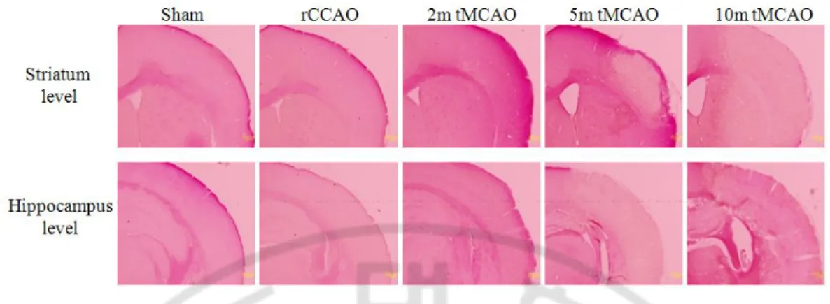
Fig. 2. A pilot experiment for induction of mild ischemia. Various ischemic conditions
were applied in female C57BL/6 mice (weighing around 20g), which were sacrificed 7 days after surgery. Acid fuchsin-stained sections showed no cerebral infarction in mice with rCCAO and those with 2m tMCAO whereas evident gross cerebral infarction was induced in mice with 5m or 10m tMCAO (magnification, x40).
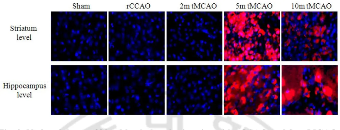
Fig. 3. No breakdown of blood brain barrier in mice with rCCAO and 2m tMCAO.
To evaluate status of the blood brain barrier in various ischemic conditions, Evan’s blue dye was infused during mice sacrificed (red, Evan’s blue; blue, DAPI; magnification, x200). The blood brain barrier was intact in mice with rCCAO or 2m tMCAO while it was impaired in those with 5m or 10m tMCAO.
Fig. 4. Decreased CBF after occlusion of cerebral arteries. Cerebral blood flow (CBF)
was measured in the ipsilateral hemisphere of mice with rCCAO (n = 3), 5m tMCAO (n = 7) and 10m tMCAO (n = 5) with laser Doppler flowmetry. About 40% decrease of CBF was shown in rCCAO-operated mice. About 70% decrease of CBF was observed after MCAO. Even after MCA was recanalized, the low CBF was lasted for 60 minutes.
C B F ( % o f B a se lin e )
Time after MCA recanalization
Pre-rCCAO Post-rCCAO Post-MCAO 5m 60m
0 20 40 60 80 100 120 rCCAO only 5m tMCAO 10m tMCAO
B. Replication study for a vascular cognitive impairment model induced by rCCAO
1. A lower recognition of novel object in rCCAO-operated mice
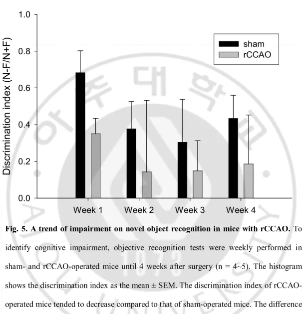
To evaluate cognitive changes after rCCAO, serial objective recognition tests were performed from week 1 to week 4 after surgery. The objects to be discriminated were made of acryl and of two different shapes and colors: 2 cubes (black) and 1 pyramid (red) of 12 cm height and 6 cm width. The object recognition test was performed before surgery, and the mice with a discrimination index less than 0.10 were excluded. Among 60 mice, 33 remained up to the final tests. On week 1, the discrimination index was lower in rCCAO-operated mice than in sham-operated mice, and the difference was marginally significant (discrimination index; 0.35 ± 0.08 for rCCAO vs 0.68 ± 0.12 for sham, p = 0.050) (Fig. 5). The index from week 2 to week 4 tended to be low in rCCAO-operated mice as compared to sham-operated mice, but the difference was not statistically significant (discrimination index in week 2; 0.14 ± 0.39 for rCCAO vs 0.38 ± 0.15 for sham, p = 0.595; week 3; 0.15 ± 0.16 for rCCAO vs 0.30 ± 0.23 for sham, p = 0.605; week 4; 0.19 ± 0.27 for rCCAO vs 0.43 ± 0.13 for sham, p = 0.434) (Fig. 5). Statistical significance of the difference in the discrimination index was maximal at week 1.
Fig. 5. A trend of impairment on novel object recognition in mice with rCCAO. To
identify cognitive impairment, objective recognition tests were weekly performed in sham- and rCCAO-operated mice until 4 weeks after surgery (n = 4~5). The histogram shows the discrimination index as the mean ± SEM. The discrimination index of rCCAO-operated mice tended to decrease compared to that of sham-rCCAO-operated mice. The difference was marginally significant at 1 week after surgery (p = 0.050), but the index was not significantly different from 2 weeks to 4 weeks after surgery.
Week 1 Week 2 Week 3 Week 4
D
is
cr
im
in
at
io
n
in
d
ex
(
N
-F
/N
+
F
)
0.0 0.2 0.4 0.6 0.8 1.0 sham rCCAO2. Occasional apoptotic cell death observed 7 days after rCCAO
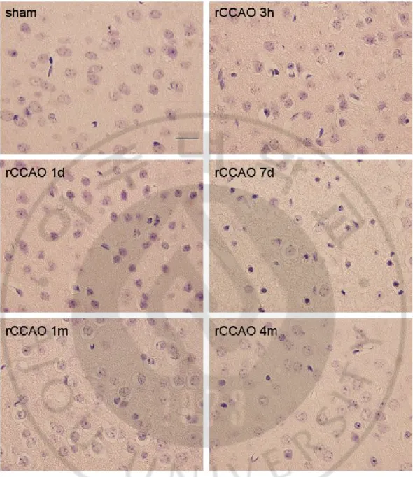
From 3 hours to 4 month after surgery serial histological analyses were performed in order to estimate whether neuronal death would be involved in rCCAO-operated mice. No evident neuronal necrosis was found on hematoxylin and eosin staining (Fig. 6). TUNEL- and caspase-3-positive cells which represented apoptotic neuronal damage were observed in one of three rCCAO-operated mice 7 days after surgery (Fig. 7 and 8). These histological changes appeared mild but were clearly evident. On the other hand, these changes were mostly observed on the core area which was revealed in mice with 5m or 10m tMCAO. The corpus callosum was not related to the apoptotic changes. These findings suggested that a small portion of rCCAO-operated mice may have some neuronal death via apoptotic pathway.
Fig. 6. No necrotic cells in rCCAO-operated mice. Sections of the right sensory cortex
were stained by hematoxylin and eosin (magnification, x1000; bar, 20 μm). Eosin-stained cells were not found in rCCAO-operated mice. Note small purple cells increased in one of three rCCAO-operated brains 7 days after surgery.
Fig. 7. TUNEL-positive cells in one rCCAO-operated mouse 7 days after surgery.
Sections of the right sensory cortex were stained by TUNEL (magnification, x1000; bar, 20 μm). TUNEL-positive cells were not found in most rCCAO-operated mice. Note that TUNEL-positive cells were gathered in one of three rCCAO-operated brains 7 days after surgery.
Fig. 8. Representative photograph of apoptotic cell death observed in an rCCAO-operated mouse 7 days after surgery. Immunohistochemical staining for activated
caspase-3 was performed in sham- and rCCAO-operated mice (right sensory cortex; magnification, x200). No significant findings were revealed in most rCCAO-operated mice. One mouse which was shown in the TUNEL staining sacrificed 7 days after rCCAO had cleaved caspase-3-positive apoptotic cells.
3. Hypometabolism in FDG microPET for an rCCAO-operated mouse
A pilot study was performed to evaluate chronic metabolic status in the brain of rCCAO-operated mouse 4 months after surgery. FDG was used as a radioactive isotope for microPET imaging. The glucose metabolism appeared to decrease in the right hemisphere (Fig. 9) and the location of metabolic deficit was accordant with the area in which the cerebral infarction was induced in mice with 5m or 10m tMCAO.
Fig. 9. Hypometabolism in the right cortex of an rCCAO-operated mouse. On the
coronal view of FDG microPET for an rCCAO-operated mouse brain with 4-month duration after surgery, metabolic deficit compared to the contralateral side was shown in the right hemisphere in which the core area was revealed in mice with 5m or 10m tMCAO.
C. Tg 2576 treated with chronic mild ischemia
1. Consequences of surgery
A total of 90 mice which consisted of Tg2576 mice (Tg+) and their littermates (Tg-) were used in this study (Table 1). Since rCCAO and 2m tMCAO in mice brought about hypoperfusion without gross infarction in our preliminary studies, they were used for induction of CCH. Among Tg- mice, 8 were used for sham-operation, 9 for rCCAO and 17 for 2m tMCAO. Among Tg+ mice, 12 were used for sham-operation, 13 for rCCAO and 21 for 2m tMCAO. All sham- and rCCAO-operated mice were survived but many mice with 2m tMCAO were dead during experimental period. Mortality rate of Tg+ 2m tMCAO group was 81.0% whereas that of Tg- 2m tMCAO group was 17.6% which is generally tolerable ratio as for tMCAO-operation in mice. The rate was significantly different between Tg- and Tg+ 2m tMCAO groups (χ2 test, p < 0.001). High mortality in our mice with 2m tMCAO might be related to cerebral amyloid angiopathy which is mostly shown in middle to old Tg2576 mice. It is known that cerebral amyloid angiopathy is a common cause of brain hemorrhage. Almost all Tg2576 mice revealed thioflavin S-stained vessels on their brain sections (Fig. 10). Because of the high mortality rate, behavioral data of both 2m tMCAO groups were excluded in the following analyses for comparison.
Table 1. Mortality after surgery.
Group Total (n) Survived (n) Dead (n) Mortality (%)
Tg- Sham 8 8 0 0 rCCAO 9 9 0 0 2m tMCAO 17 14 3 17.6* Tg+ Sham 12 12 0 0 rCCAO 13 13 0 0 2m tMCAO 21 4 17 81.0* * χ2 test, p < 0.001
Fig. 10. Cerebral amyloid angiopathy observed in Tg2576 mice. A Tg+ mouse with
thioflavin S staining shows amyloid-contained vessels as well as amyloid plaques (magnification, x200). Arrows indicate amyloid-contained vessels.
2. Behavioral tests
(1) Morris water maze test
Morris water maze test was performed 7-8 weeks after surgery to evaluate learning and memory. When we assessed the latency to escape to a hidden platform in the water maze, we observed a significant difference in the latency among the groups (repeated measures ANOVA, F = 8.700, p < 0.001) (Fig. 11). This latency to escape decreased in the Tg+ mice with or without rCCAO compared to the Tg- sham mice (post hoc, Bonferroni’s test; Tg+ sham vs Tg- sham, p = 0.008; and Tg+ rCCAO vs Tg- sham, p = 0.001). The escape performance was not statistically different between the Tg+ rCCAO and Tg+ sham group (post hoc, Bonferroni’s test, p = 1.000); however, the Tg+ rCCAO group showed an impairment of the learning curve (repeated measures ANOVA for a session analysis of the Tg+ rCCAO group, F = 0.884, p = 0.420) whereas the Tg+ sham group had a preserved learning curve (repeated measures ANOVA for a session analysis of the Tg+ sham group, F = 6.647, p = 0.003). A probe test showed a statistical difference among the groups in terms of the time spent in the target quadrant in which the platform had been removed (mean time spent in the target quadrant (s) ± SEM; Tg- sham, 15.39 ± 1.25; Tg- rCCAO, 14.00 ± 1.80; Tg+ sham, 8.94 ± 1.92; Tg+ rCCAO, 7.66 ± 2.33; one was ANOVA, F = 3.500, p = 0.025) (Fig. 12). The memory retention of Tg+ mice was more impaired than the Tg- mice (post hoc, t-test; Tg+ sham vs Tg- sham, p = 0.017; Tg+ rCCAO vs Tg- sham, p = 0.021). However, it was not different between Tg+ rCCAO- and sham-operated mice (post hoc, Scheffe’s test; Tg+ rCCAO vs Tg+ sham, p = 0.972).
Tg- Tg+ T im e in t he t ar ge t q ua dr an t (s ) 0 2 4 6 8 10 12 14 16 18 sham rCCAO p < 0.05
B
Fig. 11. Impairment of learning curve on Morris water maze test in Tg+ rCCAO-operated mice. A Morris water maze test indicates that the latency to escape to the
platform was prolonged in the Tg+ mice compared to the Tg- mice (repeated measures ANOVA, F = 8.700, p < 0.001) but it was not different between Tg+ rCCAO- and sham-operated mice (post hoc, Bonferroni’s test, p = 1.000). To evaluate a presence of learning ability, additional repeated measures ANOVAs for a session effect of single group were performed. The Tg+ rCCAO-operated mice had an impaired learning ability (* F = 0.884, p = 0.420) whereas the Tg+ sham-operated mice showed a preserved learning curve (** F = 6.647, p = 0.003). Blocks of 3 trials 1d 2d 3d 4d 5d L a ta n cy ( s) 20 30 40 50 60 70 Tg- sham Tg- rCCAO Tg+ sham Tg+ rCCAO
*
**
p < 0.05Fig. 12. Impairment of memory retention in Tg+ mice. A probe test showed that the
time spent in the target quadrant decreased in the Tg+ mice compared to the Tg- mice (ANOVA, F = 3.500, p = 0.025) but it was not different between Tg+ rCCAO- and sham-operated mice (post hoc, Scheffe’s test; Tg+ rCCAO vs Tg+ sham, p = 0.972).
Tg- Tg+ T im e in t h e ta rg e t q u ad ra n t (s ) 0 2 4 6 8 10 12 14 16 18 sham rCCAO p < 0.05
(2) Object recognition test
We evaluated the non-spatial working memory by the object recognition test 6 weeks after surgery. The discrimination index was significantly different among the groups (mean of the discrimination index ± SEM; 0.78 ± 0.10 for Tg- sham, 0.03 ± 0.20 for Tg- rCCAO, 0.42 ± 0.20 for Tg+ sham, and -0.01 ± 0.19 for Tg+ rCCAO; one way ANOVA, F = 3.269, p = 0.035) (Fig. 13). The Tg- or Tg+ rCCAO-operated mice had low index compared to that of the Tg- sham mice (post hoc, t-test, Tg- rCCAO vs Tg- sham, p = 0.005; Tg+ rCCAO vs Tg- sham, p = 0.007). There was no significant difference between the Tg+ rCCAO and Tg+ sham mice (post hoc, t-test; p = 0.146). It might be result from the relatively decreased baseline index of the Tg+ sham mice even though the indices were not significantly different between the Tg+ sham and Tg- sham group (post hoc, t-test; p = 0.202).
Discrimination index -0.4 -0.2 0.0 0.2 0.4 0.6 0.8 1.0 Tg+ Tg-rCCAO sham p < 0.05
Fig. 13. Decreased discrimination index on rCCAO-operated mice. The object
recognition test was performed 6 weeks after surgery. The index of both Tg+ and Tg- rCCAO-operated mice significantly decreased when it was compared to the Tg- sham mice (one way ANOVA, F = 3.269, p = 0.035: post hoc, t-test, Tg- rCCAO vs Tg- sham, p = 0.005; Tg+ rCCAO vs Tg- sham, p = 0.007).
3. Histopathological tests
(1) Cellular features
Mice were sacrificed 8 weeks after surgery for histological studies. No cell death was observed in the brain sections of the Tg+ rCCAO and Tg- rCCAO mice when they were examined with acid fuchsin (Fig. 14) and TUNEL staining (Fig. 15). Axonal or demyelinating damage on white matter including corpus callosum was not found in sections stained with pan-axonal neurofilament (Fig. 16) or Luxol fast blue (Fig. 17). The extent of free radicals and inflammation did not seem to be different examined by 8-OHdG and Iba-1 staining, respectively (data not shown).
(2) Amyloid plaques
Amyloid plaques were detected with thioflavin S fluorescence staining and plaque burden on the right brain sections was quantified. There was no statistical difference in the plaque burden between the Tg+ rCCAO and Tg+ sham mice (mean area ± SEM; sham, 39971.35 ± 10380.34 μm2 vs rCCAO, 39023.56 ± 11797.44 μm2; t-test, p = 0.952) (Fig. 18).
Fig. 14. No evident necrotic cell death in the brain sections. Right hemisphere
including the cortex and hippocampus was examined by acid fuchsin staining 8 weeks after surgery (coronal section at the level of the hippocampus, magnification x40, bar = 500 μm). Cerebral infarction or necrotic neuronal death was not observed regardless of rCCAO operation.
Fig. 15. No evident apoptotic cell death in the brain sections. Right cortex was
examined by TUNEL assay 8 weeks after surgery (coronal section at the level of the hippocampus, magnification x200, bar = 100 μm). Apoptotic neuronal death was not observed regardless of rCCAO operation.
Fig. 16. No axonal injury in the brain sections. Right medial corpus callosum was
immunostained with monoclonal antibody to pan-axonal neurofilament 8 weeks after surgery (coronal section at the level of the hippocampus, magnification x400, bar = 50 μm). Axonal damage was not observed in the corpus callosum.
Fig. 17. No myelin damage in the brain sections. Right corpus callosum was examined
by Luxol fast blue staining 8 weeks after surgery (coronal section at the level of the hippocampus, magnification x200, bar = 100 μm). Demyelinating damage was not observed in the corpus callosum.
Tg+ sham Tg+ rCCAO B ur d en o f a m yl oi d p la q ue ( u m 2 ) 0 10000 20000 30000 40000 50000 60000
Fig. 18. No increased burden of amyloid plaques in the Tg+ mice with rCCAO.
Amyloid plaques stained with thioflavin S were frequently observed in the cortex and hippocampus 8 weeks after surgery. The plaque burden in the right hemisphere was not different between Tg+ sham- and rCCAO-operated mice (t-test, p = 0.952).
IV. DISCUSSION
A. Cognitive deterioration induced by rCCAO in Tg2576 mice
Alzheimer- and VCI-type cognitive impairments were observed in the Tg+ mice and rCCAO model, respectively. In addition, we found an impairment of learning curve in Tg+ rCCAO-operated mice. From the clinical point of view, cognitive manifestations between patients with AD and patients with vascular dementia are somewhat different. In the early stages, episodic memory is impaired in most patients with AD, whereas a memory deficit is usually mild in patients with VCI (Looi and Sachdev, 1999). Frontal lobe syndrome such as attention deficit and impairment on working memory is more common in patients with VCI than in those with AD (Erkinjuntti, 1987; Kertesz and Clydesdale, 1994; Looi and Sachdev, 1999). In summary, patients with AD have hippocampal dysfunction while patients with VCI have dysfunction on the frontal-subcortical circuit. In this animal study, the Tg+ rCCAO-operated mice exhibited both these distinct characteristics of AD and VCI. Moreover, a learning impairment became evident after rCCAO surgery in Tg+ mice. Tg2576 mouse is a good Alzheimer mouse model in that amyloid plaques are induced and cognitive deficit accompany the pathological changes. However, a learning ability is preserved even though the mice show impaired performance on the escape latency of Morris water maze test (Routtenberg, 1997). Several reports showed that learning curves of Tg+ mice were intact even though the mice were old (Hsiao, et al., 1996; Westerman, et al., 2002). We had similar results in that Tg+ mice without rCCAO revealed lower performance in the escape latency but
showed a preserved learning ability. However, Tg+ mice had an impairment of the learning curve in case that the mice were rCCAO-operated. It is thought that the learning impairment might result from a synergistic effect from both vascular and Alzheimer pathomechanisms because each single abnormality did not cause the learning impairment. Therefore, there must be more than coexistence between AD and VCI. It can be hypothesized that CCH deteriorates cognitive abnormality of AD or vice versa.
Our study failed to demonstrate an aggravating effect of CCH on the amyloid plaque burden. Some previous studies suggested that Aβ burden is associated with ischemia. The size of the cerebral infarction was enlarged in FVB/N mice expressing the amyloid precursor protein with Swedish mutation compared to control mice when the middle cerebral artery was occluded (Zhang, et al., 1997). Amyloid precursor protien/presenilin 1 Tg mice with a transient occlusion of both CCA’s for 45 minutes had a greater cognitive deficit in the Morris water maze test compared to sham-operated mice (Sadowski, et al., 2004). In these 2 studies, amyloid beta (Aβ) deposition tended to increase in the TG mice with CCAO; however, statistical significance was not observed (Zhang, et al., 1997; Sadowski, et al., 2004). On the other hand, Tg- rats with permanent bilateral CCAO had increased levels of Aβ compared to sham-operated rats (Zhiyou, et al., 2009). The Morris water maze test showed an accompanying cognitive impairment in the bilateral CCAO-operated rats. These studies suggested that cerebral hypoperfusion might lead to deterioration of cognitive function and the pathological condition, e.g., Aβ deposition and amyloid plaque formation. However, CCH induced by rCCAO for up to 8 weeks had little effect on pathology of Tg+ brains. First, it is thought that the hypoperfusion was too weak to increase amyloid plaques. In our preliminary study apoptosis was shown in Tg- mice
with rCCAO; however, it was revealed only in one mouse brain at 7 days after surgery (Lee, et al., 2009). Second, 8 weeks might be too short to induce amyloid plaque formation from chronic hypoperfusion in mice with rCCAO. Amyloid plaque is considered to increase about 3 months after Aβ burden has grown up. Further study to maintain CCH for longer duration in Tg mice is warranted. From our experiences and a review of literatures, rCCAO for 8 weeks appears to induce an ischemic insult too mild and too short to cause histological change. These two reasons can explain why the additional rCCAO insult to the Tg2576 mice did not lead to an increase of amyloid plaque deposition.
The presence of cognitive impairment with little neuronal death in rCCAO-operated mice was questioned. There could be two possible explanations. First, an early ischemic insult might cause neuronal damage, which is very subtle but has permanent consequences. Our preliminary study identified a small amount of apoptotic cells at 7 days after rCCAO surgery in Tg- mice (Lee, et al., 2009). The findings were mild, and they were observed in only a small proportion of mice; in addition, the changes were not evident after 1 month. An early ischemic insult could be a causal factor for the induction of VCI. If there was a greater amount of apoptosis, rarefaction might be observed in the chronic phase after rCCAO. Second, cerebral hypometabolism can be induced by rCCAO. Regardless of neuronal death, dendritic arborization and synaptic contact might be affected (Farkas, et al., 2007). A decline in the levels of the microtubule-associated protein 2, which plays a role in dendritic branching, remodeling, or plasticity, and a reduction in the levels of the synaptophysin protein, which plays a role in synaptic conditioning, have been reported to be associated with CCH (Liu, et al., 2005; Thiel,
1993). These factors might affect cognitive dysfunction without evident neuronal death with a status between benign oligemia and ischemic penumbra. Regardless of a pilot study nature, our PET study supported this assumption. Tg- mice with rCCAO which showed dissociation between cognitive impairment and absence of significant neuronal death did have hypometabolism in the right cortex on FDG microPET study.
Tg+ 2m tMCAO group had great mortality rate compared to Tg- 2m tMCAO group. The brains of dead Tg+ mice could not be easily examined since those got rapidly rotten. Nonetheless, cerebral amyloid angiopathy shown in Tg2576 mice can be explained for those phenomena. Cerebral amyloid angiopathy is known to be associated with brain hemorrhage. Growing amount of amyloid is contained into cerebral vessels as the mice get old (Kawarabayashi, et al., 2001; Westerman, et al., 2002). Second, Tg+ mice are susceptible to ischemic insult. When MCA is occluded for 24 hours, cerebral infarction was larger in Tg+ mice than in Tg- mice (Zhang, et al., 1997). However, this possibility is less likely in our experiments because neuronal death was not caused in survived mice with 2m tMCAO.
In summary, our results can infer that mixed dementia can be more than AD plus VCI. Tg+ rCCAO-operated mice had both cognitive deficits on spatial memory and non-spatial working memory induced in AD and VCI models, respectively. Moreover, an impairment of learning curve was synergistically induced from both pathomechanisms.
B. Vascular cognitive impairment induced by rCCAO
Cognitive impairment observed in rCCAO-operated mice might be associated with vascular damage in the cortical-subcortical circuit. It has been suggested that in human patients with subcortical ischemic vascular dementia, the area vulnerable to hypoperfusion is associated with impairment of the cortical-subcortical circuit (Román, 2004). Among patients with white matter lesions, those who had cognitive impairment showed decreased levels of regional CBF as compared to those without cognitive impairment (Kato, et al., 2008). Similarly, animal models of cerebral hypoperfusion showed behavioral change that reflects damage to the cortical-subcortical circuit. In one study, spatial working memory examined by an eight-arm radial maze test was selectively impaired in a mouse model with bilateral CCA stenosis (Shibata, et al., 2004). In another study, nonspatial working memory examined by the object recognition test was impaired in a rat model with bilateral CCA occlusion (Sarti, et al., 2002).
In our study for rCCAO mouse model, dissociation between abnormality on non-spatial working memory and absence of significant neuronal damage was also shown like the previous study. The rCCAO in mice has been proposed as a model to bring about VCI via CCH without acute lesion (Yoshizaki, et al., 2008). The authors suggested that the cognitive impairment was associated with decreased fiber density on the medial corpus callosum. However, our results contradict that study. Damage or rarefaction on the corpus callosum was not observed in our study. Instead, apoptotic cells were observed in some rCCAO-operated mice in the early phase, and this feature was prominent on the watershed zone of the right CCA. The cortical area was similarly involved in another
mouse model, in which cognitive impairment was induced by varying degrees of bilateral CCA stenosis using microcoils (Miki, et al., 2009). Moderate stenosis on the right CCA and then severe stenosis on the left CCA were induced. The model revealed that cerebral infarction was maximal in the cortex and rarefaction was observed in the hippocampus and corpus callosum. In addition, our pilot microPET study for an rCCAO-operated mouse showed a defect of glucose metabolism in the same area. Regardless of the contradiction, it is thought that the impairment on non-spatial working memory in our study was also related to damage on subcortical circuit. Damage to the cortical-subcortical circuit may indicate any cortical lesion in the circuit as well as white matter lesion such as the corpus callosum. Hypoperfusion injury on the watershed area of unilateral CCA may affect the cortical-subcortical circuit. VCI induced by rCCAO in our study is possibly associated with hypoperfusion to the area.
An early ischemic insult from rCCAO seems to affect the cognitive changes apart from the chronic effect of cerebral hypoperfusion. In our serial object recognition tests, the difference in performance between the rCCAO- and sham-operated mice was significant at the earliest point, i.e., 1 week after surgery. It is logical to postulate that blood flow to the brain is suddenly disturbed in the course of surgery for rCCAO. Then collateral circulations, primarily abetted by the Circle of Willis, develop quickly, and hypoperfusion recovers to some extent in the chronic stage (Tamaki, et al., 2006). On the other hand, a tendency of decreased cognition in rCCAO-operated mice at chronic stage might be affected under sustained oligemic status due to rCCAO. There was a mismatch in our results in that cognition was impaired but neuronal death was not observed. Injury due to rCCAO seemed to be relatively mild because neuronal death was not observed at
chronic stage. Hypometabolism, which was shown in an rCCAO-operated mouse 4 months after surgery, can be a possible explanation for the mismatch. Our results suggest the ipsilalateral brain in rCCAO-operated mice is in the status between benign oligemia and ischemic penumbra.
C. A review of vascular cognitive impairment
VCI, which has a wide range of cognitive levels, is a syndromatic term encapsulating abnormal cognition and vascular etiology in the brain (Bowler, et al., 1999; O’Brien, et al., 2003; Román, et al., 2004). Cerebrovascular disease is highly prevalent in old people, so the accompanying cognitive impairment is very common (O’Brien, et al., 2003). VCI is found at a high frequency in Asian populations (Ikeda, et al., 2001) compared to Western populations (Rockwood et al., 2000). Vascular dementia was more commonly than AD in persons with dementia aged 65 years and older in Japan (Ikeda, et al., 2001), whereas AD was found to be more frequent than VD in a Canadian study (Rockwood et al., 2000).
1. Subtypes of VCI
VCI encompasses various levels of cognitive decline and includes several types (Fig. 13). Severe vascular dementia with impaired activities of daily living is one extreme, while VCI with no dementia or vascular mild cognitive impairment represent the other extreme. Amongst the subtypes of VCI, multi-infarct dementia was the first to be identified (Hachinski et al., 1974) (Fig. 19A); it is also known as poststroke vascular dementia or cortical vascular dementia (Erkinjuntti and Gauthier, 2009). A focal neurological deficit with an abrupt onset and a stepwise deterioration of cognition are characteristic of multi-infarct dementia. Cortical infarction with or without concomitant subcortical infarction and watershed zone infarction are mainly involved (Erkinjuntti and Gauthier, 2009). Its prevalence is not thought to be higher than any other subtype of VCI
(O’Brien, et al., 2003).
There is a growing interest in subcortical ischemic vascular dementia (SIVD) because white matter lesions are frequently found on an MRI and SIVD is associated with cognitive impairment. SIVD also has various subtypes, but Binswanger’s disease (Fig. 19C) and the lacunar state (Fig. 19D) are representative examples (Román, et al., 2002). A common risk factor of SIVD is hypertension resulting in arteriosclerosis. Binswanger’s disease is related to chronic hypoperfusion revealed by white matter hyperintensities on an MRI, whereas the lacunar state is related to multiple lacunar infarctions of the white matter (Román, et al., 2002).
VCI with no dementia is included in the subtypes of VCI as an evolving entity. It is regarded as the mildest stage of VCI and it is comparable to mild cognitive impairment which is regarded as a prodrome of AD (Román, et al., 2004; Stephan et al., 2009). There is no deterioration in the quality of life of patients with dementia (Román, et al., 2004). As mild cognitive impairment can progress to AD, it is thought that the milder subtypes of VCI prevent further progression to obvious vascular dementia.
Fig. 19. Brain lesions of VCI subtypes. (A) Multi-infarct dementia, (B) strategic infarct