Int J Clin Exp Pathol 2015;8(7):7998-8007 www.ijcep.com /ISSN:1936-2625/IJCEP0009622
Original Article
Squash smear cytology of Langerhans cell histiocytosis
Ji Hae Nahm1*, Gun Yoon2*, Sung-Im Do3, Hyun-Soo Kim11Department of Pathology, Severance Hospital, Yonsei University College of Medicine, Seoul, Republic of
Korea; 2Department of Gynecology and Obstetrics, Pusan National University Yangsan Hospital, Pusan National
University School of Medicine, Yangsan, Republic of Korea; 3Department of Pathology, Kangbuk Samsung
Hospital, Sungkyunkwan University School of Medicine, Seoul, Republic of Korea. *Equal contributors. Received April 26, 2015; Accepted June 22, 2015; Epub July 1, 2015; Published July 15, 2015
Abstract: Squash smear cytology of Langerhans cell histiocytosis (LCH) has rarely been reported. We described squash cytological findings of cranial LCH. Additionally, based on recent data that suggests an association of LCH with either viral infection or genetic alteration, we investigated the presence of several viruses or mutation of TP53 and BRAF in LCH tissue samples. Intraoperative squash smears of a small tissue fragment excised from the lesion demonstrated a mixed population of eosinophils, neutrophils, small lymphocytes and a high content of histiocytes. The histiocytes possessed abundant dense cytoplasm with round cell shape and eccentrically located nuclei with fine chromatin, delicate nuclear membranes and prominent nuclear grooves, indentations and pseudoinclusions. The cytologic features were consistent with Langerhans cells (LCs). Subsequent histopathologic examination con-firmed the diagnosis of LCH. Immunohistochemically, the LCs were positive for S-100, CD1a and langerin, but nega-tive for adenovirus, CMV, EBV, HHV-8, HPV, HSV, SV 40 and p53. BRAF V600E mutation was absent. Our findings did not support the role of viruses and genetic abnormalities in the pathogenesis of LCH. In summary, the presence of a mixed population of inflammatory cells and a high content of histiocytes with characteristic cytomorphology, along with radiologic evidence and appropriate clinical findings, is highly suggestive of LCH on the intraoperative squash smears. Awareness of characteristic cytological features of LCH is necessary for rapid and accurate diagnosis. Squash smear cytology is a potentially useful tool in the intraoperative diagnosis of LCH.
Keywords: Langerhans cell histiocytosis, squash cytological preparation, virus, Epstein-Barr virus, TP53, BRAF
Introduction
Langerhans cell histiocytosis (LCH) is a rare dis-ease that accounts for less than 1% of all tumor-like lesions of bone mainly affecting the skull, femur, mandible and ribs [1]. In the past, the disease was referred to as histiocytosis X with 3 variants: eosinophilic granuloma, Hand-Schuller-Christian disease and Letterer-Siwe syndrome that represent different expressions of the same disease, now known as LCH [2]. There is a wide spectrum of clinical manifesta-tions of LCH that depends on the location and extent of the proliferation of Langerhans cell (LCs). LC is a major type of nonlymphoid mono-nuclear cell involved in both inflammatory responses and neoplastic processes. There is an ongoing debate on whether the pathogene-sis of LCH includes a reactive or neoplastic pro-cess. Inflammatory reaction to viral infection is reportedly the potential etiology at disease onset [3-7]. However, no convincing evidence
has emerged for an infectious cause of LCH. There are several reports on immune dysregula-tion and aberrant cytokine expression in patients with LCH, indicating that the nature of LCH is predominantly immune or inflammatory [8-10]. However, the role of cytokines and immune system regulators in the pathogenesis of LCH remains controversial. Meanwhile, the discovery of frequently recurring oncogenic
BRAF mutations in LCH, along with TP53
muta-tion, supports the classification of this disease as a neoplasm [11]. However, there is also a debate about the source of pathologic LCs in LCH lesions. Furthermore, the presence of BRA For TP53 mutation alone may not be sufficient for tumorigenesis. Even though the identifica-tion of genetic alteraidentifica-tions in LCH strongly sup-ports the hypothesis of its neoplastic nature, several questions remain unanswered.
Squash smear cytology is of great value in intra-operative pathologic consultations. It provides
a rapid and reliable intraoperative diagnosis and guidance to the surgeon during surgical resection and lesion targeting. It also helps the surgeon to monitor and modify the surgical approach. Using a very small amount of mate-rial, this technique makes it possible to detect cellular details and establish a provisional pathologic diagnosis [12]. In published series and case reports of LCH, its diagnosis has been established using routine histopathologic methods [4, 12-14]; the morphology of LCH is well recognized in processed biopsy or excised material. In contrast, the squash cytology description of LCH is limited. Knowledge of its cytomorphology, nevertheless, is of diagnostic importance [12, 15-17]. The appearance on squash smears has certain characteristic and diagnostic features. We described squash smear cytological findings for rapid and correct intraoperative diagnosis of cranial LCH in chil-dren. The findings of this study will increase the
accuracy of the pathologists’ cytological diag-nosis and guide appropriate management. Additionally, we investigated a possible rela-tionship between LCH and viral infection or
TP53 and BRAF mutation. The presence and
cellular localization of viruses, as well as p53 expression status, was examined using immu-nohistochemical staining and in situ hybridiza-tion. The presence of BRAF V600E mutation was also examined using polymerase chain reaction (PCR).
Case presentation
Case 1
A 12-year-old boy presented to the neurosur-gery outpatient department with a painless swelling of the frontal part of the head of 6 weeks duration. The lesion was not accompa-nied by fever or tenderness. There was no
his-Figure 1. Imaging findings of LCH. Case 1: (A) A plain skull roentgenogram revealed a punched-out osteolytic lesion in the mid-portion of frontal bone (white arrow). (B) A sagittal view of T1-weighted TFE MR image showed a well-demarcated enhancing lesion (white arrow), which abuts the meninges. (C) An axial view of T2-weighted TSE MR im-age revealed that the mass (white arrow) involved the inner table of skull bone. Case 2: A plain skull roentgenogram (D, anteroposterior view; E, lateral view) revealed a well-demarcated osteolytic lesion in the left frontal bone (white arrow). (F) CT scan showed a localized osteolysis (white arrow), involving the inner and outer tables.
Squash cytomorphology of LCH
tory of trauma, headache or vomiting. Local examination revealed a firm and slightly mobile swelling of approximately 1.5 cm in diameter over the upper forehead. The overlying skin appeared normal. Systemic examination was otherwise unremarkable, and the patient was neurologically normal. He had no lymphade-nopathy, organomegaly, skin rash or any other abnormality. Complete routine laboratory tests including hematological and biochemical inves-tigations revealed no abnormality. A plain skull roentgenogram revealed a sharply demarcat-ed, punched-out osteolytic lesion in the most anterior midline point on the frontal bone, approximately 3 centimeters above the glabella (Figure 1A). Sagittal (Figure 1B) and axial (Figure 1C) view of magnetic resonance imag-ing (MRI) scan demonstrated an osteolytic lesion in the frontal bone. Surgical excision with intraoperative frozen section and squash cyto-logical preparations was performed. Subse- quent permanent section diagnosis, followed by immunohistochemical staining, Epstein-Barr virus (EBV)-encoded RNA in situ hybridization (EBER-ISH) and analysis for BRAF V600E muta-tion detecmuta-tion, was also performed. The prima-ry cytological diagnosis of LCH was made using squash preparations, and confirmed by the fro-zen and permanent sections. The postopera-tive course was uneventful, and the patient left the hospital 5 days later. Fourteen months after surgery, MRI scan showed no evidence of recurrence. Whole body positron emission tomographic scan showed no abnormal hyper-metabolic lesion.
Case 2
A 6-year-old girl was referred to our hospital presenting with painful swelling over the left
side of forehead for a month. There was no his-tory of trauma, headache or vomiting. On exam-ination, a fluctuant, ill-defined soft tissue mass of approximately 2 cm in diameter was identi-fied on the left frontal region. The underlying bone appeared to be indented on palpation, and the overlying skin appeared normal. She had no significant past medical history. Lymphadenopathy or skin rash was not found. The liver and spleen were not palpable. Other physical examination and laboratory tests were unremarkable. A plain skull roentgenogram (Figure 1D, anteroposterior view; Figure 1E, lat-eral view) revealed a well-demarcated osteo-lytic lesion in the left frontal bone. Computed tomographic (CT) scan also showed a localized osteolysis of the frontal bone with erosion of the inner and outer tables, along with suspect-ed involvement of the adjacent soft tissue (Figure 1F). As in Case 1, surgery with intraop-erative frozen section and squash smears was performed. The primary cytological diagnosis of LCH was further confirmed by the tissue sec-tions. The ancillary tests, including immunohis-tochemistry, EBER-ISH and BRAF V600E muta-tional analysis, were performed. The postoper-ative course was uneventful, and the patient left the hospital 4 days later. No recurrence over the last 6 months was apparent.
Materials and methods
Squash cytological preparation
For squash cytology, biopsy samples obtained at the time of surgery were transported imme-diately in small vials containing isotonic saline for processing in the pathology departments’ cryolaboratory. One to 2 millimeters of the biop-Table 1. Antibodies used for immunohistochemical staining
Antibody Source Clone Dilution
Adenovirus Millipore Corporation, Bedford, MA, USA 20/11 & 2/6 1:500 CD1a DakoCytomation, Glostrup, Denmark 10 1:50 CD3 DakoCytomation, Glostrup, Denmark Polyclonal 1:200 CD20 Novocastra Laboratories, Newcastle upon Tyne, UK L26 1:400 CMV DakoCytomation, Glostrup, Denmark CCH2/DDG9 1:40 HHV-8 Novocastra Laboratories, Newcastle upon Tyne, UK 13B10 1:50 HPV DakoCytomation, Glostrup, Denmark K1H8 1:100 HSV Novocastra Laboratories, Newcastle upon Tyne, UK 20.7.1 1:3,000 p53 Invitrogen, Carlsbad, CA, USA BP53.12 1:5,000 S-100 DakoCytomation, Glostrup, Denmark Polyclonal 1:5,000 SV 40 Calbiochem, Merck KGaA, Darmstadt, Germany PAb416 1:100
sy material were crushed between 2 glass slides with just enough pressure to spread the tissue into a thin film; the smear was subse-quently fixed in 95% alcohol and stained by hematoxylin and eosin. The residual tissue sample was stored at -20°C or fixed in 10% for-malin solution and embedded in the paraffin block. Formalin-fixed, paraffin-embedded sec-tions were stained with hematoxylin and eosin for light microscopy.
Immunohistochemistry
Immunohistochemical staining was performed by using standard reagents and techniques, as previously described [18-24]. Briefly, the sec-tions were deparaffinized with Bond Dewax solution (Leica Biosystems, Bannockburn, IL, USA) and an antigen retrieval procedure was
performed using Bond Epitope Retrieval solu-tion 1 (Leica Biosystems) for 30 min at 100°C. Endogenous peroxidases were blocked by incu-bation with hydrogen peroxide for 5 min and the sections were incubated for 15 min at ambient temperature with primary antibodies (Table 1). Staining was performed using a biotin-free polymeric horseradish peroxidase-linker anti-body conjugate system with a Leica Bond-Max automated immunostainer (Leica Microsys- tems), and the slides were visualized with 3, 3’-diaminobenzidine (DAB) solution (1 mm DAB, 50 mm Tris-HCl buffer [pH 7.6] and 0.006% H2O2). In addition, the nuclei were counter-stained with hematoxylin, and the slides were dehydrated through a series of graded alcohols (70%, 90% and 100%), cleared in xylene (Sigma-Aldrich, St. Louis, MO, USA) and sealed
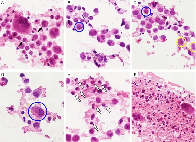
Figure 2. Squash cytological findings of LCH. A. LCs displayed centrally or eccentrically located, oval or retiform, grooved or contorted nuclei (black arrows), and were admixed with inflammatory background. B. They had abun-dant, dense cytoplasm and round or oval cell shape. Their nuclei possessed fine granular chromatin and delicate nuclear membranes and intranuclear cytoplasmic pseudoinclusion (blue circle). C. A prominent folded configuration manifested as nuclear grooves (yellow circle) and pseudoinclusions (blue circle). D. An eccentrically located nucleus showed a large pseudoinclusion (blue circle). E. The background consisted predominantly of numerous eosinophils with bilobed nuclei (white arrows). F. Some neutrophils, scattered small lymphocytes and multinucleated giant cells were accompanied.
Squash cytomorphology of LCH
with coverslips. Positive and negative controls were done according to manufacturer’s instruction.
EBV-encoded RNA in situ hybridization and CD3/CD20 double staining
The formalin-fixed, paraffin-embedded sec-tions obtained from excised specimens were used for EBER-ISH and CD3 or CD20 double staining. The sections of LCH tissue were depa-raffinized with xylene (Sigma-Aldrich), pretreat-ed with proteinase K for 20 min and incubatpretreat-ed with fluorescein isothiocyanate (FITC)-conju- gated EBER oligonucleotide probes (Novocastra Laboratories, Newcastle upon Tyne, UK) at 55°C for 2 h. The sections were rinsed in water and incubated with horseradish peroxidase-conjugated anti-FITC antibody for 15 min before adding fresh 3, 3-DAB color substrate to pro-duce an alcohol-insoluble brown intranuclear stain in EBV-positive cells. Immediately
follow-ing the EBER-ISH, immunohistochemical stain-ing with CD3 and CD20 was performed on the Leica Bond-maX autostainer (Leica Micro sys-tems) using standard reagents and techniques to produce fast red membranous stain in either CD3- or CD20-positive cells. We used EBV-negative lymphoid tissues processed using the hybridization mixture without EBER oligonucle-otides as negative controls.
Detection of BRAF V600E mutation
For the detection of BRAF V600E mutation, nucleic acids from fresh tissue were isolated using a DNA extraction kit (Qiagen, Hilden, Germany) according to the manufacturer’s pro-tocol. Briefly, isolated nucleic acids were mixed with a PCR master mix from a Seeplex-
BRAF V600E ACE detection kit (Seegene Inc.,
Seoul, Republic of Korea). The mixed samples were immediately placed in a preheated (94°C) thermal cycler for 15 min, and PCR was carried
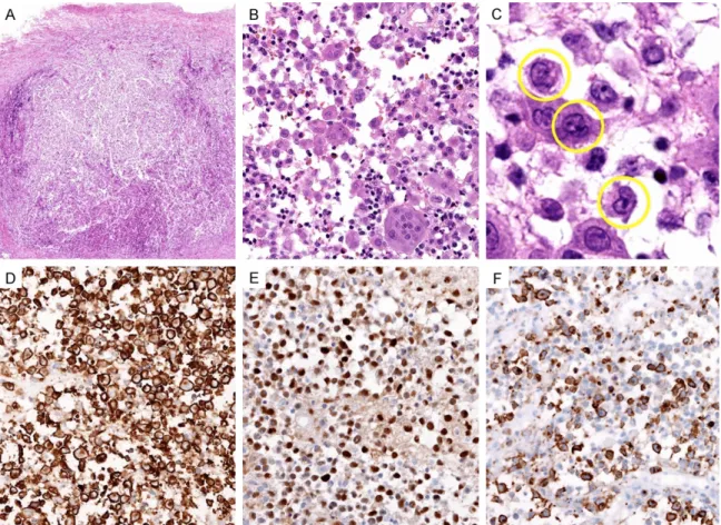
Figure 3. Histological (A-C) and immunohistochemical (D-F) findings of LCH. (A) At low power, diffuse infiltration of LCs was noted. (B) LCs were admixed with eosinophils, neutrophils, lymphocytes and multinucleated giant cells (C) Consistent with squash cytological findings, characteristic nuclear indentations (yellow circles) and grooves were detected. Immunohistochemically, the LCs were strongly reactive for (D) CD1a, (E) S-100 and (F) langerin.
out using the recommended program in a GeneAmp PCR 9700 system (Applied Biosystems, Foster City, CA, USA). The cycling amplification program consisted of 35 cycles: denaturation for 30 s at 94°C, annealing for 30 s at 63°C and extension for 1 min at 72°C. The amplified PCR products were loaded onto a 2% agarose gel and visualized with Safe View Stain (Applied Biological Materials Inc., Richmond, BC, Canada). The BRAF V600E mutation was detected with a Gel Documentation system (Bio-Rad Laboratories Inc., Hercules, CA, USA). Results
Smears were highly cellular and showed numer-ous histiocytes as the predominant cell type scattered singly or in loosely cohesive clusters with an admixture of a polymorphic population of inflammatory cells (Figure 2A). The histio-cytes were large cells with abundant, dense
cytoplasm and round or oval cell shape (Figure 2B). Their nuclei, eccentrically or centrally located, had characteristic features, consisting of fine granular chromatin, delicate nuclear membranes and a distinctive, prominent folded configuration manifested as nuclear grooves (with a coffee bean appearance; Figure 2C), indentations and pseudoinclusions (Figure 2D). The nucleoli were inconspicuous to small, but occasionally, were prominent. Mitotic figures were rare. The cytological features of these his-tiocytes were consistent with LCs. The back-ground of the smears consisted of numerous eosinophils with bilobed nuclei (Figure 2E), neutrophils, scattered small lymphocytes, plas-ma cells and multinucleated giant cells (Figure 2F). Cytological diagnosis of LCH was made. Macroscopically, the specimen revealed a gray-white, soft mass that involved the skull bone. Microscopically, a diffuse infiltrate of LCs
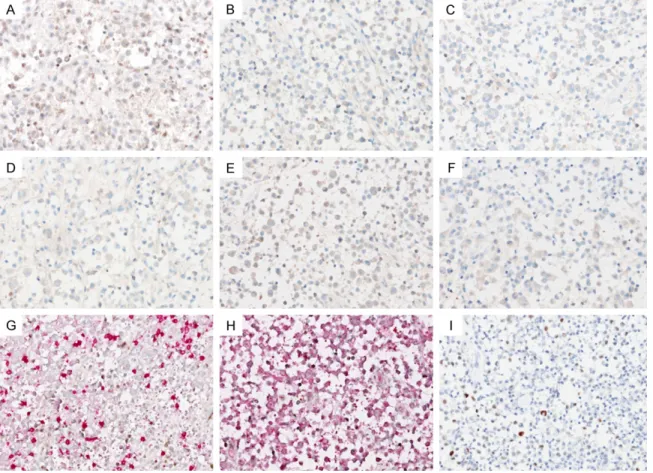
Figure 4. Findings of immunohistochemical staining for viruses (A-F and I) and EBER-ISH (G and H). LCs did not demonstrate immunoreactivity for (A) adenovirus, (B) CMV, (C) HHV-8, (D) HPV, (E) HSV and (F) SV 40. Double stain-ing for EBER-ISH and CD3 or CD20 showed that neither LCs, (G) CD3-positive T-lymphocytes nor (H) CD20-positive B-lymphocytes exhibited EBV positivity. (I) LCs displayed scattered, weak to moderate nuclear p53 immunoreactivity, indicating wild-type expression pattern (absence of TP53 mutation).
Squash cytomorphology of LCH
(Figure 3A) was admixed with eosinophils, poly-morphonuclear leukocytes, lymphocytes and multinucleated giant cells (Figure 3B). The LCs were large-sized and polygonal, with abundant glassy eosinophilic cytoplasm. Round to oval, grooved nuclei displayed finely granular or slightly vesicular chromatin (Figure 3C). Multinucleation was evident. Mitotic figures were rarely seen. No necrosis was detected. Immunohistochemically, the LCs were strongly reactive for CD1a (Figure 3D), S-100 (Figure 3E) and langerin (Figure 3F), confirming the diagnosis of LCH.
Based on the recent data that suggests that viral infection is associated with the pathophys-iology of LCH [3-6, 25], we further investigated whether viruses were present in the LCH lesions. However, LCs did not demonstrate immunoreactivity for any of the following anti-bodies: adenovirus (Figure 4A), cytomegalovi-rus (CMV; Figure 4B), human herpesvicytomegalovi-rus-8 (HHV-8; Figure 4C), human papilloma virus (HPV; Figure 4D), herpes simplex virus (HSV; Figure 4E) and simian virus 40 (SV 40; Figure 4F). We also performed double staining for EBER-ISH and CD3/CD20 to investigate wheth-er EBV infects bystandwheth-er T- or B-lymphocytes. However, neither LCs, T-lymphocytes (Figure 4G) nor B-lymphocytes (Figure 4H) exhibited EBV positivity.
We assessed the status of 2 known molecular abnormalities in LCH, i.e., p53 protein expres-sion and the presence of BRAF V600E muta-tion in order to find evidence of neoplastic pathogenesis [11, 26]. However, no mutant pattern of p53 expression was observed in LCs (Figure 4I). No LCH tissue samples exhibited
BRAF V600E mutation.
Discussion
The diagnosis of LCH in our patients was made on the basis of characteristic cytomorphology on squash cytological preparations. This was corroborated by appropriate radiological and clinical findings. In the present cases, both MRI and CT scans showed lytic lesions in the skull bones with sharp borders and punched-out appearances. In Case 2, destruction of both inner and outer tables resulted in a double-con-tour or a beveled-edge appearance, which is a typical roentgenographic finding in the diagno-sis of LCH [4, 27].
This report highlights the usefulness of squash cytological preparations for rapid intraopera-tive diagnosis. Intraoperaintraopera-tive frozen section is an important component of surgical manage-ment of tumors. Critical decision regarding treatment and the extent of surgical resection can sometimes depend on an appropriate intraoperative cytological diagnosis [28]. The cytological findings were similar to those described in fine needle aspiration samples of LCH in other organs [29-34]. In the present cases, the squash smears displayed a high cel-lularity consisting of isolated or loosely aggre-gated LCs with eccentrically located, oval to reniform nuclei showing delicate nuclear mem-branes and prominent nuclear grooves, inden-tations and pseudoinclusions. These were admixed with a polymorphic population of inflammatory cells with a predominance of eosinophils. The key to the diagnosis is identifi-cation of LCs through characteristic nuclear features, i.e., nuclear grooves and pseudoinclu-sions. However, occasionally, the LCs are few or nuclear grooves not very prominent. Further- more, they can show variable degree of pleo-morphism and mitotic activity [31, 32, 35, 36]. Similarly, the degree of eosinophilic infiltration varies in different areas of the LCH lesion, thus their number can vary from scant to abundant in cytology smears [35]. Nevertheless, the presence of eosinophils can help attract atten-tion to the diagnosis of LCH. There have been several case reports on the cytologic features of LCH, but most of these cases were taken from the bone or thyroid as aspiration cytology [2, 29-34]. There is only one previous Japanese case demonstrating squash cytologic features of LCH of the skull [12]. Together with previous reports, we demonstrated that squash cyto-morphology closely reflects histocyto-morphology. The cytological diagnosis may be missed due to lack of familiarity with its cytomorphology among pathologists or due to the lack of char-acteristic cytological findings resulting from a sampling error. Therefore it is prudent for the pathologist to consider this diagnosis only in an appropriate clinical and radiological setting. It is also necessary to be familiar with cytological features of other differential diagnoses. The most common differential diagnoses of cranial LCH include Ewing sarcoma, non-Hodgkin lym-phoma and osteomyelitis with abundant histio-cytes. Ewing sarcoma and non-Hodgkin
lym-phoma are characterized by a monomorphic population of small round blue cells. In acute osteomyelitis, numerous neutrophils forma prominent component of inflammatory lesion. The observed reactive histiocytes can be easily distinguished due to the absence of character-istic features of LCs. Plasma cells and lympho-cytes predominate in chronic osteomyeltis. Neutrophils and plasma cells can be present, but infrequent in LCH.
A possible relationship between the develop-ment of LCH and expression of some human herpes viruses has been reported [25]. The granulomatous nature of LCH lesions and the generally benign morphology of pathologic LCs have suggested possible infectious, environ-mental or autoimmune pathogeneses [3, 5]. In particular, EBV infects monocytes and LCs and is associated with the pathophysiology of LCH according to some studies [3, 5-7, 25]. However, other studies failed to replicate these findings, and the possible causative role of EBV in LCH is debated [37-39]. Shimakage and colleagues [25] showed positive signals for EBER-ISH in 17 cases of LCH, in contrast, McClain and col-leagues [37] failed to find evidence of genomes for EBV in 56 LCHs. Slacmeulder and col-leagues [40] demonstrated the absence of HHV-8 DNA in 12 LCH biopsy specimens. Similarly, herpes viruses such as EBV, CMV and HHV-6 have been detected in clinical samples [41], but differences in the overall prevalence or titers of antibodies against these viruses were not significant between LCH patients and controls. Consistent with earlier reports that were unable to document an association between herpes viruses and LCH, we did not observe immunoreactivity for all viruses exam-ined [37-40, 42]. Double staining for EBER-ISH and CD3 or CD20 revealed the absence of EBV in B- and T-lymphocytes. However, we could not definitively exclude an association of EBV infec-tion with LCH because of a number of EBV-infected cells infiltrating LCH lesion, suggestive of an EBV-infected B-lymphocytes and LC inter-action that might contribute to the develop-ment of LCH [40, 41]. Jeziorski and colleagues [41] observed that EBV-positive cells were labeled with CD20 or CD79a, but not with CD1a, CD3 or CD68, indicating that EBV infects bystander B-lymphocytes and not LCs. Thus, EBV-infected B-lymphocytes in the lesion may trigger the activation of LCs and eventually the development of LCH in some cases.
We did not observe the mutation pattern (either diffuse and strong positivity or complete absence of immunoreactivity) of p53 protein expression. BRAF V600E mutation was absent. Some previous studies have shown TP53 muta-tion and p53 overexpression in LCH [11, 43], raising the possibility that p53 may be a prima-ry contributor to a hyperproliferative, trans-formed phenotype through its inability to induce apoptosis [26]. Furthermore, the demonstra-tion of BRAF V600E mutademonstra-tion in more than a half of LCH samples examined provides the recurrent genomic abnormality that is required, in addition to clonality, to assign LCH a neoplas-tic origin [11]. However, p53 overexpression can be a normal secondary response to a prolif-erative stimulus. There are several examples of benign, reactive proliferative disorders that are characterized by high levels of p53 expression [44-46]. Likewise, BRAF mutation alone may be necessary but not sufficient for the develop-ment of LCH; several additional genetic abnor-malities are required for tumorigenesis. Much effort is required to identify additional genetic abnormalities in LCH and to understand the molecular basis of its pathogenesis.
In conclusion, the presence of a mixed popula-tion of inflammatory cells, including abundant eosinophils, and a high content of histiocytes with characteristic cytomorphology on the intraoperative squash smears, along with radio-logic evidence and appropriate clinical findings, is highly suggestive of LCH. Nuclear features including grooves and pseudoinclusions were evident in squash cytological smears, closely reflecting histomorphology of LCH. Squash smear cytology can serve as a useful tool for the intraoperative diagnosis of LCH. Awareness of cytological features of LCH, its differential diagnoses and causes of diagnostic pitfall is necessary. In addition, our results did not sup-port an association between LCH and viral infection or genetic abnormalities. Although the precise etiology remains to be elucidated, our understanding of LCH has been greatly enhanced by the application of advanced genomic technologies to the analysis of primary human material. Further investigation is required to clarify the molecular pathogenesis of LCH.
Acknowledgements
This study was supported by a faculty research grant of Yonsei University College of Medicine for 2015 (6-2015-0072).
Squash cytomorphology of LCH
Disclosure of conflict of interest None.
Address correspondence to: Dr. Hyun-Soo Kim, Department of Pathology, Severance Hospital, Yonsei University College of Medicine, 50-1, Yonsei-ro, Seodaemun-gu, Seoul 03722, Republic of Korea. Tel: +82-2-2228-2648; Fax: +82-2-2227-7939; E-mail: hyunsookim@yuhs.ac; Dr. Sung-Im Do, Department of Pathology, Kangbuk Samsung Hospital, Sungkyunkwan University School of Medi- cine, 29 Saemunan-ro, Jongno-gu, Seoul 03181, Republic of Korea. Tel: 2-2001-2393; Fax: +82-2-2001-2398; E-mail: sungim.do@samsung.com
References
[1] Laman JD, Leenen PJ, Annels NE, Hogendoorn PC, Egeler RM. Langerhans-cell histiocytosis ‘insight into DC biology’. Trends Immunol 2003; 24: 190-196.
[2] Kumar N, Sayed S, Vinayak S. Diagnosis of Langerhans cell histiocytosis on fine needle aspiration cytology: a case report and review of the cytology literature. Pathology Res Int 2011; 2011: 439518.
[3] Chen CJ, Ho TY, Lu JJ, Sheu LF, Lee SY, Tien CH, Cheng SN. Identical twin brothers concordant for Langerhans’ cell histiocytosis and discor-dant for Epstein-Barr virus-associated haemo-phagocytic syndrome. Eur J Pediatr 2004; 163: 536-539.
[4] Chen HC, Shen WC, Chou DY, Chiang IP. Langerhans cell histiocytosis of the skull com-plicated with an epidural hematoma. AJNR Am J Neuroradiol 2002; 23: 493-495.
[5] Sakata N, Toguchi N, Kimura M, Nakayama M, Kawa K, Takemura T. Development of Langerhans cell histiocytosis associated with chronic active Epstein-Barr virus infection. Pediatr Blood Cancer 2008; 50: 924-927. [6] Tugizov S, Herrera R, Veluppillai P, Greenspan
J, Greenspan D, Palefsky JM. Epstein-Barr vi-rus (EBV)-infected monocytes facilitate dis-semination of EBV within the oral mucosal epi-thelium. J Virol 2007; 81: 5484-5496.
[7] Walling DM, Ray AJ, Nichols JE, Flaitz CM, Nichols CM. Epstein-Barr virus infection of Langerhans cell precursors as a mechanism of oral epithelial entry, persistence, and reactiva-tion. J Virol 2007; 81: 7249-7268.
[8] Egeler RM, Favara BE, van Meurs M, Laman JD, Claassen E. Differential In situ cytokine profiles of Langerhans-like cells and T cells in Langerhans cell histiocytosis: abundant ex-pression of cytokines relevant to disease and treatment. Blood 1999; 94: 4195-4201. [9] Emile JF, Tartour E, Brugieres L, Donadieu J, Le
Deist F, Charnoz I, Fischer A, Fridman WH,
Brousse N. Detection of GM-CSF in the sera of children with Langerhans’ cell histiocytosis. Pediatr Allergy Immunol 1994; 5: 162-163. [10] Coury F, Annels N, Rivollier A, Olsson S, Santoro
A, Speziani C, Azocar O, Flacher M, Djebali S, Tebib J, Brytting M, Egeler RM, Rabourdin-Combe C, Henter JI, Arico M, Delprat C. Langerhans cell histiocytosis reveals a new IL-17A-dependent pathway of dendritic cell fu-sion. Nat Med 2008; 14: 81-87.
[11] Badalian-Very G, Vergilio JA, Degar BA, MacConaill LE, Brandner B, Calicchio ML, Kuo FC, Ligon AH, Stevenson KE, Kehoe SM, Garraway LA, Hahn WC, Meyerson M, Fleming MD, Rollins BJ. Recurrent BRAF mutations in Langerhans cell histiocytosis. Blood 2010; 116: 1919-1923.
[12] Kobayashi TK, Ueda M, Nishino T, Bamba M, Echigo T, Oka H, Hino A, Fuse I, Fujimoto M, Katsumori T, Kaneko C. Langerhans cell histio-cytosis of the skull on cytologic squash prepa-rations. Diagn Cytopathol 2007; 35: 154-157. [13] Fung KM, Schwalb JM, Riina HA, Kurana JS,
Mindaxy JM, Grady MS, Lavi E. February 2002: 29-year-old woman with a skull mass for 2 months. Brain Pathol 2002; 12: 393-394, 397. [14] Lunardi P, Farah JO, Qhaso R, Puzzilli F,
Siciliano P, Di Stefano D. Primary eosinophilic granuloma invading the skull base: case report and critical review of the literature. Tumori 1996; 82: 397-400.
[15] Chandekar SA, Shah VB, Kavishwar V. Cytological diagnosis of Langerhans cell histio-cytosis with cutaneous involvement. J Cytol 2013; 30: 81-83.
[16] Lee SR, Suh JH, Cha HJ, Kim YM, Choi HJ. Fine needle aspiration cytology of Langerhans cell histiocytosis of mandible. Korean J Pathol 2010; 44: 106-109.
[17] Chandekar SA, Shah VB, Kavishwar V. Cytological diagnosis of Langerhans cell histio-cytosis with cutaneous involvement. J Cytol 2013; 30: 81-83.
[18] Kim HS, Do SI, Noh BJ, Jeong YI, Park SJ, Kim YW. Expression of phosphorylated extracellular signal-regulated kinase at the invasive front of hepatic colorectal metastasis. Oncol Lett 2015; 9: 1261-1265.
[19] Kim HS, Kim GY, Lim SJ, Kim YW. Loss of Raf-1 kinase inhibitory protein in pancreatic ductal adenocarcinoma. Pathology 2010; 42: 655-660.
[20] Kim HS, Kim GY, Lim SJ, Kim YW. Raf-1 kinase inhibitory protein expression in thyroid carcino-mas. Endocr Pathol 2010; 21: 253-257. [21] Kim HS, Kim GY, Lim SJ, Kim YW. Expression of
Raf-1 kinase inhibitory protein in extrahepatic bile duct carcinoma. Korean J Pathol 2010; 44: 234-242.
[22] Kim HS, Kim GY, Lim SJ, Park YK, Kim YW. Reduced expression of Raf-1 kinase inhibitory protein is a significant prognostic marker in pa-tients with gallbladder carcinoma. Hum Pathol 2010; 41: 1609-1616.
[23] Kim HS, Park SJ, Lee KY, Park YK, Kim YW. Reduced Raf-1 kinase inhibitor protein expres-sion predicts less favorable outcomes in pa-tients with hepatic colorectal metastasis. Oncol Rep 2012; 28: 161-171.
[24] Kim HS, Won KY, Kim GY, Kim SC, Park YK, Kim YW. Reduced expression of Raf-1 kinase in-hibitory protein predicts regional lymph node metastasis and shorter survival in esophageal squamous cell carcinoma. Pathol Res Pract 2012; 208: 292-299.
[25] Shimakage M, Sasagawa T, Kimura M, Shimakage T, Seto S, Kodama K, Sakamoto H. Expression of Epstein-Barr virus in Langerhans’ cell histiocytosis. Hum Pathol 2004; 35: 862-868.
[26] Badalian-Very G, Vergilio JA, Fleming M, Rollins BJ. Pathogenesis of Langerhans cell histiocyto-sis. Annu Rev Pathol Mech Dis 2008; 8: 1-20. [27] Hermans R, De Foer B, Smet MH, Leysen J,
Feenstra L, Fossion E, Baert AL. Eosinophilic granuloma of the head and neck: CT and MRI features in three cases. Pediatr Radiol 1994; 24: 33-36.
[28] Jaiswal S, Vij M, Jaiswal AK, Srivastava AK, Behari S. Squash cytology of subependymal gi-ant cell astrocytoma: report of four cases with brief review of literature. Diagn Cytopathol 2012; 40: 333-336.
[29] Dey P, Luthra UK, Sheikh ZA. Fine needle aspi-ration cytology of Langerhans cell histiocytosis of the thyroid. A case report. Acta Cytol 1999; 43: 429-431.
[30] Kilpatrick SE. Fine needle aspiration biopsy of Langerhans cell histiocytosis of bone: are an-cillary studies necessary for a “definitive diag-nosis”? Acta Cytol 1998; 42: 820-823. [31] Musy JP, Ruf L, Ernerup I, Baltisser-Bielecka I.
Cytopathologic diagnosis of an eosinophilic granuloma of bone by needle aspiration biop-sy. Acta Cytol 1989; 33: 683-685.
[32] Pohar-Marinsek Z, Us-Krasovec M. Cytomor- phology of Langerhans cell histiocytosis. Acta Cytol 1996; 40: 1257-1264.
[33] Shabb N, Fanning CV, Carrasco CH, Guo SQ, Katz RL, Ayala AG, Raymond AK, Cangir A. Diagnosis of eosinophilic granuloma of bone by fine-needle aspiration with concurrent insti-tution of therapy: a cytologic, histologic, clini-cal, and radiologic study of 27 cases. Diagn Cytopathol 1993; 9: 3-12.
[34] Zhu H, Hu DX. Langerhans cell histiocytosis of the thyroid diagnosed by fine needle aspiration cytology. A case report. Acta Cytol 2004; 48: 278-280.
[35] Akhtar M, Ali MA, Bakry M, Sackey K, Sabbah R. Fine-needle aspiration biopsy of Langerhans histiocytosis (histiocytosis-X). Diagn Cytopathol 1993; 9: 527-533.
[36] Elsheikh T, Silverman JF, Wakely PE Jr, Holbrook CT, Joshi VV. Fine-needle aspiration cytology of Langerhans’ cell histiocytosis (eosinophilic granuloma) of bone in children. Diagn Cytopathol 1991; 7: 261-266.
[37] McClain K, Jin H, Gresik V, Favara B. Langerhans cell histiocytosis: lack of a viral eti-ology. Am J Hematol 1994; 47: 16-20.
[38] Brousset P. Epstein-Barr virus and Langerhans cell histiocytosis. Hum Pathol 2004; 35: 1573-1574; author reply 1574.
[39] Schenka AA, De Angelo Andrade LA, Amstalden EM, Cintra ML, Vassallo J, Cardinalli IA, de Azevedo AC, Brandalise SR, Soares FA. Langerhans cell histiocytosis and its relation-ship with Epstein-Barr virus. Hum Pathol 2006; 37: 1508-1509; author reply 1509-1511. [40] Slacmeulder M, Geissmann F, Lepelletier Y,
Fournet JC, Brousse N, Thomas C, Donadieu J, Gessain A. No association between Langerhans cell histiocytosis and human herpes virus 8. Med Pediatr Oncol 2002; 39: 187-189. [41] Jeziorski E, Senechal B, Molina TJ, Devez F,
Leruez-Ville M, Morand P, Glorion C, Mansuy L, Gaudelus J, Debre M, Jaubert F, Seigneurin JM, Thomas C, Joab I, Donadieu J, Geissmann F. Herpes-virus infection in patients with Langerhans cell histiocytosis: a case-con-trolled sero-epidemiological study, and in situ analysis. PLoS One 2008; 3: e3262.
[42] Jenson HB, McClain KL, Leach CT, Deng JH, Gao SJ. Evaluation of human herpesvirus type 8 infection in childhood langerhans cell histio-cytosis. Am J Hematol 2000; 64: 237-241. [43] Weintraub M, Bhatia KG, Chandra RS, Magrath
IT, Ladisch S. p53 expression in Langerhans cell histiocytosis. J Pediatr Hematol Oncol 1998; 20: 12-17.
[44] Castren K, Vahakangas K, Heikkinen E, Ranki A. Absence of p53 mutations in benign and pre-malignant male genital lesions with over-expressed p53 protein. Int J Cancer 1998; 77: 674-678.
[45] Porcelli B, Frosi B, Terzuoli L, Arezzini L, Marinello E, Vernillo R, De Martino A, Vatti R, Minacci C. Expression of p185 and p53 in be-nign and malignant colorectal lesions. Histochem J 2001; 33: 51-57.
[46] Rohan TE, Hartwick W, Miller AB, Kandel RA. Immunohistochemical detection of c-erbB-2 and p53 in benign breast disease and breast cancer risk. J Natl Cancer Inst 1998; 90: 1262-1269.