https://doi.org/10.3340/jkns.2019.0027 pISSN 2005-3711 eISSN 1598-7876
Mechanistic Target of Rapamycin Pathway in Epileptic
Disorders
Jang Keun Kim, B.S.,
1Jeong Ho Lee, M.D., Ph.D.
1,2Biomedical Science and Engineering Interdisciplinary Program,1 Korea Advanced Institute of Science and Technology (KAIST), Daejeon, Korea
Graduate School of Medical Science and Engineering,2 Korea Advanced Institute of Science and Technology (KAIST), Daejeon, Korea
The mechanistic target of rapamycin (mTOR) pathway coordinates the metabolic activity of eukaryotic cells through environmental
signals, including nutrients, energy, growth factors, and oxygen. In the nervous system, the mTOR pathway regulates fundamental
biological processes associated with neural development and neurodegeneration. Intriguingly, genes that constitute the mTOR
pathway have been found to be germline and somatic mutation from patients with various epileptic disorders. Hyperactivation of
the mTOR pathway due to said mutations has garnered increasing attention as culprits of these conditions : somatic mutations, in
particular, in epileptic foci have recently been identified as a major genetic cause of intractable focal epilepsy, such as focal cortical
dysplasia. Meanwhile, epilepsy models with aberrant activation of the mTOR pathway have helped elucidate the role of the mTOR
pathway in epileptogenesis, and evidence from epilepsy models of human mutations recapitulating the features of epileptic
patients has indicated that mTOR inhibitors may be of use in treating epilepsy associated with mutations in mTOR pathway genes.
Here, we review recent advances in the molecular and genetic understanding of mTOR signaling in epileptic disorders. In particular,
we focus on the development of and limitations to therapies targeting the mTOR pathway to treat epileptic seizures. We also discuss
future perspectives on mTOR inhibition therapies and special diagnostic methods for intractable epilepsies caused by brain somatic
mutations.
Key Words : mTORC1 ∙ mTORC2 ∙ Epilepsy ∙ Malformation of cortical development.
• Received : January 22, 2019 • Revised : March 11, 2019 • Accepted : March 12, 2019 • Address for reprints : Jeong Ho Lee, M.D., Ph.D.
Biomedical Science and Engineering Interdisciplinary Program, Korea Advanced Institute of Science and Technology (KAIST), 291 Daehak-ro, Yuseong-gu, Daejeon 34141, Korea
Tel : +82-42-350-4246, Fax : +82-42-350-4240, E-mail : jhlee4246@kaist.ac.kr
This is an Open Access article distributed under the terms of the Creative Commons Attribution Non-Commercial License (http://creativecommons.org/licenses/by-nc/4.0) which permits unrestricted non-commercial use, distribution, and reproduction in any medium, provided the original work is properly cited.
INTRODUCTION
The discovery of the mechanistic target of rapamycin
(mTOR) began with the identification of new antimicrobial
agents in soil samples from Rapa Nui (also known as Easter
Island)
93). This new antimicrobial agent, called rapamycin
(clinically called sirolimus), was found to exhibit
immunosup-pressive and cancer potential and to be of use as an
anti-epileptic drug
43,47,144). For two decades, endeavors to define the
function of mTOR revealed that mTOR coordinates
environ-mental signals and metabolic activity by forming two distinct
complexes, mTOR complex 1 (mTORC1) and mTOR complex
2 (mTORC2)
130). mTOR was first found in brain lysates,
sug-gesting the importance of the brain-specific function of
mTOR
125). Indeed, the mTOR pathway has been found to
reg-ulate a variety of functions in the brain from brain
develop-ment to degeneration
85).
Human genetic studies of epileptic patients and epilepsy
models of aberrant activation of the mTOR pathway have
demonstrated the importance of activating mutations in the
mTOR pathway in neurodevelopmental disorders with
epi-lepsy
66). With the discovery of mutations activating the mTOR
pathway as genetic causes of medically intractable epilepsy
4),
mTOR inhibitors, such as sirolimus and everolimus, have
emerged as being of potential medical use in treating
intracta-ble epilepsy patients
33). However, with the limited efficacy and
significant drawbacks of clinically available mTOR inhibitors,
new drugs that are more effective and tolerable based on the
understanding of pathogenic mechanisms are required. Key
discoveries in research on the role of the mTOR pathway in
epilepsy are summarized in Supplementary Box 1.
THE MTOR PATHWAY
mTOR is a serine/threonine protein kinase that forms the
core subunit of two functionally distinct protein complexes :
mTORC1 and mTORC2
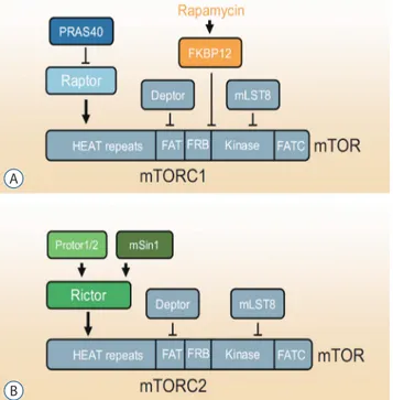
130)(Fig. 1). mTORC1 comprises three
core components : mTOR, Raptor (regulatory protein
associ-ated with mTOR), and mLST8 (mammalian lethal with Sec13
protein 8, also known as GßL)
130). In response to
environmen-tal signals, mTORC1 acts to control the balance between
anabolism and catabolism
130). mTORC2 controls proliferation,
survival, and cytoskeleton organization
130). In a nutrient-rich
environment, cells convert energy sources into
macromole-cules, such as proteins, lipids, and nucleotides. In
nutrient-starved environments, however, cells downregulate the
pro-duction of macromolecules and rely on catabolic pathways,
such as autophagy, for energy.
The mTOR pathway receives various inputs from upstream
signaling pathways in response to growth factors, amino
ac-ids, energy, oxygen, and stress
130), and the upstream pathways
of mTOR can be divided as energy-sensing
PI3K-PTEN-AKT-TSC and amino acid-sensing GATOR2-GATOR1-Rag GTPase
pathways (Fig. 2) : phosphatidylinositol-3-kinase (PI3K) is
critical to integrating insulin signaling for growth and
surviv-al
130). PTEN antagonizes the action of PI3K. Akt is activated by
PI3K and is a positive regulator of mTORC1 via inhibition of
Tuberous Sclerosis Complex (TSC). TSC is a heterotrimeric
complex comprising TSC1, TSC2, and TBC1D7
38). TSC
inhib-its mTORC1 by acting as a GTPase activating protein for Ras
homolog enriched in brain (Rheb)
67). Rheb is a small GTPase
that activates mTORC1 by directly binding to mTORC1 on
the surface of lysosomes
88). Meanwhile, Rag GTPase, a
com-ponent of the amino acid sensing pathway
127), activates
mTORC1 by promoting translocation of mTORC1 to the
lyso-somal surface. Upstream regulators of Rag GTPase in amino
acid signaling are the GATOR1 and GATOR2 complexes
10).
The GATOR1 complex, consisting of DEPDC5, Nprl2, and
Nprl3, inhibits the mTORC1 pathway by acting as a guanine
exchange factor for Rag GTPase. The GATOR2 complex,
con-sisting of Mios, WDR24, WDR59, Seh1L, and Sec13, is a
posi-tive regulator of the mTORC1 pathway by inhibiting
GA-TOR1. KICSTOR, which is composed of four proteins, KPTN,
ITFG2, C12orf66, and SZT2, recruits GATOR1 to the
lyso-some to inhibit Rag GTPase
150). Leucyl-tRNA synthetase,
which is another amino acid sensor, functions as a GTPase
ac-Fig. 1. mTORC1 and mTORC2. A : The components of mTORC1 and respective binding site on mTOR. B : The components of mTORC2 and respective binding site on mTOR. PRAS40 : proline-rich Akt substrate of 40 kDa, Raptor : regulatory protein associated with mTOR, FKBP12 : FK506 binding protein 12, Deptor : DEP domain-containing mTOR-interacting protein, mLST8 : mammalian lethal with Sec13 protein 8, mTOR : mechanistic target of rapamycin, mTORC1 : mTOR complex 1, mSin1 : mammalian stress-activated protein kinase-interacting protein, Rictor : rapamycin-insensitive companion of mammalian target of rapamycin, mTORC2 : mTOR complex 2.
A
tivating protein for Rag GTPase
57). mTORC1 senses amino
ac-ids in an intra-lysosome fashion. Lysosomal amino acid
regu-lates Rag GTPase via v-ATPase, which increases the guanine
exchange factor activity of Ragulator towards Rag GTPase
159).
SLC38A9 is a sensor of lysosomal arginine and activates
mTORC1
69). Additional novel mTOR upstream regulators,
in-cluding a methionine sensor, have recently been discovered
1,55).
For macromolecule metabolism, mTORC1 regulates
trans-lation through inhibitory eukaryotic initiation factor 4E
(eIF4E)-binding protein 1/2/3 (4E-BPs) and the S6 kinases
(S6Ks)
21,50). Translational control occurs predominantly at the
initiation step, which commences with the binding of the
eu-karyotic translation initiation factor 4F (eIF4F) complex to the
5’cap
52,135). As the limiting component of the eIF4F complex,
eIF4E is considered to be a critical determinant in translation
of mRNA
37). Facilitating eIF4F formation and the progression
of translation, mTORC1 phosphorylates (inactivates) the
4E-BPs, leading to their dissociation from eIF4E
51,60). The S6Ks
activate the eukaryotic translation initiation factor 4B (eIF4B),
which is an activator of the eukaryotic translation initiation
factor 4A, leading to an increase in the helicase activity of
eI-F4A and the initiation of translation
39,61).
Stimulating lipid synthesis, mTORC1 interacts with the
ste-rol responsive element binding proteins transcription
fac-tors
116). For sufficient supply of nucleotides during growth,
mTORC1 promotes purine and pyrimidine nucleotide
bio-synthesis through MTHFD2 and the carbamoyl-phosphate
synthetase
17). Through increased translation of the HIF1α
transcription factor that drives the expression of glycolytic
en-zymes, mTORC1 further promotes growth by changing
glu-cose metabolism from oxidative phosphorylation to
glycoly-sis
130). The mTORC1 pathway also activates the transcriptional
coactivator PGC1α for increased mitochondrial
biosynthe-sis
34). Meanwhile, mTORC1 inhibits autophagy, which plays
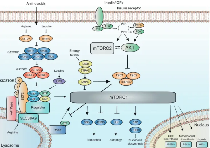
Fig. 2. Upstream and downstream of mTORC1 and mTORC2. The signaling network of mTORC1 and mTORC2. Positive regulators of mTORC1 signaling are shown in blue to green. Negative regulators of mTORC1 signaling are shown in red to yellow.
an important role in scavenging damaged and harmful
cellu-lar structures and sustains energy homeostasis, through
ULK1
118). Recently, it was also demonstrated that mTORC1
regulates ribosomal protein degradation through NUFIP1
151).
mTORC2 largely functions as a primary effector of the
in-sulin/PI3K signaling
130)(Fig. 2). Generally, the role of the
mTORC2 pathway is thought to overlap with the insulin
sig-naling pathway, considering that phenotypes caused by
per-turbation of the mTORC2 pathway are very similar to those
elicited by perturbation of insulin signaling
105). Upon
phos-phorylation by mTORC2
128), Akt promotes cell survival and
proliferation. mTORC2 further regulates cellular proliferation
and survival through the AGC (PKA/PKG/PKC) family of
protein kinases.
PHYSIOLOGICAL ROLES OF THE MTOR
PATH-WAY IN THE BRAIN
The mTOR pathway plays important roles in development
and aging of the central nervous system (CNS) : the mTORC1
pathway is a core regulator of neural development, cortical
ar-chitecture, neuronal morphology, circuit formation, synaptic
plasticity, and neurodegeneration
85). Perturbation of the mTOR
pathway has been found to disrupt several developmental
pro-cesses. Defective telencephalic development was reported in
mTOR mutant rats, called “flat-top” mutants
59). The loss of function
of tuberous sclerosis complex 2 (Tsc2), which is an upstream
inhibi-tor of mTOR, was found to disrupt neuroepithelial growth
119).
Altogether, these studies demonstrated that the mTOR pathway
is important for brain development by regulating neural stem
cells. Meanwhile, activation of the mTOR pathway via
condi-tional deletion of Tsc2 was found to lead to the disruption of
cortical layering
147). Additionally, increased activity of the mTOR
pathway due to mTOR mutations, loss of Tsc1/2, or Pten, was
described as eliciting hypertrophy of soma, fewer dendritic
spine, increased axon length, and increased dendritic
complexi-ty in cortical neurons
53,77,78,137,138).
Perturbation of the mTOR pathway has also been found to
adversely affect neural circuit formation. The mTOR pathway
regulates axon length
53)and axon guidance in response to
en-vironmental signals
156). Among neurons, local protein
synthe-sis in synapses distant from the soma is mediated by mTOR
and is critical for the formation of the neural circuit. The
ex-pression levels of synaptic proteins, such as the Arc and
Syn-apsin, are increased by activation of mTOR
81,117).
Hyperactiva-tion of the mTOR pathway by loss of Pten has been shown to
increase glutamatergic and GABAergic signals
148). In addition
to neurons, the mTOR pathway has been found to regulate
glial cells during neural circuit formation. Deletion of the core
component of the mTORC1 or the mTORC2, Raptor or
Ric-tor, was reported to result in defective myelination and
oligo-dendrocyte maturation
18,146). Conditional Tsc1 knockout in the
astrocyte reportedly disrupted electrical signaling in the
mouse brain by abrogating neural circuitry
141). Interestingly,
researchers have discovered crosstalk between neurons and
glial cells as evidenced by a loss of myelination upon loss of
TSC1 in neurons
96). In TSC patients, the critical role of the
mTOR pathway in neural circuit formation was echoed by
re-ductions in white matter and cortical connectivity
112).
Neuronal synapses are crucial to storing memories.
Obser-vations of impaired memory formation and synaptic function
in response to rapamycin treatment suggest the important
role of the mTOR pathway in memory formation
25). Indeed,
the mTOR pathway has been found to play a crucial role in
both long-term potentiation (LTP) and long-term depression
(LTD) : activation of the mTOR pathway via Tsc1+/-, Tsc2+/-,
or 4E-BP2 knockout (KO), results in a decrease in LTP
thresh-old
8,41,145). Meanwhile, reduced mTOR pathway activity
dis-rupts LTD through metabotropic glutamate receptors
(mGluRs) that disturb local protein synthesis
62,64).
ACTIVATION OF MTOR PATHWAY IN
NEURODE-VELOPMENTAL DISORDERS WITH EPILEPSY
Neurodevelopmental disorders, such as malformations of
cortical development (MCD), Cowden syndrome (CS),
PIK-3CA-related overgrowth spectrum (PROS), and hamartoma
tumor syndromes, commonly present cortical abnormalities
and are highly associated with epilepsy, developmental delay,
and autism-spectrum disorders
4,12,70,80,97,106,133). These
neurode-velopmental disorders with epilepsy are a common cause of
drug-resistant epilepsy in children requiring surgery for
treat-ment
4,12). However, about 50% of these individuals continue to
experience seizures after surgical resection of the epileptic
fo-cus
94). In addition to clinical intractability, refractory epileptic
seizures also pose tremendous socioeconomic burden, based
on poor quality of life, to patients and their caregivers
28).
Aberrant activation of the mTOR pathway has been
identi-fied in brain lesions from patients with neurodevelopmental
disorders with epilepsy, especially in MCD
15,86,100). With these
results, researchers have suspected genetic mutations as
cul-prits of the observed mTOR pathway activation in
neurode-velopmental disorders
32). Indeed, a number of
neurodevelop-mental disorders with epilepsy has been shown to be caused
by germline or somatic mutations in the mTOR pathway :
these mutations can be categorized as those affecting the
en-ergy-sensing PI3K-PTEN-AKT-TSC pathway and those
af-fecting the amino acid-sensing GATOR2-GATOR1-Rag
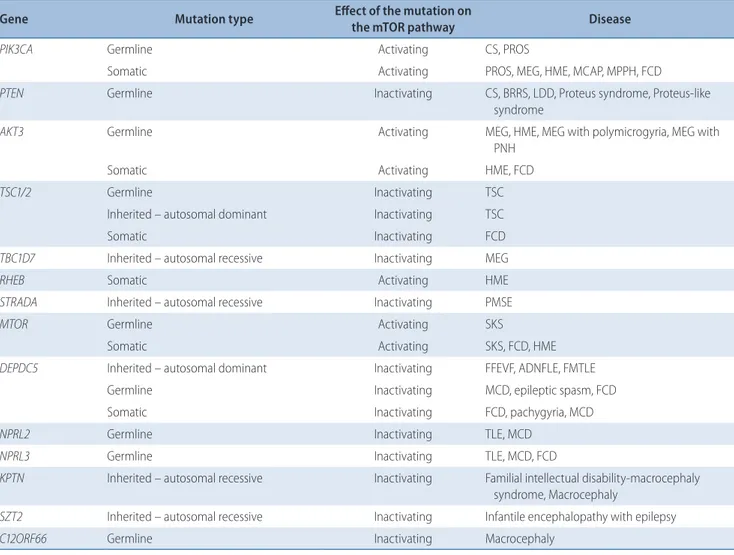
GT-Pase pathway (Table 1).
MUTATIONS IN THE ENERGY-SENSING
PI3K-PTEN-AKT-TSC PATHWAY
Germline mutations in the PIK3CA gene, which activates
the mTOR pathway, have been reported in CS
106).
Character-Table 1. Mutations in the mTOR pathway in the neurodevelopmental disorders with epilepsy Gene Mutation type Effect of the mutation on
the mTOR pathway Disease
PIK3CA Germline Activating CS, PROS
Somatic Activating PROS, MEG, HME, MCAP, MPPH, FCD
PTEN Germline Inactivating CS, BRRS, LDD, Proteus syndrome, Proteus-like
syndrome
AKT3 Germline Activating MEG, HME, MEG with polymicrogyria, MEG with
PNH
Somatic Activating HME, FCD
TSC1/2 Germline Inactivating TSC
Inherited – autosomal dominant Inactivating TSC
Somatic Inactivating FCD
TBC1D7 Inherited – autosomal recessive Inactivating MEG
RHEB Somatic Activating HME
STRADA Inherited – autosomal recessive Inactivating PMSE
MTOR Germline Activating SKS
Somatic Activating SKS, FCD, HME
DEPDC5 Inherited – autosomal dominant Inactivating FFEVF, ADNFLE, FMTLE
Germline Inactivating MCD, epileptic spasm, FCD
Somatic Inactivating FCD, pachygyria, MCD
NPRL2 Germline Inactivating TLE, MCD
NPRL3 Germline Inactivating TLE, MCD, FCD
KPTN Inherited – autosomal recessive Inactivating Familial intellectual disability-macrocephaly syndrome, Macrocephaly
SZT2 Inherited – autosomal recessive Inactivating Infantile encephalopathy with epilepsy
C12ORF66 Germline Inactivating Macrocephaly
mTOR : mechanistic target of rapamycin, CS : cowden syndrome, PROS : PIK3CA-related overgrowth spectrum, MEG : megalencephaly, HME : hemimegalencephaly, MCAP : megalencephaly-capillary malformation, MPPH : megalencephaly-polydactylyl-polymicrogyria-hydrocephalus, FCD : focal cortical dysplasia, PTEN : phosphatase and tensin homolog, BRRS : Bannayan-Riley-Ruvalcaba syndrome, LDD : Lhermitte-Duclos disease, PNH : periventricular nodular heterotopia, TSC : tuberous sclerosis, RHEB : Ras homolog enriched in brain, STRADA : STE20-related kinase adaptor α, PMSE : polyhydramnios, megalencephaly, and symptomatic epilepsy syndrome, MTOR : mechanistic target of rapamycin, SKS : Smith-Kingsmore syndrome, FFEVF : familial focal epilepsy with variable foci, ADNFLE : autosomal dominant nocturnal epilepsy, FMTLE : familial mesial temporal lobe epilepsy, TLE : temporal lobe epilepsy
ized by hamartomatous overgrowth of tissues, CS is also
ac-companied by epileptic seizure in a subset of patients
98,113).
There are also other brain and body overgrowth disorders
with epilepsy that are caused by post-zygotic mutations in the
PIK3CA pathway, which are termed PROS
70). Additionally,
mosaic or brain somatic mutations in PIK3CA have been
re-ported in several other neurodevelopmental disorders with
epilepsy, including the megalencephaly (MEG),
hemimegalen-cephaly (HME), megalenhemimegalen-cephaly-capillary malformation
(MCAP),
megalencephaly-polydactyly-polymicrogyria-hy-drocephalus syndromes, and focal cortical dysplasia (FCD)
type IIa/IIb
36,68,79,99,122,132).
Classically, germline PTEN mutation has been reported in
hamartoma tumor syndromes, including CS,
Bannayan-Ri-ley-Ruvalcaba syndrome, Lhermitte-Duclos disease, Proteus
syndrome, and Proteus-like conditions that share the
patho-logical phenotypes of macrocephaly and megalencephaly
97).
Epileptic seizures have been reported in patients with
germ-line mutation in PTEN
29,30,90).
Brain somatic mutations in AKT3 have primarily been
de-tected in MCD, including the HME and FCD. Brain somatic
gain of function mutations in AKT3 were first demonstrated
to cause HME
79), followed by identification of somatic AKT3
mutations in FCD
68). Germline AKT3 mutations were also
re-ported in MEG
104). To date, germline AKT3 mutations have
been reported in HME, MEG, MEG with polymicrogyria,
and MEG with periventricular nodular heterotopia
2).
TSC1 and TSC2 germline mutations have been identified in
TSC patients
142). Brain somatic mutations in TSC1 and TSC2
have also been discovered in FCD type IIb
82). Homozygous
TBC1D7 loss of function mutation have been identified in
MEG patients
3,22). More recently, brain somatic mutation in
RHEB were identified in an HME patient
126).
STE20-related kinase adaptor α (STRADA) is a negative
regu-lator of mTOR, by activating AMPK
107). A loss-of-function
mu-tation in STRADA was reported in autosomal recessive disease
polyhydramnios, megalencephaly, and symptomatic epilepsy
syndrome, also called Pretzel syndrome (PMSE)
111).
Germline or mosaic mutation in MTOR has been described
in the Smith-Kingsmore syndrome (SKS) OMIM #616638,
which is characterized by epileptic seizure and
neurodevelop-mental defect
54). SKS is a rare disease for which only 23
pa-tients have been reported as of 2018
54,101,102). Brain somatic
mu-tations in MTOR have also been described in FCD and HME,
with varying degrees of mutational burden
91). All of these
tations have been found to be missense gain-of-function
mu-tations that activate the mTOR pathway.
MUTATIONS IN THE AMINO ACID-SENSING
GATOR2-GATOR1-RAG
GTPASE PATHWAY
Amino acid-sensing pathway is mediated by the
GATOR2-GATOR1-Rag GTPase pathway. Since GATOR1 is a negative
regulator of the mTORC1 pathway, researchers proposed that
loss-of-function of GATOR1 would lead to
neurodevelop-mental disorders with epilepsy via hyperactivation of the
mTORC1 pathway. Germline mutations in DEPDC5, NPRL2,
and NPRL3 have been reported in familial focal epilepsy with
variable foci, autosomal dominant nocturnal epilepsy,
tempo-ral lobe epilepsy, and familial mesial tempotempo-ral lobe
epilep-sy
13,121). In addition, germline mutations in DEPDC5, NPRL2,
and NPRL3 have also been discovered in MCD and epileptic
spasms patients
24,131,134). Additionally, mosaicism and brain
so-matic mutation in GATOR1 have been reported in MCD
36),
pachygyria
26)and familial focal epilepsy with focal cortical
dysplasia
14). Interestingly, it was recently demonstrated that
epilepsy patients with focal cortical dysplasia carry a germline
mutation in one allele of DEPDC5 and a brain somatic
muta-tion in the other allele, suggesting a two-hit hypothesis as a
disease mechanism
14,120). Autosomal recessive mutations in the
KICSTOR complex, including KPTN
9,108), SZT2
11,143), and the
genomic locus that contains C12orf66
95), have been identified
in neurodevelopmental disorders with epilepsy. Until now,
mutations in GATOR2 have not been identified in epilepsy
patients.
EPILEPSY ANIMAL MODELS OF
HYPERACTIVA-TION OF THE MTOR PATHWAY
For extensive research into epilepsy, genetic animal models
of mutations activating the mTOR pathway in epilepsy
pa-tients have been developed (Table 2). The first genetic model
of epilepsy was the Pten KO mouse reported in 2001
6,78).
Al-though Eker rats with germline mutations in the Tsc2 gene
were the first genetic animal model of mTOR hyperactivation,
the model was not suitable for analyzing epileptic seizures due
to early phase lethality
155). In 2002, the first TSC model with
epilepsy, which was the astrocyte-specific Tsc1 KO model, was
reported
141); neuron-specific KO of Tsc1 was also found to lead
to spontaneous seizure in the mouse model
96). After these, the
Tsc2 GFAP KO mouse model was developed and shown to
lead to epilepsy
157). A mouse model expressing human PI3KCA
mutation in developing neural progenitors was generated to
recapitulate pathological features of PROS syndrome,
includ-ing epilepsy
123). A knock-in mouse model with
gain-of-func-tion mutagain-of-func-tion in Akt3 has also been found to elicit
spontane-ous seizure
140).
Recently, epilepsy models of brain somatic mutations,
which recapitulate focal malformations of cortical
develop-ment (FMCD), have been reported. A mouse model of FMCD
was first generated by in utero electroporation, thereby
pro-viding a small portion of neurons to express known mTOR
mutations in human FMCD patients
7,83,110); these models
de-veloped spontaneous seizures. Additionally, FMCD models of
brain somatic mutation in mTOR pathway genes, such as
RHEB or TSC, have consistently recapitulated spontaneous
seizures
63,82). Heterozygote GATOR1 gene (Depdc5, Nprl2, and
Nprl3) KO animals have not been found to recapitulate
epi-leptic seizures observed in patients with germline mutations
in the GATOR1 genes
40,65,72,92). Interestingly, FMCD mouse
model caused by a biallelic two-hit mutation (brain somatic
and germline) in Depdc5 was shown to lead to spontaneous
seizure
120). Homozygous KO of Szt2, a component of the
KIC-STOR complex, has been reported as leading to low seizure
threshold in mice
46).
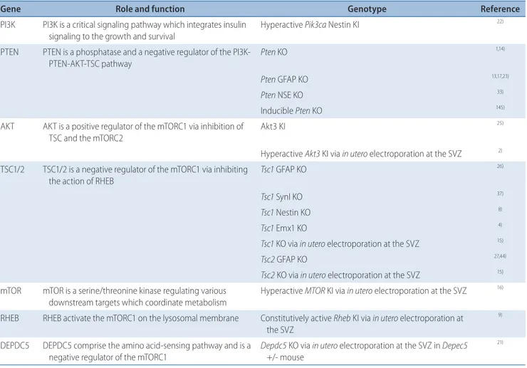
Table 2. Epilepsy animal models with genetic mTOR pathway hyperactivation
Gene Role and function Genotype Reference
PI3K PI3K is a critical signaling pathway which integrates insulin signaling to the growth and survival
Hyperactive Pik3ca Nestin KI 22)
PTEN PTEN is a phosphatase and a negative regulator of the
PI3K-PTEN-AKT-TSC pathway Pten KO
1,14)
Pten GFAP KO 13,17,23)
Pten NSE KO 33)
Inducible Pten KO 145)
AKT AKT is a positive regulator of the mTORC1 via inhibition of TSC and the mTORC2
Akt3 KI 25)
Hyperactive Akt3 KI via in utero electroporation at the SVZ 2) TSC1/2 TSC1/2 is a negative regulator of the mTORC1 via inhibiting
the action of RHEB Tsc1 GFAP KO
26)
Tsc1 SynI KO 37)
Tsc1 Nestin KO 8)
Tsc1 Emx1 KO 4)
Tsc1 KO via in utero electroporation at the SVZ 15)
Tsc2 GFAP KO 27,44)
Tsc2 KO via in utero electroporation at the SVZ 15) mTOR mTOR is a serine/threonine kinase regulating various
downstream targets which coordinate metabolism
Hyperactive MTOR KI via in utero electroporation at the SVZ 16) RHEB RHEB activate the mTORC1 on the lysosomal membrane Constitutively active Rheb KI via in utero electroporation at
the SVZ
9)
DEPDC5 DEPDC5 comprise the amino acid-sensing pathway and is a negative regulator of the mTORC1
Depdc5 KO via in utero electroporation at the SVZ in Depec5 +/- mouse
21)
mTOR : mechanistic target of rapamycin, PI3K : phosphatidylinositol-3-kinase, KI : knock-in, PTEN : phosphatase and tensin homologon chromosome 10, GFAP : glial fibrillary acidic protein, KO : knock-out, NSE : neuron specific enonlase, SVZ : subventricular zone, TSC : tuberous sclerosis, SynI : synapsin I, RHEB : Ras homologue enriched in brain
MTOR PATHWAY ACTIVATION IN
EPILEPTO-GENESIS
Epileptogenesis refers to the process leading to the first
spontaneous seizure after pro-epileptogenic insult (e.g.,
genet-ic defect, brain injury, status epileptgenet-icus, or cancer).
Epilepto-genesis is a brain-wide circuit rewiring process comprising
re-organization of microcircuits to long-range circuits, as well as
gliosis, blood-brain barrier damage, inflammation, and
neu-rodegeneration
114).
Since epilepsy is the consequence of electrical changes,
elec-trophysiological characteristics in animal models and epilepsy
patients have been analyzed in an attempt to understand the
epileptogenic mechanism. Studies have indicated that
high-frequency oscillations on electroencephalogram are
charac-teristic of FCD
20). Organotypic brain slice analysis of resected
FCD lesions has revealed an electrographic firing pattern of
ictal-like discharges after 4-aminopyridine treatment
5), which
decreased with treatment with GABA
Areceptor antagonist.
In brain slice cultures of hyperactive mTOR signaling,
re-searchers have found both the frequency and amplitude of the
miniature excitatory postsynaptic current to be increased in
glutamatergic synaptic transmission of the hippocampus
89,138).
In GABAergic neurons, hyperactive mTOR signaling increased
evoked synaptic responses in the hippocampus
148). In a subset of
auditory cortical neurons of Pten conditional KO, synaptic inputs
from the long-range connections, including the contralateral
au-ditory cortex and thalamus, and local connections were
in-creased
152).
In an epilepsy animal model of brain somatic mutations in
the mTOR pathway, mutation-carrying cortical neurons
showed an increased capacitance, increased cell size, reduced
input resistance indicative of a higher current needed to reach
the voltage threshold of the action potential, and increased
gain of firing frequency
120). Also, spontaneous excitatory
post-synaptic current frequency was decreased in the mTOR
path-way mutation-carrying cortical neurons
84,120).
There have been tremendous advances in the identification
of the contribution of mTOR downstream functions to
neu-ronal hypertrophy, dendritic branching, axon length, and
neuronal migration. Although numerous epilepsy animal
models have been utilized to unravel the path to
epileptogene-sis, the downstream mechanism of mTOR signaling that
ac-counts for epileptogenesis remains unclear. The complexity of
the downstream output of the mTOR pathway has hampered
attempts to understand the epileptogenic mechanism. Using
genetic manipulation techniques, normalizing major
down-stream outputs of hyperactive mTOR signaling, including
translation via overexpressing constitutively active 4E-BP or
knockdown of S6Ks
84)and autophagy via knockdown of
OFD1
110), have failed to prevent or even reduce epileptogenesis.
Further studies defining the downstream pathway of mTOR
in epileptogenesis will be necessary.
INHIBITION OF THE MTOR PATHWAY IN
TREAT-MENT OF EPILEPTIC DISORDERS
Researchers have hypothesized that epilepsy resulting from
mTOR pathway activation could be treated by mTOR
inhibi-tion, and mTOR inhibition therapies have been evaluated in
several clinical trials for epilepsy (Table 3). Rapalogs,
includ-ing sirolimus and everolimus, have been reported as
promis-ing new anti-epileptics because they can penetrate the blood
brain-barrier
71). Rapalogs have been extensively tested and
have gained the US Food and Drug Administration (FDA)
ap-proval as anti-epileptics in TSC
27,47,73).
The first study to show the beneficial effect of the rapalogs
on epilepsy did so in a mouse model of TSC
158). In this study,
early treatment with sirolimus prevented the development of
epilepsy. The anti-epileptic effect of rapalogs has also been
demonstrated in other genetic models of epilepsy with mTOR
pathway hyperactivation, including Pten
87)KO and Strada
KO
111). Recently, intractable epilepsy mouse models with brain
somatic mutation in the mTOR pathway were found to be
cured by rapalogs
63,82,83). Interestingly, epileptic seizure was
al-most completely suppressed by mTOR inhibitors in mouse
models of TSC KO or mTOR activating mutation, but only
partially suppressed in Pten KO mouse models
87). These
re-sults suggest that an independent mechanism of
epileptogene-sis distinct from that in TSC KO or mTOR mutation is present
in the Pten KO model. In nongenetic seizure models,
includ-ing the pilocarpine-induced epilepsy model, kainic
acid-in-duced epilepsy model, and absence epilepsy model, rapalogs
partially reduced seizures. Citraro et al.
31)reported a
represen-tative studies of the anti-epileptic effect of rapalogs in an
ani-mal model of intractable epilepsy.
Ta bl e 3 . C lini ca l s tu di es w ith t he m TO R i nh ib ito rs f or t he a nt i-e pi le pt ic e ffe ct s Ty pe o f s tu dy D ise ase D rug a nd do se N um be r a nd a ge o f p at ie nt (s ) D ur at io n o f tr ea tm en t An ti-ep ilep tic e ffec t Re fs Pr os pe ct iv e, o pen -lab el , p has e I / II cl in ic al t rial TS C Ev er oli m us 4.7 –5 .6 m g/ m 2 /d ay 16 p at ien ts ; 3 y ear -o ld o r o ld er M ed ian d ur at io n : 21 .5 m on th s (ra ng e, 4 .7– 34 .4 ) Re du ct io n i n s ei zur e f re qu en cy i n 9 /1 6 pa tie nt s 11 ) Cas e r ep or t TS C Ev er oli m us 4. 5 m g/m 2 /d ay 1 p at ien t; 1 0-ye ar -o ld m an 12 m on th s Ce ss at io n o f s ei zur e 20) Pr os pe ct iv e, m ul tic en ter , o pen -lab el , p has e I /II cl in ic al t rial TS C Ev er oli m us 5 m g/m 2 /d ay 20 p at ien ts ; m ed ian a ge : 8 y ear s (a ge r an ge , 2 –2 1) 12 w ee ks Re du ct io n i n s ei zur e f re qu en cy i n 1 7/ 20 pa tien ts , 4 o f t he se p at ien ts w er e sei zur e-fre e a t 1 2 w ee ks 24 ) Pr os pe ct iv e, d oub le -b lin d, par al le l-gr oup , p la ceb o-co nt ro lle d, m ul tic en ter p has e I II TS C Ev er oli m us 4. 5 m g/m 2 /d ay 8 p at ien ts , ch ildr en un der t he ag e o f 3 35 m on th s (ran ge , 3 3– 38 ) Ce ss at io n o f s ei zur es i n 1 p at ien t, sign ifi can t ( at l eas t a 5 0% ) r ed uc tio n i n th e n um ber o f s ei zur es i n 2 p at ien ts 10 ) Cas e s tu dy s er ie s TS C Ev er oli m us 5– 7 m g/ da y 6 p at ien ts ; m ed ian a ge : 5 y ear s (a ge r an ge , 2 –1 2) 36 we ek s Re du ct io n i n s ei zur e f re qu en cy i n 4 /6 pa tie nt s 29) O pen -lab el , s in gl e c en ter c as e ser ie s TS C Ev er oli m us 5 m g/d ay 1 p at ien ts ; 1 4-ye ar -o ld f em al e 18 m on th s 25 –5 0% s ei zur e r ed uc tio n 3) Cas e s tu dy TS C Ev er oli m us 5 m g/m 2 /d ay 1 p at ien t; 1 3. 5-ye ar -o ld f em al e 12 d ay s Sei zur e a ggr av at io n 30) Cas e s tu dy TS C Ev er oli m us 5 m g/d ay 1 p at ien t; 1 3-ye ar -o ld f em al e 1.5 y ear Re du ct io n i n s ei zur e f re qu en cy 28) Pr os pe ct iv e, o pen -lab el , p has e I / II cl in ic al t rial TS C Ev er oli m us 5 m g/m 2 /d ay 14 p at ien ts ; m ed ian a ge : 8 y ear s (a ge r an ge , 2 .0 –2 1.3 ) 48 m ont hs Re du ct io n i n s ei zur e f re qu en cy i n 1 3/ 14 pa tien ts ( ov er 5 0% s ei zur e r ed uc tio n) 12 ) Co re p has e, p has e I II, ran do m ize d, d oub le -b lin d, pl ace bo -co nt ro lle d TS C Ev er oli m us Lo w e xp os ur e gr oup ; 5 .2 m g/ m 2 /d ay (ra ng e, 1 .3– 14 .5 ) Hi gh e xp os ur e gr oup ; 7 .5 m g/ m 2 /d ay (ra ng e, 1 .4 –2 4.4 ) 36 6 p at ien ts ( in cl udi ng p la ceb o gr oup ); m ed ian a ge ; 1 0.1 y ear s (a ge r an ge , 2 .2 –5 6. 3) 18 w ee ks Re sp on se r at e; 1 5.1 % w ith p la ceb o 28 .2 % w ith l ow -e xp os ur e e ver ol im us 40. 0% w ith h igh -e xp os ur e e ver ol im us 7) Ex ten sio n p has e, p has e I II, ran do m ize d, d oub le -b lin d, pl ace bo -co nt ro lle d TS C Ev er oli m us 5– 9 m g/ m 2 /d ay 29 4 p at ien ts ; m ed ian a ge : 8 .7 y ear s (a ge r an ge , 2 .2 –1 8. 0) 1 y ear Su st ai ne d s ei zur e r ed uc tio n i n 4 8. 05 % ; M ed ian p er cen ta ge r ed uc tio n i n s ei zur e fre qu en cy : 4 8. 2% 5)
subependymal giant astrocytoma, kidney tumors, and partial
epilepsy in TSC patients. The first trial to test the efficacy of
everolimus to treat epileptic seizure was reported in 2010 for
TSC patients
74). In this study, seizure frequency was reduced in
nine of 16 patients with everolimus. Following studies
sup-ported the anti-epileptic effects of everolimus or sirolimus
(Table 3).
The prospective, randomized, multicenter,
placebo-con-trolled study testing the seizure suppression effect of
everoli-mus on seizure frequency in TSC patients with drug-resistant
epilepsy reported positive results
49). Recently, long-term results
of the prospective, open-label, non-randomized study of
everolimus for treating epileptic seizure in TSC indicated that
reduction in seizure frequency is sustained for up to 4 years
75).
This effect has been confirmed in the long-term follow-up of
the EXIST3 trial
35). Large-scale studies on the anti-epileptic
effect of everolimus have described response rates for
everoli-mus and a median seizure frequency reduction in respondents
of about 40%
35,75). In 2017 and 2018, respectively, both the
Eu-ropean Medicinal Agency in Europe and the US FDA
ap-proved everolimus as an adjunctive therapy in partial-onset
seizures in TSC patients aged 2 years and older. However,
there are reports of non-responders, seizure aggravation, or an
withdrawal after everolimus administration
35,149). Meanwhile,
clinical trials for treating epileptic seizure in FCD type II with
everolimus have recently started (clinicaltrials.gov identifier
NCT03198949).
The first report of the anti-epileptic effect of sirolimus in
TSC patients, which appears to be similar to that of
everolim-us
23), was in 2009
103). In HME patients with brain somatic
mu-tation in mTOR, sirolimus administration reduced seizures
153).
Interestingly, sirolimus has been shown to prevent epilepsy in
PMSE patients
111).
Animal and human treatment studies reported that
with-drawal of rapalogs provokes seizure recurrence
73). In
electro-physiological analysis of brain slices of resected epileptic foci,
everolimus was found to reduce spontaneous excitatory
post-synaptic activity, burst discharges, and epileptiform activity in
TSC, FCD, and HME cases; effects were subtle in epilepsy
pa-tients without mTOR mutations
27).
While rapalogs have been approved for use in various
hu-man diseases, concerns have been raised for their long-term
use and their adverse effect profile, including potentially
seri-ous adverse effects. The adverse effects of rapalogs include
im-Ta bl e 3 . C on tinu ed Ty pe o f s tu dy D ise ase D rug a nd d ose N um be r a nd a ge o f p at ie nt (s ) D ur at io n o f tr ea tm en t An ti-ep ilep tic e ffec t Re fs Cas e s tu dy s er ie s TS C Sir ol im us 1.5 m g/ kg/ da y 3 p at ien ts ; 1 5 y ear s ( ag e r an ge , 5. 5– 21 y ear s) M ed ian 4 m on th s (ran ge , 3 –5 ) Re du ct io n i n s ei zur e f re qu en cy i n 2 /3 pa tie nt s 6) Cas e r ep or t TS C Sir ol im us 0.15 m g/ kg /d ay 1 p at ien t; 9 -y ear -o ld f em al e 10 m on th s Re du ct io n i n s ei zur e f re qu en cy 18 ) Cas e s tu dy s er ie s PM SE Sir ol im us 1-5 m g/ m 2 /d ay 6 p at ien ts ; m ed ian a ge : 3 y ear s (a ge r an ge , 5 m on th s– 5 y ear ) 6 m ont hs Re du ct io n i n s ei zur e f re qu en cy 19 ) O pen -lab el , s in gl e c en ter c as e TS C Sir ol im us 1 m g/m 2 /d ay 6 p at ien ts ; m ed ian a ge : 6 y ear s (a ge r an ge , 3 –1 7) M ed ian d ur at io n : 18 m on th s (ran ge , 6 –3 6) O ver 5 0% r ed uc tio n i n s ei zur e f re qu en cy in 5 /6 p at ien ts 3) Cas e r ep or t HM E Sir ol im us 1 m g/m 2 /d ay 1 p at ien t; 3 -m on th -o ld m an 3 m ont hs Sei zur e r ed uc tio n ( ov er 5 0% ) 31) m TO R : m ech an ist ic t ar ge t o f r ap am yci n, T SC : t ub er ou s s cl er os is, P M SE : p ol yh ydr am ni os , m eg al en cep hal y, an d s ym pt om at ic ep ilep sy s yn dr om e, H M E : h em im eg al en cep hal y
munosuppression, mucositis, hyperlipidemia, hyperglycemia,
diabetes-like syndrome, and fatal pneumonitis
42,76,109,139). In a
phase III clinical trial with everolimus, over 90% of 111
pa-tients experienced an adverse effects of any grade
48). With
everolimus treatment for over 1 year, grade 3 or 4 adverse
events, which are severe adverse events, were reported in 45%
of younger patients and 38% of older patients among 150
pa-tients
35). We should stress that developmental delay caused by
rapalogs
63)should be a concern, considering that the onset of
intractable epilepsy with mTOR pathway mutation is usually
before adolescence
4). Additionally, treatment with rapalogs has
been found to induce adverse nervous system-specific effects
altering sociability
129), learning and memory
16,25), and anxiety
56)in mouse models.
For epilepsy caused by brain somatic mutations, therapeutic
regimens must seek to avoid perturbations outside of the CNS,
and in this regard, antisense oligonucleotide (ASO) drugs appear
to be promising therapeutic options. ASO is a complementary
oligonucleotide sequence of sense mRNA sequences that
ham-pers normal gene expression processes, including splicing,
tran-scription, and translation
136). The characteristics of the
blood-brain barrier prevent ASO from permeating outside of the CNS
when administered into the cerebrospinal fluid via intrathecal
in-jection. Moreover, the half-life of ASO drugs is more than 3
months
19). Recently, ASO drugs have been approved by the FDA
for treating various types of neurodegenerative disorders
45). We
suspect that intrathecal injection of ASO drugs targeting mTOR
itself, mTOR mutation, or downstream targets of mTOR will
prove effective in treating intractable epilepsies while avoiding the
adverse effects of rapalogs outside of the CNS.
CONCLUSION
Over the last two decades, extensive research to outline the
roles of the mTOR pathway, to identify mutations in the
mTOR pathway in human epilepsy patients, and to develop
mTOR inhibitors have led to the discovery of novel strategies
for diagnosing and treating intractable epilepsy. The
overarch-ing goal of this clinical research is attainoverarch-ing seizure-free status
with few to no adverse effects in epilepsy patients. While
rapa-logs have proven to be effective in controlling seizures,
com-plete seizure cessation has not been recorded in most of
pa-tients
35,75). Also, about half of all TSC patients fail to respond
to rapalogs. These discrepancies suggest that there might be
an unknown biological mechanism at play. Considering about
40% of patients treated with rapalogs experience a severe
ad-verse effect, it will be necessary to develop more potent and
tolerable therapies.
While FMCD patients with drug-resistant epilepsy typically
undergo surgical resection of the epileptic focus for treating
the seizure, up to 50% of patients do not respond to therapy.
Based on preclinical and clinical studies of TSC with rapalogs,
it may no longer be necessary to perform highly invasive
sur-gical resection for seizure treatment. However, there is a
prob-lem in that diagnosing FMCD patients with low-frequency
brain somatic mutation in the mTOR pathway requires
ana-lyzing DNA from brain tissue after surgical resection. To
alle-viate this issue, minimally invasive diagnosis through
bio-markers in patient cerebrospinal fluid would prove invaluable.
CONFLICTS OF INTEREST
J.H.L is a co-founder of SoVarGen, Inc. that develops new
diagnostics and therapeutics for brain disorders. The
remain-ing authors declare no competremain-ing financial interests.
INFORMED CONSENT
Informed consent was obtained from all individual
partici-pants included in this study.
●
Acknowledgements
This work was supported by grants from the Suh Kyungbae
Foundation (to J.H.L.), Korean Health Technology R&D Project,
Ministry of Health & Welfare, Republic of Korea (H15C3143 and
H16C0415 to J.H.L.), and Cheong-Am Science Fellow supported
by POSCO Cheong-Am Foundation (to J.K.K).
●
Supplementary materials
The online-only data supplement is available with this
arti-cle at https://doi.org/10.3340/jkns.2019.0027.
References
1. Abu-Remaileh M, Wyant GA, Kim C, Laqtom NN, Abbasi M, Chan SH, et al. : Lysosomal metabolomics reveals V-ATPase- and mTOR-dependent regulation of amino acid efflux from lysosomes. Science 358 : 807-813, 2017
2. Alcantara D, Timms AE, Gripp K, Baker L, Park K, Collins S, et al. : Mutations of AKT3 are associated with a wide spectrum of developmental disorders including extreme megalencephaly. Brain 140 : 2610-2622, 2017 3. Alfaiz AA, Micale L, Mandriani B, Augello B, Pellico MT, Chrast J, et al. :
TBC1D7 mutations are associated with intellectual disability, macrocrania, patellar dislocation, and celiac disease. Hum Mutat 35 : 447-451, 2014 4. Aronica E, Becker AJ, Spreafico R : Malformations of cortical
develop-ment. Brain Pathol 22 : 380-401, 2012
5. Avoli M, Bernasconi A, Mattia D, Olivier A, Hwa GG : Epileptiform discharges in the human dysplastic neocortex: in vitro physiology and pharmacology. Ann Neurol 46 : 816-826, 1999
6. Backman SA, Stambolic V, Suzuki A, Haight J, Elia A, Pretorius J, et al. : Deletion of Pten in mouse brain causes seizures, ataxia and defects in soma size resembling Lhermitte-Duclos disease. Nat Genet 29 : 396-403, 2001
7. Baek ST, Copeland B, Yun EJ, Kwon SK, Guemez-Gamboa A, Schaffer AE, et al. : An AKT3-FOXG1-reelin network underlies defective migra-tion in human focal malformamigra-tions of cortical development. Nat Med 21 : 1445-1454, 2015
8. Banko JL, Poulin F, Hou L, DeMaria CT, Sonenberg N, Klann E : The translation repressor 4E-BP2 is critical for eIF4F complex formation, synaptic plasticity, and memory in the hippocampus. J Neurosci 25 : 9581-9590, 2005
9. Baple EL, Maroofian R, Chioza BA, Izadi M, Cross HE, Al-Turki S, et al. : Mutations in KPTN cause macrocephaly, neurodevelopmental delay, and seizures. Am J Hum Genet 94 : 87-94, 2014
10. Bar-Peled L, Schweitzer LD, Zoncu R, Sabatini DM : Ragulator is a GEF for the rag GTPases that signal amino acid levels to mTORC1. Cell 150 : 1196-1208, 2012
11. Basel-Vanagaite L, Hershkovitz T, Heyman E, Raspall-Chaure M, Kakar N, Smirin-Yosef P, et al. : Biallelic SZT2 mutations cause infantile encepha-lopathy with epilepsy and dysmorphic corpus callosum. Am J Hum Genet 93 : 524-529, 2013
12. Bast T, Ramantani G, Seitz A, Rating D : Focal cortical dysplasia: preva-lence, clinical presentation and epilepsy in children and adults. Acta Neurol Scand 113 : 72-81, 2006
13. Baulac S : MTOR signaling pathway genes in focal epilepsies in Ros-signol E, Carmant L, Lacaille JC (eds) : Progress in Brain Research. Amsterdam : Elsevier, 2016, Vol 226, pp61-79
14. Baulac S, Ishida S, Marsan E, Miquel C, Biraben A, Nguyen DK, et al. : Familial focal epilepsy with focal cortical dysplasia due to DEPDC5 muta-tions. Ann Neurol 77 : 675-683, 2015
15. Baybis M, Yu J, Lee A, Golden JA, Weiner H, McKhann G 2nd, et al. : mTOR cascade activation distinguishes tubers from focal cortical dyspla-sia. Ann Neurol 56 : 478-487, 2004
16. Beaumont V, Zhong N, Fletcher R, Froemke RC, Zucker RS : Phosphoryla-tion and local presynaptic protein synthesis in calcium- and calcineurin-dependent induction of crayfish long-term facilitation. Neuron 32 : 489-501, 2001
17. Ben-Sahra I, Howell JJ, Asara JM, Manning BD : Stimulation of de novo pyrimidine synthesis by growth signaling through mTOR and S6K1. Sci-ence 339 : 1323-1328, 2013
18. Bercury KK, Dai J, Sachs HH, Ahrendsen JT, Wood TL, Macklin WB : Conditional ablation of raptor or rictor has differential impact on oligo-dendrocyte differentiation and CNS myelination. J Neurosci 34 : 4466-4480, 2014
19. Bishop KM : Progress and promise of antisense oligonucleotide thera-peutics for central nervous system diseases. Neuropharmacology 120 : 56-62, 2017
20. Blümcke I : Neuropathology of focal epilepsies: a critical review. Epi-lepsy Behav 15 : 34-39, 2009
21. Brown EJ, Beal PA, Keith CT, Chen J, Shin TB, Schreiber SL : Control of p70 S6 kinase by kinase activity of FRAP in vivo. Nature 377 : 441-446, 1995
22. Capo-Chichi JM, Tcherkezian J, Hamdan FF, Décarie JC, Dobrzeniecka S, Patry L, et al. : Disruption of TBC1D7, a subunit of the TSC1-TSC2 protein complex, in intellectual disability and megalencephaly. J Med Genet 50 : 740-744, 2013
23. Cardamone M, Flanagan D, Mowat D, Kennedy SE, Chopra M, Lawson JA : Mammalian target of rapamycin inhibitors for intractable epilepsy and subependymal giant cell astrocytomas in tuberous sclerosis com-plex. J Pediatr 164 : 1195-1200, 2014
24. Carvill GL, Crompton DE, Regan BM, McMahon JM, Saykally J, Zemel M, et al. : Epileptic spasms are a feature of DEPDC5 mTORopathy. Neurol Genet 1 : e17, 2015
25. Casadio A, Martin KC, Giustetto M, Zhu H, Chen M, Bartsch D, et al. : A transient, neuron-wide form of creb-mediated long-term facilitation can be stabilized at specific synapses by local protein synthesis. Cell 99 : 221-237, 1999
26. Cen Z, Guo Y, Lou Y, Jiang B, Wang J, Feng J : De novo mutation in DEPDC5 associated with unilateral pachygyria and intractable epilepsy. Seizure 50 : 1-3, 2017
27. Cepeda C, Levinson S, Yazon VW, Barry J, Mathern GW, Fallah A, et al. : Cellular antiseizure mechanisms of everolimus in pediatric tuberous scle-rosis complex, cortical dysplasia, and non-mTOR-mediated etiologies. Epilepsia Open 3(Suppl Suppl 2) : 180-190, 2018
28. Chen HH, Chen C, Hung SC, Liang SY, Lin SC, Hsu TR, et al. : Cognitive and epilepsy outcomes after epilepsy surgery caused by focal cortical dysplasia in children: early intervention maybe better. Child’s Nerv Syst 30 : 1885-1895, 2014
29. Cheung KM, Lam CW, Chan YK, Siu WK, Yong L : Atypical focal cortical dysplasia in a patient with Cowden syndrome. Hong Kong Med J 20 : 165-167, 2014
30. Child ND, Cascino GD : Mystery case: Cowden syndrome presenting with partial epilepsy related to focal cortical dysplasia. Neurology 81 : e98-e99, 2013
31. Citraro R, Leo A, Constanti A, Russo E, De Sarro G : mTOR pathway inhibition as a new therapeutic strategy in epilepsy and epileptogenesis. Pharmacol Res 107 : 333-343, 2016
32. Crino PB : Focal brain malformations: a spectrum of disorders along the mTOR cascade. Novartis Found Smyp 288 : 260-272; discussion 272-281, 2007
33. Crino PB : The mTOR signalling cascade: paving new roads to cure neu-rological disease. Nat Rev Neurol 12 : 379-392, 2016
34. Cunningham JT, Rodgers JT, Arlow DH, Vazquez F, Mootha VK, Puig-server P : mTOR controls mitochondrial oxidative function through a YY1-PGC-1a transcriptional complex. Nature 450 : 736-740, 2007 35. Curatolo P, Franz DN, Lawson JA, Yapici Z, Ikeda H, Polster T, et al. :
Adjunctive everolimus for children and adolescents with treatment-refractory seizures associated with tuberous sclerosis complex: post-hoc analysis of the phase 3 EXIST-3 trial. Lancet Child Adolesc Health 2 : 495-504, 2018
36. D’Gama AM, Geng Y, Couto JA, Martin B, Boyle EA, LaCoursiere CM, et al. : Mammalian target of rapamycin pathway mutations cause hemi-megalencephaly and focal cortical dysplasia. Ann Neurol 77 : 720-725, 2015
37. De Benedetti A, Joshi-Barve S, Rinker-Schaeffer C, Rhoads RE : Expres-sion of antisense RNA against initiation factor eIF-4E mRNA in HeLa cells results in lengthened cell division times, diminished translation rates, and reduced levels of both eIF-4E and the p220 component of eIF-4F. Mol Cell Biol 11 : 5435-5445, 1991
38. Dibble CC, Elis W, Menon S, Qin W, Klekota J, Asara JM, et al. : TBC1D7 is a third subunit of the TSC1-TSC2 complex upstream of mTORC1. Mol Cell 47 : 535-546, 2012
39. Dorrello NV, Peschiaroli A, Guardavaccaro D, Colburn NH, Sherman NE, Pagano M : S6K1- and ßTRCP-mediated degradation of PDCD4 promotes protein translation and cell growth. Science 314 : 467-471, 2006 40. Dutchak PA, Laxman S, Estill SJ, Wang C, Wang Y, Wang Y, et al. :
Regulation of hematopoiesis and methionine homeostasis by mTORC1 inhibitor NPRL2. Cell Rep 12 : 371-379, 2015
41. Ehninger D, Han S, Shilyansky C, Zhou Y, Li W, Kwiatkowski DJ, et al. : Reversal of learning deficits in a Tsc2+/- mouse model of tuberous
sclero-sis. Nat Med 14 : 843-848, 2008
42. Eisen T, Sternberg CN, Robert C, Mulders P, Pyle L, Zbinden S, et al. : Targeted therapies for renal cell carcinoma: review of adverse event management strategies. J Natl Cancer Inst 104 : 93-113, 2012 43. Eng CP, Sehgal SN, Vézina C : Activity of rapamycin (AY-22,989) against
transplanted tumors. J Antibiot (Tokyo) 37 : 1231-1237, 1984 44. European Chromosome 16 Tuberous Sclerosis Consortium :
Identifica-tion and characterizaIdentifica-tion of the tuberous sclerosis gene on chromosome 16. Cell 75 : 1305-15, 1993
45. Finkel RS, Mercuri E, Darras BT, Connolly AM, Kuntz NL, Kirschner J, et al. : Nusinersen versus sham control in infantile-onset spinal muscular atrophy. N Engl J Med 337 : 1723-1732, 2017
46. Frankel WN, Yang Y, Mahaffey CL, Beyer BJ, O’Brien TP : Szt2, a novel gene for seizure threshold in mice. Genes Brain Behav 8 : 568-576, 2009 47. Franz DN, Agricola K, Mays M, Tudor C, Care MM, Holland-Bouley K,
et al. : Everolimus for subependymal giant cell astrocytoma: 5-year final analysis. Ann Neurol 78 : 929-938, 2015
48. Franz DN, Belousova E, Sparagana S, Bebin EM, Frost M, Kuperman R, et al. : Everolimus for subependymal giant cell astrocytoma in patients with tuberous sclerosis complex: 2-year open-label extension of the ran-domised EXIST-1 study. Lancet Oncol 15 : 1513-1520, 2014
49. French JA, Lawson JA, Yapici Z, Ikeda H, Polster T, Nabbout R, et al. : Ad-junctive everolimus therapy for treatment-resistant focal-onset seizures associated with tuberous sclerosis (EXIST-3): a phase 3, randomised, double-blind, placebo-controlled study. Lancet 388 : 2153-2163, 2016 50. Gingras A, Kennedy SG, O’Leary MA, Sonenberg N, Hay N : 4E-BP1, a
repressor of mRNA translation, is phosphorylated by the Akt(PKB) sig-naling pathway. Genes Dev 12 : 502-513, 1998
51. Gingras AC, Gygi SP, Raught B, Polakiewicz RD, Abraham RT, Hoekstra MF, et al. : Regulation of 4E-BP1 phosphorylation : a novel two-step mechanism. Genes Dev 13 : 1422-1437, 1999
52. Gingras AC, Raught B, Sonenberg N : eIF4 initiation factors : effectors of mRNA recruitment of translation. Annu Rev Biochem 68 : 913-963, 1999 53. Gong X, Zhang L, Huang T, Lin T V., Miyares L, Wen J, et al. : Activat-ing the translational repressor 4E-BP or reducActivat-ing S6K-GSK3ß activity prevents accelerated axon growth induced by hyperactive mTOR in vivo. Hum Mol Genet 24 : 5746-5758, 2015
54. Gordo G, Tenorio J, Arias P, Santos-Simarro F, García-Miñaur S, Moreno JC, et al. : mTOR mutations in Smith-Kingsmore syndrome: four addi-tional patients and a review. Clin Genet 93 : 762-775, 2018
55. Gu X, Orozco JM, Saxton RA, Condon KJ, Liu GY, Krawczyk PA, et al. : SAMTOR is an S-adenosylmethionine sensor for the mTORC1 pathway. Science 358 : 813-818, 2017
56. Hadamitzky M, Herring A, Kirchhof J, Bendix I, Haight MJ, Keyvani K, et al. : Repeated systemic treatment with rapamycin affects behavior and amygdala protein expression in rats. Int J Neuropsychopharmacol 21 : 592-602, 2018
57. Han JM, Jeong SJ, Park MC, Kim G, Kwon NH, Kim HK, et al. : Leucyl-tRNA synthetase is an intracellular leucine sensor for the mTORC1-signaling pathway. Cell 149 : 410-424, 2012
58. Heitman J, Movva NR, Hall MN : Targets for cell cycle arrest by the im-munosuppressant rapamycin in yeast. Science 253 : 905-909, 1991 59. Hentges K, Thompson K, Peterson A : The flat-top gene is required for
the expansion and regionalization of the telencephalic primordium. De-velopment 126 : 1601-9, 1999
60. Hiremath LS, Webb NR, Rhoads RE : Immunological detection of the mes-senger RNA cap-binding protein. J Biol Chem 260 : 7843-7849, 1985 61. Holz MK, Ballif BA, Gygi SP, Blenis J : mTOR and S6K1 mediate assembly
of the translation preinitiation complex through dynamic protein inter-change and ordered phosphorylation events. Cell 123 : 569-580, 2005 62. Hou L, Klann E : Activation of the phosphoinositide 3-kinase-Akt-mam-malian target of rapamycin signaling pathway is required for metabo-tropic glutamate receptor-dependent long-term depression. J Neurosci 24 : 6352-6361, 2004
63. Hsieh LS, Wen JH, Claycomb K, Huang Y, Harrsch FA, Naegele JR, et al. : Convulsive seizures from experimental focal cortical dysplasia occur
in-dependently of cell misplacement. Nat Commun 7 : 11753, 2016 64. Huber KM, Kayser MS, Bear MF : Role for rapid dendritic protein
synthe-sis in hippocampal depression. Science 288 : 1254-1257, 2000 65. Hughes J, Dawson R, Tea M, McAninch D, Piltz S, Jackson D, et al. :
Knockout of the epilepsy gene Depdc5 in mice causes severe embryonic dysmorphology with hyperactivity of mTORC1 signalling. Sci Rep 7 : 12618, 2017
66. Iffland PH 2nd, Crino PB : Focal cortical dysplasia: gene mutations, cell signaling, and therapeutic implications. Annu Rev Pathol 12 : 547-571, 2017
67. Inoki K, Li Y, Xu T, Guan KL : Rheb GTPase is a direct target of TSC2 GAP activity and regulates mTOR signaling. Genes Dev 17 : 1829-1834, 2003
68. Jansen LA, Mirzaa GM, Ishak GE, O’Roak BJ, Hiatt JB, Roden WH, et al. : PI3K/AKT pathway mutations cause a spectrum of brain malformations from megalencephaly to focal cortical dysplasia. Brain 138(Pt 6) : 1613-1628, 2015
69. Jung J, Genau HM, Behrends C : Amino acid-dependent mTORC1 regula-tion by the lysosomal membrane protein SLC38A9. Mol Cell Biol 35 : 2479-2494, 2015
70. Keppler-Noreuil KM, Parker VE, Darling TN, Martinez-Agosto JA : Somatic overgrowth disorders of the PI3K/AKT/mTOR pathway & thera-peutic strategies. Am J Med Genet C Semin Med Genet 172 : 402-421, 2016
71. Klawitter J, Gottschalk S, Hainz C, Leibfritz D, Christians U, Serkova NJ : Im-munosuppressant neurotoxicity in rat brain models: oxidative stress and cellular metabolism. Chem Res Toxicol 23 : 608-619, 2010
72. Kowalczyk MS, Hughes JR, Babbs C, Sanchez-Pulido L, Szumska D, Sharpe JA, et al. : Nprl3 is required for normal development of the car-diovascular system. Mamm Genome 23 : 404-415, 2012
73. Krueger DA, Care MM, Agricola K, Tudor C, Mays M, Franz DN : Evero-limus long-term safety and efficacy in subependymal giant cell astrocy-toma. Neurology 80 : 574-580, 2013
74. Krueger DA, Care MM, Holland K, Agricola K, Tudor C, Mangeshkar P, et al. : Everolimus for subependymal giant-cell astrocytomas in tuberous sclerosis. N Engl J Med 369 : 1801-1811, 2010
75. Krueger DA, Wilfong AA, Mays M, Talley CM, Agricola K, Tudor C, et al. : Long-term treatment of epilepsy with everolimus in tuberous sclerosis. Neurology 87 : 2408-2415, 2016
76. Kuegler PB, Zimmer B, Waldmann T, Baudis B, Ilmjärv S, Hescheler J, et al. : Markers of murine embryonic and neural stem cells, neurons and astrocytes: reference points for developmental neurotoxicity testing. ALTEX 27 : 17-42, 2010
77. Kumar V, Zhang MX, Swank MW, Kunz J, Wu G : Regulation of dendritic morphogenesis by Ras-PI3K-Akt-mTOR and Ras-MAPK signaling path-ways. J Neurosci 25 : 11288-11299, 2005
78. Kwon CH, Zhu X, Zhang J, Knoop LL, Tharp R, Smeyne RJ, et al. : Pten regulates neuronal soma size: a mouse model of Lhermitte-Duclos dis-ease. Nat Genet 29 : 404-411, 2001
79. Lee JH, Huynh M, Silhavy JL, Kim S, Dixon-Salazar T, Heiberg A, et al. : De novo somatic mutations in components of the PI3K-AKT3-mTOR
path-way cause hemimegalencephaly. Nat Genet 44 : 941-945, 2012 80. Lee SK, Kim DW : Focal cortical dysplasia and epilepsy surgery. J
Epi-lepsy Res 3 : 43-47, 2013
81. Li N, Lee B, Liu RJ, Banasr M, Dwyer JM, Iwata M, et al. : mTOR-dependent synapse formation underlies the rapid antidepressant effects of NMDA antagonists. Science 329 : 959-964, 2010
82. Lim JS, Gopalappa R, Kim SH, Ramakrishna S, Lee M, Kim WI, et al. : Somatic mutations in TSC1 and TSC2 cause focal cortical dysplasia. Am J Hum Genet 100 : 454-472, 2017
83. Lim JS, Kim WI, Kang HC, Kim SH, Park AH, Park EK, et al. : Brain so-matic mutations in MTOR cause focal cortical dysplasia type II leading to intractable epilepsy. Nat Med 21 : 395-400, 2015
84. Lin TV, Hsieh L, Kimura T, Malone TJ, Bordey A : Normalizing translation through 4E-BP prevents mTOR-driven cortical mislamination and ame-liorates aberrant neuron integration. Proc Natl Acad Sci U S A 113 : 11330-11335, 2016
85. Lipton JO, Sahin M : The neurology of mTOR. Neuron 84 : 275-291, 2014
86. Ljungberg MC, Bhattacharjee MB, Lu Y, Armstrong DL, Yoshor D, Swann JW, et al. : Activation of mammalian target of rapamycin in cytomegalic neurons of human cortical dysplasia. Ann Neurol 60 : 420-429, 2006 87. Ljungberg MC, Sunnen CN, Lugo JN, Anderson AE, D’Arcangelo G :
Rapamycin suppresses seizures and neuronal hypertrophy in a mouse model of cortical dysplasia. Dis Model Mech 2 : 389-398, 2009 88. Long X, Lin Y, Ortiz-Vega S, Yonezawa K, Avruch J : Rheb binds and
regulates the mTOR kinase. Curr Biol 15 : 702-713, 2005
89. Luikart BW, Schnell E, Washburn EK, Bensen AL, Tovar KR, Westbrook GL : Pten knockdown in vivo increases excitatory drive onto dentate granule cells. J Neurosci 31 : 4345-4354, 2011
90. Marchese M, Conti V, Valvo G, Moro F, Muratori F, Tancredi R, et al. : Autism-epilepsy phenotype with macrocephaly suggests PTEN, but not GLIALCAM, genetic screening. BMC Med Genet 15 : 26, 2014 91. Marsan E, Baulac S : Review: mechanistic target of rapamycin (mTOR)
pathway, focal cortical dysplasia and epilepsy. Neuropathol Appl Neurobiol 44 : 6-17, 2018
92. Marsan E, Ishida S, Schramm A, Weckhuysen S, Muraca G, Lecas S, et al. : Depdc5 knockout rat: a novel model of mTORopathy. Neurobiol Dis 89 : 180-189, 2016
93. Martel RR, Klicius J, Galet S : Inhibition of the immune response by ra-pamycin, a new antifungal antibiotic. Can J Physiol Pharmacol 55 : 48-51, 1977
94. Martinez-Lizana E, Fauser S, Brandt A, Schuler E, Wiegand G, Doostkam S, et al. : Long-term seizure outcome in pediatric patients with focal cortical dysplasia undergoing tailored and standard surgical resections. Seizure 62 : 66-73, 2018
95. Mc Cormack A, Sharpe C, Gregersen N, Smith W, Hayes I, George AM, et al. : 12q14 microdeletions: additional case series with confirmation of a macrocephaly region. Case Rep Genet 2015 : 192071, 2015 96. Meikle L, Talos DM, Onda H, Pollizzi K, Rotenberg A, Sahin M, et al. :
A mouse model of tuberous sclerosis: neuronal loss of Tsc1 causes dys-plastic and ectopic neurons, reduced myelination, seizure activity, and