Impact of statin treatment
on strut coverage
after drug-eluting stent implantation
Yongsung Suh
Department of Medicine
Impact of statin treatment
on strut coverage
after drug-eluting stent implantation
Directed by Professor Myeong-Ki Hong
The Master's Thesis
submitted to the Department of Medicine,
the Graduate School of Yonsei University
in partial fulfillment of the requirements for the
degree of Master of Medical Science
Yongsung Suh
June 2014
This certifies that the Master's
Thesis of Yongsung Suh is
approved.
---
Thesis Supervisor: Myeong-Ki Hong
---
Thesis Committee Member: Jong Yun Won
---
Thesis Committee Member: Byeong-Keuk Kim
The Graduate School
Yonsei University
ACKNOWLEDGEMENTS
First of all, I really appreciate my thesis supervisor,
professor Myeong-Ki Hong, for his supervision and
encouragement to study this subject. Thanks to
professor Hong, I had this great opportunity to
experience this study.
I also appreciate professors Jong Yoon Won and
Byeong-Keuk Kim who gave me experienced advices
and warm supports
I am grateful to my family members, especially my
parents. They have tolerated and supported the years of
my study with patience and love. Also thanks to my
little girl, Jeong-in. I give my love and admiration to
<TABLE OF CONTENTS>
ABSTRACT ... 1
I. INTRODUCTION ... 3
II. MATERIALS AND METHODS ... 4
1. Study design and patient enrollment ... 4
2. Coronary intervention and quantitative coronary angiography analysis 6 3. OCT imaging and cross-sectional analysis ... 6
4. Statistical analysis ... 7 III. RESULTS ... 7 IV. DISCUSSION ... 14 1. Study limitation ... 16 V. CONCLUSION ... 16 REFERENCES ... 17 ABSTRACT(IN KOREAN) ... 22
LIST OF FIGURES
Figure 1. Flowchart of study ... 5
Figure 2. Relationship between the percentage of uncovered struts
versus follow-up low-density lipoprotein (LDL) cholesterol level
(A) or the level of LDL cholesterol reduction (B) ... 16
LIST OF TABLES
Table 1. Baseline clinical and angiographic characteristics between
pitavastatin- and pravastatin-treated groups ... 9
Table 2. Optical coherence tomographic findings of pitavastatin and
pravastatin-treated groups ... 16
1
<ABSTRACT>
Impact of statin treatment on strut coverage
after drug-eluting stent implantation
Yongsung Suh
Department of Medicine
The Graduate School, Yonsei University
(Directed by Professor Myeong-Ki Hong)
Background:
There is no human data to evaluate impact of statin treatment on strut coverage after drug-eluting stent (DES) implantation.
Methods:
A total of 60 patients were randomly treated with pitavastatin 2mg or pravastatin 20mg for 6 months after DES implantation. Selection of DES was also randomly assigned to sirolimus-eluting stents (SES) or biolimus-eluting stents (BES). The degree of strut coverage was assessed with 6-month follow-up optical coherence tomography (OCT) which was performed in 52 DES-treated patients.
Results:
2
patients (n=25) and 19.1±15.2% in pravastain-treated patients (n=27) (p=0.927); 23.3±16.6% in SES-treated patients (n=28) and 14.5±10.9% in BES-treated patients (n=24) (p=0.026). Lower percentage of uncovered struts was significantly correlated with lower level of follow-up low-density lipoprotein (LDL) cholesterol (r=0.486, p=0.009) and greater reduction of LDL cholesterol level (r=-0.456, p=0.015) in SES-treated patients, but not BES-treated patients. In SES-treated patients, the percentage of uncovered struts was significantly lower in those who follow-up LDL cholesterol level less than 70mg/dL at 6 months follow-up (10.1±12.4% vs. 26.9±15.6%, p=0.025); but there were no significant differences of percentage of uncovered struts between patients who had 6-month follow-up LDL cholesterol level less vs. greater than 70mg/dL in BES-treated patients (14.6±12.7% vs. 14.4±10.5%, p=0.971).
Conclusion:
lower level of follow-up LDL cholesterol, especially less than 70mg/dL, might have a protective effect against delayed strut coverage after DES implantation. This vascular healing effect of lower LDL cholesterol level could be different according to types of DES.
--- Key words : Stents, Optical coherence tomography, Statins
3
Impact of statin treatment on strut coverage
after drug-eluting stent implantation
Yongsung Suh
Department of Medicine
The Graduate School, Yonsei University
(Directed by Professor Myeong-Ki Hong)
I. INTRODUCTION
Neointimal coverage over stent struts has been emerged as a clinical issue since it was reported that incomplete strut coverage might be associated with occurrence of late stent thrombosis following drug-eluting stent (DES) implantation.1-4 Optical coherence tomography (OCT) is one of the useful tools among currently available intravascular imaging devices, which has excellent resolution to demonstrate neointima and stent strut coverage.5,6 Although several characteristics such as sirolimus-eluting stent (SES)7,8 and acute coronary syndrome 9,10 were reported as risk factors for delayed strut coverage in previous OCT studies, the relationship of specific medication (i.e. statin) with strut coverage have not been fully understood. Statin is the most widely used lipid-lowering agent in patients with coronary artery disease, which has the pleotropic effect to reduce inflammation and enhance endothelial function.11 Previous in vitro and animal studies reported that statin regulate migration and proliferation of smooth muscle cells, and control neointimal formation.12-15 However, there has been no human data to assess the impact of statin treatment on strut coverage after DES
4
the effect of statin treatment on DES strut coverage.
II. MATERIALS AND METHODS 1. Study design and patient enrollment.
This study is a subgroup analysis to evaluate impact of statin treatment on strut coverage in a previous randomized OCT study16. The previous OCT study was performed to compare strut coverage at 6-month follow-up after the Nobori biolimus-eluting stent (N-BES, Nobori®, Terumo Corporation, Tokyo, Japan; n=60 patients) versus SES (CypherTM, Cordis Corp, Miami Lakes, FL, USA; n=60 patients) implantation. Inclusion and exclusion criteria of that OCT study were provided in the previous report.16 Written informed consents were obtained from all participants and the institutional review boards of our institute approved this study. A total of sixty patients were enrolled in this study and randomly treated with pitavastatin 2mg or pravastatin 20mg from the day of stent implantation. SES or N-BES was also randomly allocated. Thus, four groups were classified as follows: pitavastatin-N-BES group (12 patients), pitavastatin-SES group (17 patients), pravastatin-N-BES group (18 patients) and pravastatin-SES group (13 patients). Post-intervention OCT examination was performed in all patients immediately after DES implantation. Among 60 patients, 6-month follow-up angiogram was not performed in 3 patients; the OCT catheter could not be passed through lesion due to severe angulation in 3 patients; and there was poor image quality in 2 patients. Therefore, follow up OCT evaluation was available in 52 patients as follows: pitavastatin-N-BES group (10 patients), pitavastatin-SES group (15 patients), pravastatin-N-BES group (14 patients) and pravastatin-SES group (13 patients) (Figure 1). Blood samples to evaluate lipid profiles were obtained at the time of stent implantation and follow up angiography. All patients were clinically followed up 1, 3, 6 months after stent implantation.
5
Figure 1. Flowchart of study is shown.
2. Coronary intervention and quantitative coronary angiography analysis. All patients received at least 75mg of aspirin and a loading dose of 300mg of clopidogrel at least 12 hours pre-intervention. Unfractionated heparin was administered to maintain the activated clotting time >250 seconds. All percutaneous coronary interventions were performed according to current standard techniques. Post-intervention, dual antiplatelet therapy with aspirin 100mg and clopidogrel 75mg daily was prescribed for 12 months.
Quantitative coronary angiography analysis was performed before and after stent implantation and at follow-up using an off-line quantitative coronary angiographic system (CASS system, Pie Medical Instruments, Maastricht, Netherlands) in an independent core laboratory (Cardiovascular Research
6
Center, Seoul, Korea). Using the guiding catheter for magnification-calibration, reference vessel diameters and minimal luminal diameter were measured from diastolic frames in a single, matched view showing the smallest minimal luminal diameter. Late loss was defined as the difference between post-procedure and follow-up minimal luminal diameter.
3. OCT imaging and cross-sectional analysis
Post-intervention and at six months, OCT imaging of the target lesion was performed using a frequency-domain OCT system (C7-XR OCT imaging system, LightLab Imaging, Inc., St. Jude Medical, St. Paul, MN). In this study, OCT cross-sectional images were generated at a rotational speed of 100 frames/s while the fiber was withdrawn at a speed of 20 mm/s within the stationary imaging sheath. All OCT images were analyzed at a core laboratory (Cardiovascular Research Center, Seoul, Korea) by analysts who were blinded to patient and procedural information.
Cross-sectional OCT images were analyzed at 0.2mm longitudinal intervals. Stent and luminal cross-sectional areas (CSAs) were measured; neointimal hyperplasia (NIH) CSA was calculated as the stent minus luminal CSA. NIH thickness was measured as the distance between the endoluminal surface of the neointima and the strut.8 An uncovered strut was defined as having a NIH thickness=0µm.8 A malapposed strut was defined as a strut that had detached from the vessel wall by ≥130μm (N-BES) or≥160μm (SES).17,18
The percentage of uncovered or malapposed struts was calculated as the ratio of uncovered or malapposed struts to total struts in all OCT cross-sections. Intra-stent thrombi were defined as an irregular mass protruding into the lumen by more than 250μm at the thickest point.19
7
Statistical analysis was performed with the Statistical Analysis System software (SAS; v. 9.1.3., SAS Institute, Cary, NC) and R version 2.15.1 (R Development Core Team, Vienna, Austria, http://www.R-project.org). Categorical data were presented as numbers (%) and compared with Chi-square statistics or Fisher’s
paired t-test, Student t-test or the Mann-Whitney U test. To avoid problems of sample size inflation and correlated data, patients with only one target lesion were included for the study. Cross-section or strut-level analysis may not be straightforward because struts congregate within each lesion under each individual. For this analysis, we performed multilevel regression model analysis. Specifically, the patient and lesion information was incorporated as random effect components using the lme4 package with R (http://cran.r-project.org/web/packages/lme4/index.html).20 Pearson correlation analysis was performed to evaluate the relationship between low-density lipoprotein (LDL) cholesterol level and percentage of uncovered struts. A value of p<0.05 denoted statistical significance.
III. RESULTS
Baseline clinical and angiographic characteristics were similar between pitavastatin and pravastatin-treated groups (Table 1). Significant reduction of LDL cholesterol was observed at 6-month follow- up (24mg/dL in pitavastin-treated group, p<0.001 and 21mg/dL in pravastatin-pitavastin-treated group, p=0.003). Follow-up LDL cholesterol level less than 70mg/dL was achieved in 10 (34.5%) patients in pitavastatin-treated group and 5 (16.1%) patients in pravastatin-treated group (p=0.101). OCT findings were also similar between pitavastatin- and pravastatin-treated groups (Table 2). Percentage of uncovered struts at 6-month follow-up OCT was 19.4±14.7% in pitavastain-treated patients (n=25) and 19.1±15.2% in pravastain-treated patients (n=27) (p=0.927); 23.3±16.6% in
8
SES-treated patients (n=28) and 14.5±10.9% in BES-treated patients (n=24) (p=0.026).
Table 1. Baseline clinical and angiographic characteristics between pitavastatin- and pravastatin-treated groups
Pitavastatin (n=29) Pravastatin (n=31) p Age, years 60.1±8.4 60.1±9.3 0.979 Male gender, n (%) 22 (75.9) 26 (83.9) 0.527 Hypertension, n (%) 14 (48.3) 12 (38.7) 0.455 Diabetes mellitus, n (%) 5 (17.2) 11 (35.5) 0.148 Current smoker, n (%) 6 (20.7) 10 (32.3) 0.311 Clinical presentation 0.344 Stable angina, n (%) 24 (82.8) 21 (67.7) Unstable angina, n (%) 3 (10.3) 8 (25.8)
Acute myocardial infarction, n (%) 2 (6.9) 2 (6.5)
Medication at hospital discharge
Aspirin, n (%) 29 (100) 31 (100)
9
Beta-blocker, n (%) 14 (48.3) 13 (41.9) 0.622
Angiotensin converting enzyme inhibitor/angiotensin receptor blocker, n (%)
8 (27.6) 14 (45.2) 0.158
Calcium-channel blocker, n (%) 11 (37.9) 9 (29) 0.465
Initial laboratory profiles
Total cholesterol (mg/dL) 185±41 168±55 0.208
Low density lipoprotein cholesterol (mg/dL)
111±25 106±43 0.654
High density lipoprotein cholesterol (mg/dL)
42±7 44±9 0.475
Triglyceride (mg/dL) 149±58 121±59 0.093
High-sensitivity C-reactive protein (mg/dL)
7.5±25.7 2.7±5.9 0.479
Follow-up laboratory profiles
Total cholesterol (mg/dL) 156±34 159±25 0.655
Low density lipoprotein cholesterol (mg/dL)
86±26 86±18 0.894
Low density lipoprotein cholesterol less than 70mg/dL, n (%)
10 (34.5) 5 (16.1) 0.101
10 lipoprotein cholesterol (mg/dL)
High density lipoprotein cholesterol (mg/dL)
43±6 42±9 0.611
Triglyceride (mg/dL) 125±66 146±61 0.264
High-sensitivity C-reactive protein (mg/dL)
3.1±6.7 1.2±1.0 0.278
Lesion and procedural characteristics
ACC/AHA lesion class B2 or C, n (%)
13 (44.8) 15 (48.4) 0.782
Vessel treated, left anterior descending artery, n (%)
16 (55.2) 18 (58.1)
0.821
Type of implanted drug-eluting stent,
n (%) 0.196
Sirolimus-eluting stent 17 (58.6) 13 (41.9)
Biolimus-eluting stent 12 (41.4) 18 (58.1)
Stent diameter (mm) 3.18±0.32 3.18±0.30 0.993
Stent length (mm) 20.5±7.0 20.1±6.3 0.806
Maximal inflation pressure (atm) 16.3±2.9 16.9±2.8 0.644
Quantitative angiographic analysis
11 Pre-procedural reference vessel
diameter (mm)
3.08±0.43 3.13±0.35 0.591
Pre-procedural minimum luminal diameter (mm)
1.10±0.41 1.14±0.39 0.679
Post-procedural minimum luminal diameter (mm)
2.76±0.41 2.86±0.33 0.311
Acute gain (mm) 1.66±0.43 1.72±0.50 0.645
Follow-up
Angiogram follow-up, n (%) 27 (93.1%) 30 (96.8%)
Reference vessel diameter (mm) 2.89±0.26 2.98±0.48 0.369
Minimal lumen diameter (mm) 2.54±0.42 2.67±0.48 0.285
Late loss (mm) 0.22±0.38 0.19±0.36 0.785
Values are presented as n (%) or mean ± SD. ACC, American College of Cardiology; AHA, American Heart Associatio
12
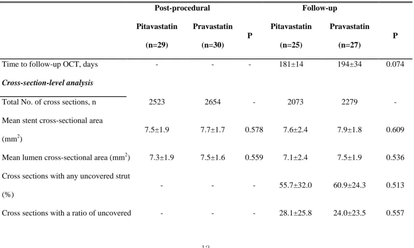
Table 2. Optical coherence tomographic findings of pitavastatin and pravastatin-treated groups
Post-procedural Follow-up Pitavastatin (n=29) Pravastatin (n=30) P Pitavastatin (n=25) Pravastatin (n=27) P
Time to follow-up OCT, days - - - 181±14 194±34 0.074
Cross-section-level analysis
Total No. of cross sections, n 2523 2654 - 2073 2279 -
Mean stent cross-sectional area
(mm2)
7.5±1.9 7.7±1.7 0.578 7.6±2.4 7.9±1.8 0.609
Mean lumen cross-sectional area (mm2) 7.3±1.9 7.5±1.6 0.559 7.1±2.4 7.5±1.9 0.536
Cross sections with any uncovered strut
(%)
- - - 55.7±32.0 60.9±24.3 0.513
13 to total strut >0.3 (%)
Presence of intrastent thrombi, n (%) - - - 0 (0.0) 0 (0.0)
Strut-level analysis
Total No. of analyzable struts, n 24484 25317 - 20,223 21,908 -
Mean neointimal hyperplasia thickness,
μm - - - 63.7±41.3 55.5±24.1 0.379
Percentage of uncovered strut, % - - - 19.4±14.7 19.1±15.2 0.927
Percentage of malapposed strut, % 2.7±3.7 3.3±8.2 0.685 0.6±1.4 0.8±3.0 0.760
Both of malapposed and uncovered
strut, %
14
In 52 overall patients, there was a trend that patients with follow-up cholesterol level less than 70mg/dL had smaller percentage of uncovered struts (12.5±12.2% vs. 21.5±15.1%, p=0.058)., While the percentage of uncovered struts was significantly lower in patients who achieved follow-up LDL cholesterol level less than 70mg/dL than in patients who did not achieve in SES-treated patients (10.1±12.4% vs. 26.9±15.6%, respectively, p=0.025), there were no significant differences of percentage of uncovered struts in BES-treated patients (14.6±12.7% in patients with follow-up LDL cholesterol level less than 70mg/dL vs. 14.4±10.5% in patients with that greater than 70mg/dL, p=0.971).
Figure 2. Relationship between the percentage of uncovered struts versus follow-up low-density lipoprotein (LDL) cholesterol level (A) or the level of
LDL cholesterol reduction (B) is shown. Black and red dots represent biolimus-
and sirolimus-eluting stent, respectively.
Figure 2 indicated the relationship of follow-up LDL cholesterol level and the
15
The percentage of uncovered struts was significantly correlated with follow-up
LDL cholesterol level (r=0.486, p=0.009) and the level of LDL cholesterol
reduction (r=-0.456, p=0.015) in SES-treated patients, but not BES-treated
patients.
IV. DISCUSSION
Previous studies reported several factors to be associated with delayed strut coverage such as SES7,8, acute coronary syndrome9,10, time interval from DES implantation to follow- up OCT21, baseline high-sensitivity C-reactive protein22, stent diameter23, diabetes mellitus23, AHA/ACC type B2/C lesion23. Although several studies showed that statins have beneficial effects on both mature endothelial cells and endothelial progenitor cells24-27, there is no human data whether use of statin could be associated with acceleration of strut coverage after DES implantation. Wang TJ et al. showed that atorvastatin pretreatment can accelerate both neointimal coverage and re-endothelialization after SES implantation in the animal model using minipigs.14 Another animal study using wire-mediated vascular injury model in mice reported that fluvastatin has protective effects against impaired re-endothelialization in sirolimus-treated arteries.24 The mechanism of protective effect of statin against delayed vascular healing has been understood as the pleotropic effect to modulate smooth muscle cell proliferation and migration and to increase circulating endothelial progenitor cells.14,24 Experimental animal studies reported that each statin has different effects according to its water solubility.28,29 In this study, we hypothesized that different kinds of statin may affect different degree of the OCT-based strut coverage in DES-treated lesions; the strut coverage was compared between pitavastatin- and pravastatin-treated lesions. Compared to
16
pravastatin (a kind of hydrophilic statin), pitavastatin (a fully synthetic lipophilic statin) has a powerful efficacy comparable with atorvastatin and rosuvastatin.30,31 In the present study, significant difference in strut coverage was not observed between pitavastatin- and pravastatin-treated groups; greater DES strut coverage was significantly related with lower level of follow-up LDL cholesterol and greater reduction of LDL cholesterol in SES-treated lesions, not in BES-treated lesions.
These findings might suggest that lowering LDL cholesterol level itself has a role in strut coverage after first-generation DES implantation. However, as mentioned above, the main mechanism of statins to modify vascular healing process has been understood as the pleotropic effect rather than as the direct LDL cholesterol lowering effect. Therefore, this discrepancy might be explained that lower level of LDL cholesterol is one of indicator to show the intensity of pleotropic effect in vascular healing process. The relationship between follow-up LDL cholesterol level and strut coverage was not founded in next generation DES (i.e. BES). The possible explanation on this finding is that the vascular healing response to statin or lower LDL cholesterol level could be different according to the types of DES. The differences of polymer, drug or drug-eluting period between BES and SES may influence on the different degree of strut coverage according to LDL cholesterol lowering effects by statin treatment. Compared to SES, BES has several different characteristics; a bioresorbable polymer carrier (poly-lactic acid) as well as coating only on the abluminal stent surface to allow a direct release of lipophilic biolimus into the vessel wall are the most important ones.32,33
1. Study limitation
First, two types of DES were used in this study. Second, there is no control group without statin therapy. Therefore, we could not evaluate an effect of statin
17
therapy on strut coverage compared with the patients without statin treatment. However, the control group without statin treatment would not be ethically justified in current clinical practice for DES-treated patients with coronary artery disease.
V. CONCLUSION
This randomized study showed that a protective effect of statin against delayed strut coverage was observed in SES-treated patients with lower level of follow-up LDL cholesterol, especially less than 70mg/dL. This vascular healing effect of lower LDL cholesterol level by statin could be different according to types of DES.
REFERENCES
1. Farb A, Burke AP, Kolodgie FD, Virmani R. Pathological mechanisms of fatal late coronary stent thrombosis in humans. Circulation. 2003;108:1701-6
2. Finn AV, Joner M, Nakazawa G, Kolodgie F, Newell J, John MC, et al. Pathological correlates of late drug-eluting stent thrombosis: Strut coverage as a marker of endothelialization. Circulation. 2007;115:2435-41 3. Guagliumi G, Sirbu V, Musumeci G, Gerber R, Biondi-Zoccai G, Ikejima H,
et al. Examination of the in vivo mechanisms of late drug-eluting stent thrombosis: findings from optical coherence tomography and intravascular ultrasound imaging. J Am Coll Cardiol Intv 2012;5:12-20.
18
4. Won H, Shin DH, Kim BK, Mintz GS, Kim JS, Ko YG, et al. Optical coherence tomography derived cut-off value of uncovered stent struts to predict adverse clinical outcomes after drug-eluting stent implantation. Int J Cardiovasc Imaging DOI 10.1007/s10554-013-0223-9
5. Prati F, Regar E, Mintz GS, Arbustini E, Di Mario C, Jang IK, et al. for the expert's OCT review document. Expert review document on methodology, terminology, and clinical applications of optical coherence tomography: physical principles, methodology of image acquisition, and clinical application for assessment of coronary arteries and atherosclerosis. Eur Heart J 2010;31:401-15.
6. Lee SY, Hong MK. Stent evaluation with optical coherence tomography. Yonsei Med J 2013;54:1075-83
7. Kim JS, Jang IK, Kim TH, Takano M, Kume T, Hur NW, et al. Optical coherence tomography evaluation of zotarolimus-eluting stents at 9-month follow-up: Comparison with sirolimus-eluting stents. Heart. 2009;95:1907-12
8. Takano M, Inami S, Jang IK, Yamamoto M, Murakami D, Seimiya K, et al. Evaluation by optical coherence tomography of neointimal coverage of sirolimus-eluting stent three months after implantation. Am J Cardiol. 2007;99:1033-38
9. Kim JS, Fan C, Choi D, Jang IK, Lee JM, Kim TH, et al. Different patterns of neointimal coverage between acute coronary syndrome and stable angina after various types of drug-eluting stents implantation; 9-month follow-up optical coherence tomography study. Int J Cardiol. 2011;146:341-6
10. Kubo T, Imanishi T, Kitabata H, Kuroi A, Ueno S, Yamano T, et al. Comparison of vascular response after sirolimus-eluting stent implantation
19
between patients with unstable and stable angina pectoris: A serial optical coherence tomography study. JACC Cardiovasc Imaging. 2008;1:475-84 11. Mihos CG, Salas MJ, Santana O. The pleiotropic effects of the
hydroxy-methyl-glutaryl-coa reductase inhibitors in cardiovascular disease: A comprehensive review. Cardiol Rev. 2010;18:298-304
12. Indolfi C, Cioppa A, Stabile E, Di Lorenzo E, Esposito G, Pisani A, et al. Effects of hydroxymethylglutaryl coenzyme a reductase inhibitor simvastatin on smooth muscle cell proliferation in vitro and neointimal formation in vivo after vascular injury. J Am Coll Cardiol. 2000;35:214-21 13. Bellosta S, Bernini F, Ferri N, Quarato P, Canavesi M, Arnaboldi L, et al.
Direct vascular effects of HMG-CoA reductase inhibitors. Atherosclerosis. 1998;137 Suppl:S101-9
14. Wang TJ, Yang YJ, Xu B, Zhang Q, Jin C, Tang Y, et al. Atorvastatin accelerates both neointimal coverage and re-endothelialization after sirolimus-eluting stent implantation in a porcine model. Circ J 2012;76:2561-71
15. Jaschke B, Michaelis C, Milz S, Vogeser M, Mund T, Hengst L, et al. Local statin therapy differentially interferes with smooth muscle and endothelial cell proliferation and reduces neointima on a drug-eluting stent platform. Cardiovasc Res. 2005;68:483-92
16. Kim BK, Ha J, Mintz GS, Kim JS, Shin DH, Ko YG, et al. Randomized comparison of strut coverage between Nobori biolimus-eluting versus sirolimus-eluting stents: An optical coherence tomography analysis. EuroIntervention (in press)
17. Tanigawa J, Barlis P, Dimopoulos K, Dalby M, Moore P, Di Mario C. The influence of strut thickness and cell design on immediate apposition of drug-eluting stents assessed by optical coherence tomography. Int J Cardiol 2009;134:180-8.
20
18. Davlouros PA, Mavronasiou E, Xanthopoulou I, Karantalis V, Tsigkas G, Hahalis G, et al. An optical coherence tomography study of two new generation stents with biodegradable polymer carrier, eluting paclitaxel vs. biolimus-A9. Int J Cardiol 2012;157:341-6.
19. Kume T, Akasaka T, Kawamoto T, Ogasawara Y, Watanabe N, Toyota E, et al. Assessment of coronary arterial thrombus by optical coherence tomography. Am J Cardiol 2006; 97:1713-17
20. Doran H, Bates D, Bliese P, Dowling M. Estimating the multilevel Rasch model: With the lme4 package. J Stat Softw .2007;20:1–18.
21. Kim BK, Kim JS, Ko YG, Choi D, Jang Y, Hong MK. Major determinants for the uncovered stent struts on optical coherence tomography after drug-eluting stent implantation. Int J Cardiovasc Imaging 2012;28(4):705-14. 22. Kim BK, Kim JS, Oh C, Ko YG, Choi D, Jang Y, et al. Impact of
preprocedural high-sensitivity c-reactive protein levels on uncovered stent struts: An optical coherence tomography study after drug-eluting stent implantation. Clin Cardiol. 2011;34:97-101
23. Ishigami K, Uemura S, Morikawa Y, Soeda T, Okayama S, Nishida T, et al. Long-term follow-up of neointimal coverage of sirolimus-eluting stents--evaluation with optical coherence tomography. Circ J 2009;73(12):2300-7. 24. Fukuda D, Enomoto S, Shirakawa I, Nagai R, Sata M. Fluvastatin
accelerates re-endothelialization impaired by local sirolimus treatment. Eur J Pharmacol 2009;612:87-92.
25. Wolfrum S, Jensen KS, Liao JK. Endothelium-dependent effects of statins. Arterioscler Thromb Vasc Biol 2003;23:729–36.
26. Werner N, Priller J, Laufs U, Endres M, Bohm M, Dirnagl U, et al. Bone marrow-derived progenitor cells modulate vascular reendothelialization and neointimal formation: effect of 3-hydroxy-3-methylglutaryl coenzyme a reductase inhibition. Arterioscler Thromb Vasc Biol 2002;22:1567–72.
21
27. Walter DH, Zeiher AM, Dimmeler S. Effects of statins on endothelium and their contribution to neovascularization by mobilization of endothelial progenitor cells. Coron Artery Dis 2004;15:235–42.
28. Sakamoto T, Kojima S, Ogawa H, Shimomura H, Kimura K, Ogata Y, et al. Usefulness of hydrophilic vs lipophilic statins after acute myocardial infarction: subanalysis of MUSASHI-AMI. Circ J 2007;71:1348-53. 29. Satoh K, Ichihara K. Lipophilic HMG-CoA reductase inhibitors increase
myocardial stunning in dogs. J Cardiovasc Pharmacol 2000; 35: 256–62. 30. Saku K, Zhang B, Noda K. Randomized head-to-head comparison of
pitavastatin, atorvastatin, and rosuvastatin for safety and efficacy (quantity and quality of LDL): the PATROL trial. Circ J 2011;75:1493-505.
31. Hayashi T, Yokote K, Saito Y, Iguchi A. Pitavastatin: efficacy and safety in intensive lipid lowering. Expert opin pharmacother 2007;8:2315-27. 32. Chevalier B, Silber S, Park SJ, Garcia E, Shuler G, Suryapranata H, et al.
Randomized comparison of the Nobori biolimus A9-eluting coronary stent with the Taxus Liberte´ paclitaxel-eluting coronary stent in patients with stenosis in native coronary arteries: The NOBORI 1 trial-Phase 2. Circ Cardiovasc Interv. 2009;2:188-195.
33. Ostojic M, Sagic D, Beleslin B, Jung R, Perisic Z, Jagic N, et al. First clinical comparison of Nobori-Biolimus A9 eluting stents with Cypher-Sirolimus eluting stents: NOBORI CORE nine months angiographic and one year clinical outcomes. EuroIntervention. 2008;3:574-579.
22
< ABSTRACT(IN KOREAN)>
약물용출 스텐트삽입술 후
신생내막형성에 미치는 스타틴의 영향
<지도교수 홍명기 >
연세대학교 대학원 의학과
서용성
배 경: 약물용출 스텐트 삽입 이후 스타틴이 신생내막의 형성에 어떠한 영향을 미치는지에 대한 임상 연구는 아직 이루어지지 않았다. 방 법: 총 60 명의 관상동맥 질환자를 대상으로 약물용출 스텐트 삽입 후 피타바스타틴 2mg 혹은 프라바스타틴 20mg 을 무작위 선정하여 6 개월간 투여하였다. 약물용출스텐트는 시로리무스와 바이오리무스용출스텐트를 무작위 선정하여 삽입하였다. 이후 52 명의 환자에서 6 개월 추적 혈관광학단층촬영을 시행하여 신생내막에 의한 스트럿 비포장률을 비교하였다. 결 과: 피타바스타틴을 사용한 25 명의 환자는 19.4±14.7%, 프라바스타틴을 사용한 28 명의 환자에서는 19.1±15.2%의 스트럿 비포장률을 보여23 유의한 차이를 보이지 않았다. (p=0.927) 하지만, 시로리무스 용출 스텐트를 삽입한 환자군은 6 개월 추적 혈중 저밀도지단백콜레스테롤 농도가 낮을수록 (r=0.486, p=0.009), 스텐트 삽입 당시에 비하여 그 감소량이 클수록 (0.456, p=0.015) 낮은 스트럿 비포장률을 보였다. 이러한 경향은 바이오리무스용출 스텐트를 삽입한 환자에서는 관찰되지 않았다. 또한 시로리무스용출스텐트 환자군에서는 6 개월 추적 저밀도지단백콜레스테롤농도가 70mg/dL 이하인 환자의 경우 그렇지 않은 경우보다 유의하게 낮은 스트럿 비포장률을 나타내었지만, (각각 10.1±12.4% 과 26.9±15.6%, p=0.025) 바이오리무스용출스텐트 환자군에서는 차이가 없었다. (각각 14.6±12.7% 과 14.4±10.5%, p=0.971) 결 론: 저밀도지단백콜레스테롤 농도를 70mg/dL 이하로 유지하는 것이 약물용출스텐트삽입 후 혈관 회복지연을 예방하는 효과가 있을 것으로 생각되며 이는 약물 용출스텐트의 종류에 따라 차이가 있었다. --- 핵심되는 말 : 스텐트, 혈관내 광학단층촬영, 스타틴