pISSN 2288-0585⋅eISSN 2288-6850
Molecular Epidemiology and Characterization of
Carbapenemase-Producing Enterobacteriaceae Isolated
at a University Hospital in Korea during 4-Year Period
Sunyoung Ahn
1, Ji Yeon Sung
1, Hyunsoo Kim
2, Myung Sook Kim
1, Younjee Hwang
3, Sori Jong
1,
Younghee Seo
1, Eunjin Ha
4, Eun Suk Park
4, Jun Yong Choi
4,5, Dongeun Yong
1,4, Kyungwon Lee
11
Department of Laboratory Medicine and Research Institute of Bacterial Resistance,
Yonsei University College of Medicine,
2Department of Laboratory Medicine, National Police Hospital,
3
Brain Korea 21 PLUS Project for Medical Science, Yonsei University,
4
Department of Infection Control, Severance Hospital,
5Division of Infectious Diseases,
Department of Internal Medicine, Yonsei University College of Medicine, Seoul, Korea
Background: Carbapenemase-producingEnterobacte-riaceae (CPE) has been increasingly reported
world-wide in the past 10 years, which is an important in-fection control concern. Since the epidemiology and characteristics of these CPEs vary according to in-stitutes, we aimed to characterize CPEs in a uni-versity hospital during the recent 4 years.
Methods: From October 2011 to September 2015,
CPE isolates from clinical specimens and hospital surveillance cultures were collected. Carbapenem re-sistance was confirmed by disk diffusion method and Minimal Inhibitory Concentration (MIC) was deter-mined by agar dilution method. Carbapenemase pro-duction was tested by double disk test using amino-phenylboronic acid and dipicolic acid. PCR and
se-quence analysis were performed to detect blaKPC,
blaIMP-1, blaVIM-2, blaNDM-1-like genes and blaOXA-48 gene.
Pulsed-field gel electrophoresis (PFGE) and Multilocus sequence typing (MLST) were conducted for KPC- producing Klebsiella pneumoniae isolates.
Results: Twenty-five isolates (11%) of CPE were
identified among 222 carbapenem-resistant
Entero-bacteriacae isolates during the study period. The
most prevalent CPE was KPC-producing K.
pneumo-nia and others were IMP-1, VIM-2, NDM-1 type and
OXA-48 producing CPEs. Most of these CPEs showed resistance to carbapenems with variable MICs. The sequence types (STs) of KPC-producing
K. pneumoniae were ST307 and ST11. The PFGE of
ST11 and ST307 showed clonality in each group suggesting the possibility of in-hospital outbreak.
Conclusion: The prevalence of CPE has been
in-creasing. In our institute, KPC-producing K.
pneumo-niae was the most frequently isolated CPE in the
re-cent 4 years. CPE including KPC producers can easily transfer their resistance. Therefore continuous monitoring and more intensified infection control for CPE should be considered. (Ann Clin Microbiol
2016;19:39-47)
Key Words: Beta-lactamase KPC, Carbapenem
re-sistance, Enterobacteriaceae, Klebsiella
pneumoniae
39
Received 15 February, 2016, Revised 25 March, 2016, Accepted 31 March, 2016
Correspondence: Ji Yeon Sung, Department of Laboratory Medicine and Research Institute of Bacterial Resistance, Yonsei University College of Medicine, 50-1 Yonsei-ro, Seodaemun-gu, Seoul 03722, Korea. (Tel) 82-2-2228-2453, (Fax) 82-2-364-1583, (E-mail) [email protected]
ⓒ The Korean Society of Clinical Microbiology.
This is an Open Access article distributed under the terms of the Creative Commons Attribution Non-Commercial License (http://creativecommons.org/licenses/by-nc/4.0) which permits unrestricted non-commercial use, distribution, and reproduction in any medium, provided the original work is properly cited.
INTRODUCTION
장내세균속(Enterobacteriaceae)은 장내 세균총의 정상 무리 중 하나이지만, 지역 사회 감염 혹은 병원 감염의 가장 흔한 병 인균이기도 하다[1]. 2000년대 들어 carbapenem을 제외한 대부 분의 β-lactam을 분해할 수 있는 TEM, SHV, CTX-M 등의 ex-tended-spectrum β-lactamase (ESBL)를 생성하는
Enterobacte-riaceae가 전세계적으로 확산되었고, ESBL 생성 세균의 감염 을 치료하기 위한 carbapenem의 사용이 증가하였다[2]. Carba-penem은 광범위한 치료 효과를 가지며, ESBL을 생성하는 그 람 음성균 감염증에도 치료 효과가 우수하다. 따라서 ESBL 생 성 Enterobacteriaceae 감염 치료를 위한 주요 항균제로 사용되 었으며, 항균제의 ‘최후의 보루’로 여겨졌다. 그러나 최근 car-bapenem 사용량이 증가하면서 carcar-bapenem 내성 장내세균속
Table 1. Primers used for carbapenemase gene PCR
Gene Primer sequence Amplicon size KPC Forward TGGACACACCCATCCGTTAC 500 bp
Reverse GACGGCCAACACAATAGGTG
VIM Forward TTTGATTGATACAGCGTGG 459 bp Reverse TGCTTCCGGGTAGTG
IMP Forward CATGGTTTGGTGGTTCTTGT 448 bp Reverse ATAATTTGGCGGACTTTGGC
NDM Forward CAATATTATGCACCCGGTCG 726 bp Reverse ATCATGCTGGCCTTGGGGAA
OXA Forward TTGGTGGCATCGATTATCGG 744 bp Reverse GAGCACTTCTTTTGTGATGG
(carbapenem-resistant Enterobacteriaceae, CRE) 감염증은 전세 계적으로 발생이 증가하는 추세이며[1], 미국 질병관리본부 (Centers for Disease Control and Prevention, CDC)에서 분류한 내성 세균 중 긴급한 위협(urgent threat)으로 지정될 만큼 주요 사안이 되고 있다[3].
Carbapenem 내성의 중요한 기전은 carbapenem을 분해하는 효소인 carbapenemase에 의한 것인데, 이들의 유전자는 trans-poson을 통하여 plasmid 간에 쉽게 전파될 수 있어 carbapenem 내성 확산에 대한 우려가 커지고 있다[4]. 1993년 최초로 NmcA 형 carbapenemase 생성 장내세균속(carbapenemase-pro-ducing Enterobacteriaceae, CPE)이 발견된[1] 이후 다양한 CPE 들이 보고되고 있다. 이들에 의한 감염증은 치료 약제 선택에 제한을 가져오고, 재원 일수를 늘리며, 환자의 예후를 불량하게 하고 사망률을 증가시킨다[4]. 특히 carbapenem 내성 Klebsiella pneumoniae의 경우 의료 환경에서 빠르게 퍼지는 경향을 보여, 병원 내 집단 발병의 위험성도 존재한다[5]. 국내에서도 CPE가 지난 몇 년간 지속적으로 증가하고 있으 며[6], 2011년부터 질병관리본부는 의료관련감염병에 CRE 감 염증을 포함시켜 발생 현황을 감시하고 있다. 국내에서 발생하 는 CPE 감염에 대한 연구는 제한되어 있으며[7-9], 또한 CPE 감염의 특성과 역학은 지역 및 기관마다 다를 수 있다. 국내 CPE 균주와 감염에 대한 정보는 감염관리 및 환자 진료를 위 한 중요한 기초 자료이기에 본 연구에서는 한 대학 병원에서 최근 4년간 분리된 CPE의 분자 역학 및 특성에 대하여 고찰하 고자 한다.
MATERIALS AND METHODS
1. 대상 균주2011년 10월부터 2015년 9월까지 세브란스병원의 임상 및 감시 배양 검체에서 분리된 CPE를 대상으로 하였다. 균종 동정 은 Vitek 2 (BioMerieux, Durham, NC, USA) 혹은 matrix as-sisted laser desorption/ionization time-of-flight mass spectrome-try (MALDI-TOF MS) (Bruker Daltonics Inc., Billerica, MA, USA)를 이용하였다. 한 환자에서 여러 균주가 분리된 경우 최 초 분리된 동일 균종의 균주 1개씩을 포함하였다. 감시 배양은 대변을 채취하여 ertapenem disk가 들어있는 trypticase soy
broth (TSB)에 접종한 뒤 35oC에서 24시간 배양 후, TSB 100
μL를 MacConkey 배지에 ertapenem disk와 함께 24시간 계대
배양 하였다. Ertapenem disk 주변 21 mm 이내에 증식한 집락 중 Enterobacteriaceae로 의심되는 독립 집락을 선택하여 동정 및 감수성 시험을 진행하였다. 2. 임상역학적 자료 수집 CPE가 분리된 환자들의 의무기록을 후향적으로 조사하여 환자들의 연령, 성별, 입원 경로, 입원 시 진단, 예후 등에 대하 여 알아보았다. 분리된 CPE가 감염의 원인 병인균으로 추정되 어 치료한 경우 infection으로, 감염 증상을 일으키지 않은 집락 화 균으로 생각된 경우 colonization으로 분류하였다. 3. 항균제 감수성 검사 및 carbapenemase 생성 여부 확인
Ampicillin-sulbactam (SAM), piperacillin-tazobactam (TZP), ampicillin (AMP), cefazolin (CFZ), cefoxitin (FOX), cefotaxime (CTX), ceftazidime (CAZ), cefepime (FEP), aztreonam (ATM), amikacin (AMK), gentamicin (GEN), trimethoprim-sulfamethox-azole (TMP/SMX), levofloxacin (LVX), tigecycline (TGC), er-tapenem (ETP), meropenem (MEM)의 16종의 항균제에 대하여 Vitek 2 자동화 장비를 이용하여 감수성 검사를 실시하여 car-bapenem에 내성인 경우 ertapenem과 meropenem에 대해 disk 확산법으로 zone diameter를 확인하고, imipenem으로 modi-fied-Hodge test (MHT)와 ertapenem, imipenem으로 dipicolinic acid (DPA)/aminophenylboronic acid (APBA) double-disk syn-ergy test (DDS)를 시행하였다. DDS는 2개 carbapenem disk 중 하나라도 양성이면 양성으로 판단하였다. Dipicolinic acid DDS 에서 양성인 경우 metallo-β-lactamase (MBL) 존재 여부를 확 인하기 위해 blaNDM-1, blaIMP-1, blaVIM-2형 유전자에 대하여 PCR
을 시행하였으며, aminophenylboronic acid DDS에서 양성인 경
우 blaKPC에 대하여 PCR을 시행하였다. MHT에서 양성을 보여
carbapenemase 생성이 의심되나, 두 가지의 DDS 모두에서 음
성인 경우는 blaOXA-48에 대한 PCR을 시행하였다. PCR에 사용
된 primer 정보는 Table 1과 같다. Imipenem, meropenem 그리 고 ertapenem의 최소억제농도(minimal inhibitory concentration, MIC)는 2015년 Clinical and Laboratory Standard Institute (CLSI) 기준에 따라 한천희석법으로 확인하였으며, 정도 관리 를 위해 Escherichia coli ATCC 25922와 Pseudomonas
aerugi-nosa ATCC 277853 균주를 이용하였다[10]. K. pneumoniae에
Table 2. Gene loci included in the Klebsiella pneumoniae Multilocus sequence typing scheme and PCR primers
Locus Putative function of gene Primer sequence Size(bp) Number of alleles
rpoB Beta-subunit of RNA polymerase B (F) VIC3: GGCGAAATGGCWGAGAACCA 501 8 (R) VIC2: GAGTCTTCGAAGTTGTAACC
gapA Glyceraldehyde 3-phosphate dehydrogenase
(F) 173: TGAAATATGACTCCACTCACGG 450 6 (R) 181: CTTCAGAAGCGGCTTTGATGGCTT
mdh Malate dehydrogenase (F) 130: CCCAACTCGCTTCAGGTTCAG 477 10 (R) 867: CCGTTTTTCCCCAGCAGCAG
pgi Phosphoglucose isomerase (F) 1R: GAGAAAAACCTGCCTGTACTGCTGGC 432 6 (R) IF: CGCGCCACGCTTTATAGCGGTTAAT
phoE Phosphoporine E (F) 604.1: ACCTACCGCAACACCGACTTCTTCGG 420 14 (R) 604.2: TGATCAGAACTGGTAGGTGAT
infB Translation initiation factor 2 (F) 1F: CTCGCTGCTGGACTATATTCG 318 10 (R) 1R: CGCTTTCCAGCTCAAGAACTTC
tonB Periplasmic energy transducer (F) 1F: CTTTATACCTCGGTACATCAGGTT 414 21 (R) 2R: ATTCGCCGGCTGRGCRGAGAG Abbreviations: (F), forward; (R), reverse.
Fig. 1. The number of carbapenemase-producing Enterobacteriaceae
isolates and carbapenemase types reported annually.
였고, CLSI에 기준이 명시되어 있지 않아 The European Committee on Antimicrobial Susceptibility Testing (EUCAST) 의 기준을 적용하였다[11]. CRE는 디스크 확산법으로 하나 이 상의 carbapenem 항균제에 내성을 보이거나 carbapenemase 유 전자를 PCR로 확인한 CPE 균주로 정의하였다.
4. K. pneumoniae 균주에 대한 MLST 및 PFGE
Klebsiella pneumoniae carbapenemase (KPC) 생성 K. pneu-moniae 균주들에 대하여 multilocus sequence typing (MLST)
및 pulsed-field gel electrophoresis (PFGE)를 시행하였다. MLST 는 7개의 housekeeping 유전자인 rpoB, gapA, mdh, pgi, phoE,
infB, tonB를 PCR 후 염기서열을 분석하였다(Table 2) [12].
Sequence type (ST)은 각 유전자의 염기서열 분석 결과를 MLST database (http://www.pasteur.fr/recherche/genopol/PF8/ mlst/Kpnuemoniae.html)에 입력 후 판정하였다.
PFGE는 Bio-Rad (Hercules, CA, USA) 시험법에 따라 XbaI (Roche, Mannheim, Germany)로 DNA 절단 후 CHEF-ERⓇII
sys-tem (Bio-Rad, Hercules, CA, USA)을 이용하여 절편들을 분리 하였고, InforQuestFP (Bio-Rad, Hercules, CA, USA, Version 4.5) 프로그램으로 분석하여 dendrogram을 얻었다.
RESULTS
연구 기간 동안 총 222개의 CRE 균주가 분리되었고, 그 중 25균주(11%)가 CPE로 확인되었다. 연도별 CPE 발생 건수는 점차 증가하였고 특히 2015년에 9개월간 16건으로 크게 증가 하였다(Fig. 1). CPE가 분리된 환자군의 평균 나이는 58.2세(0-84세)이었고 남녀 비는 1.1 : 1로 비슷하였다(Table 3). 검체의 종류는 소변 (5건), 혈액(4건), 객담(4건), 담즙(2건), 기타(5건)로 다양하였 고, 병인균은 25균주 중 11균주(44%)였다. 감염 관리 감시의 목적으로 시행한 대변 배양에서도 5건(20%) 분리되었다. 대변 감시 배양은 2013년 8월부터 시행된 검사로, 2015년 9월까지 CRE 양성률은 5.06% (36/711)였고, CPE 양성률은 2.25% (16/711)였다. 25명의 환자군 중 CPE 분리 후 30일 이내 사망 한 환자는 5명(20%, Isolates 1, 2, 3, 10, 12)이었고, 이 중 2명 (Isolates 1, 3)에서는 CPE가 병인균이었다. 분리된 CPE는 K. pneumoniae (18주)가 가장 많았고, 그 다음Klebsiella oxytoca (2주), E. coli (2주), Enterobacter aerogenes
(1주), Enterobactor cloacae (1주), Serratia macescens (1주) 순 이었다(Table 4). 2013년 이전에는 IMP-1와 VIM-2형 CPE가 분리되었으나, 2014년과 2015년에는 KPC 생성 CPE가 각각 3
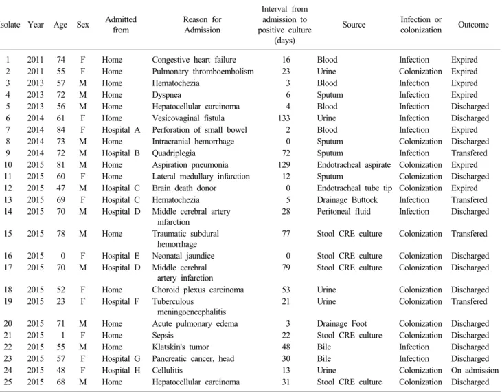
Table 3. Clinical features of 25 carbapenemase-producing Enterobacteriaceae isolates
Isolate Year Age Sex Admitted from Reason for Admission
Interval from admission to positive culture
(days)
Source Infection or colonization Outcome
1 2011 74 F Home Congestive heart failure 16 Blood Infection Expired 2 2011 55 F Home Pulmonary thromboembolism 23 Urine Colonization Expired
3 2013 57 M Home Hematochezia 3 Blood Infection Expired
4 2013 72 M Home Dyspnea 6 Sputum Infection Expired
5 2013 56 M Home Hepatocellular carcinoma 4 Blood Infection Discharged 6 2014 61 F Home Vesicovaginal fistula 133 Urine Infection Discharged 7 2014 84 F Hospital A Perforation of small bowel 2 Blood Infection Expired 8 2014 73 M Home Intracranial hemorrhage 0 Sputum Colonization Discharged 9 2014 72 M Hospital B Quadriplegia 72 Sputum Infection Transfered 10 2015 81 M Home Aspiration pneumonia 129 Endotracheal aspirate Colonization Expired 11 2015 60 F Home Lateral medullary infarction 12 Sputum Colonization Discharged 12 2015 47 M Hospital C Brain death donor 0 Endotracheal tube tip Colonization Expired 13 2015 69 F Hospital C Hematochezia 5 Drainage Buttock Infection Transfered 14 2015 70 M Hospital D Middle cerebral artery
infarction
28 Peritoneal fluid Infection Discharged
15 2015 78 M Home Traumatic subdural hemorrhage
77 Stool CRE culture Colonization Transfered
16 2015 0 F Hospital E Neonatal jaundice 0 Stool CRE culture Colonization Discharged 17 2015 70 M Hospital D Middle cerebral
artery infarction
79 Stool CRE culture Colonization Discharged
18 2015 52 F Home Choroid plexus carcinoma 53 Urine Colonization Discharged 19 2015 23 F Hospital F Tuberculous
meningoencephalitis
21 Urine Colonization Transfered
20 2015 71 M Home Acute pulmonary edema 3 Drainage Foot Colonization Discharged 21 2015 1 F Home Sepsis 22 Stool CRE culture Colonization Discharged 22 2015 55 M Home Klatskin's tumor 48 Bile Infection Discharged 23 2015 57 F Hospital G Pancreatic cancer, head 30 Bile Infection Discharged 24 2015 48 F Hospital H Cellulitis 13 Urine Colonization On admission 25 2015 68 M Home Hepatocellular carcinoma 31 Stool CRE culture Colonization Discharged
주, 14주 검출되었다. KPC형 CPE 중에서 16주가 K.
pneumo-niae였다. OXA-48 및 NDM-1형 CPE 균주는 E. coli와 K. pneumoniae로 2015년 5월 및 2015년 7월에 각각 한 주씩 분리
되었다. CPE 균주는 IMP-1, VIM-2, NDM-1 형인 경우 DPA DDS 양성을 보였고, 2균주(IMP-1형과 VIM-2형)를 제외하고 는 모두 MHT 양성을 보였다. KPC 생성 CPE 균주는 모두 MHT와 APBA DDS에서 양성을 보였다. OXA-48 생성 E. coli 는 MHT는 양성이었으나 DDS는 음성이었다.
항균제 감수성 결과는 다양하였다(Table 4). 검출된 모든 균 주가 penicillin과 대부분의 cephalosporin, carbapenem 및 fluo-roquinolone에 내성을 보였다. Aztreonam, amikacin, gentami-cin, trimethoprim-sulfamethoxazole (TMP-SMX) 내성률은 각각 84%, 20%, 48%, 및 48%였다. Tigecycline에 대한 내성 및 중간 내성도 10주(40%)에서 확인되었다. 특징적으로 2015년에 분리 된 OXA-48 생성 E. coli 한 주의 경우 자동화 장비에서 ertape-nem MIC가 4 ug/mL로 내성이어서 시행한 디스크 확산법에서
는 ertapenem과 meropenem에 모두 중간 내성을 보였고, 한천 희석법으로 확인한 MIC는 모든 carbapenem 계열 항균제에 감 수성이었다. 이 균주는 MHT에서 양성이었고 PCR 및 염기서
열 분석 결과 blaOXA-48이 확인되었다. 총 18주의 K. pneumoniae
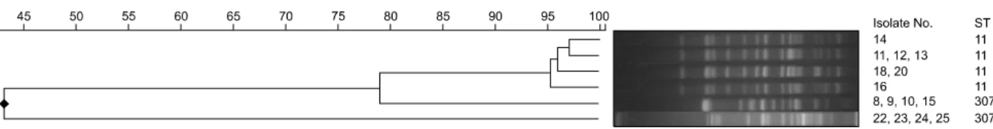
에서 시행한 colistin MIC는 16주에서 감수성이었고(MIC 0.5-1 μg/mL), 2주(Isolates 15와 21)에서 MIC가 각각 4 μg/mL와 8 μg/mL로 내성이었다. MLST를 시행한 KPC 생성 K. pneumoniae 16주는 모두 ST307 (9주) 또는 ST11 (7주)이었다. ST307은 2014년과 2015 년에 걸쳐서, ST11은 2015년에 분리되었다. ST307 8주 및 ST11 7주에 대하여 시행한 PFGE 결과 각 ST별로 연관성이 나 타났고, 특히 시기별로 PFGE 상 동일한 클론을 보여 병원 내 유행을 시사하였다. 하지만 2014년말에서 2015년초에 걸쳐 분 리된 ST307 균주들과 2015년 9월에 분리된 ST307 균주들은 PFGE에서 80% similarity를 기준으로 하였을 때, 서로 다른 클 론으로 판정되었다(Fig. 2).
Ta bl e 4 . A nt im icr obi al s us cept ibi lit y, s eque nce t ype and pl as m id c har ac ter is tics of 2 5 c ar bap ene m as e-pr oduc ing E nt er oba ct er iace ae iso late s Is ol at e Y ea r Sour ce Spe ci es M H T D D S Ca rb ap e-nem as e type M in im al inh ibitor y c on cen tra tio ns ( μ g/m L )* ETP IP M M EM SA M TZP A M P C FZ FO X C TX C A Z FEP A TM A M K G EN TM P/ SM X LV X TG C 1 2011 Bl oo d K O X − +I M P-1 ≥ 64 8 8 ≥ 32 ≥ 12 8 ≥ 32 ≥ 64 ≥ 64 ≥ 64 ≥ 64 16 2 ≤ 2 ≥ 16 ≤ 20 1 ≤ 0. 5 2 2011 U ri ne E A E + + IM P-1 64 16 4 ≥ 32 ≥ 12 8 ≥ 32 ≥ 64 ≥ 64 ≥ 64 ≥ 64 2 ≥ 64 ≤ 2 ≤ 1 ≤ 20 ≤ 0.1 2 ≤ 0. 5 3 2013 B lood K O X − +V IM -2 8 4 8 ≥ 32 ≥ 12 8 ≥ 32 ≥ 64 ≥ 64 8 16 ≤ 1 16 ≤ 2 8 ≤ 20 1 ≤ 0. 5 4 2013 Sput um E C L + + V IM -2 2 8 2 ≥ 32 ≥ 12 8 ≥ 32 ≥ 64 ≥ 64 84 ≤ 1 ≤ 1 ≥ 64 ≥ 16 ≥ 32 0 ≥ 8 1 5 2013 B lood K PN + + IM P-1 64 64 32 ≥ 32 ≥ 12 8 ≥ 32 ≥ 64 ≥ 64 ≥ 64 ≥ 64 ≥ 64 ≥ 64 ≤ 2 ≤ 1 ≤ 20 1 2 6 20 14 Ur in e SMA + + VI M-2 4 2 4 ≥ 32 ≥ 12 8 ≥ 32 ≥ 64 ≥ 64 16 16 2 2 16 ≥ 16 80 ≥ 8 2 7 2014 B lood K PN + + K PC 32 8 8 ≥ 32 ≥ 12 8 ≥ 32 ≥ 64 16 ≥ 64 ≥ 64 16 ≥ 64 ≤ 2 ≥ 16 ≥ 32 0 ≥ 8 2 8 2014 Sput um K PN + + K PC 8 4 4 ≥ 32 ≥ 12 8 ≥ 32 ≥ 64 16 ≥ 64 ≥ 64 32 ≥ 64 4 ≥ 16 ≥ 32 0 ≥ 8 2 9 2014 Sput um K PN + + K PC 32 16 8 ≥ 32 ≥ 12 8 ≥ 32 ≥ 64 16 4 16 2 ≥ 64 ≤ 2 ≤ 1 ≥ 32 0 ≥ 8 2 10 2015 E nd ot rac hea l aspir ate KP N + + K PC 32 8 8 ≥ 32 ≥ 12 8 ≥ 32 ≥ 64 ≥ 64 ≥ 64 ≥ 64 ≥ 64 ≥ 64 4 2 ≥ 32 0 ≥ 8 ≥ 8 11 2015 Sput um K PN + + K PC ≥ 12 8 32 12 8 ≥ 32 ≥ 12 8 ≥ 32 ≥ 64 ≥ 64 ≥ 64 ≥ 64 ≥ 64 ≥ 64 ≥ 64 ≥ 16 ≤ 20 ≥ 8 2 12 2015 E nd ot rac hea l tube t ip KP N + + K PC ≥ 12 8 32 12 8 ≥ 32 ≥ 12 8 ≥ 32 ≥ 64 ≥ 64 ≥ 64 ≥ 64 ≥ 64 ≥ 64 ≥ 64 ≥ 16 ≤ 20 ≥ 8 1 13 2015 D ra ina ge But tock KP N + + K PC ≥ 12 8 32 ≥ 128 ≥ 32 ≥ 12 8 ≥ 32 ≥ 64 ≥ 64 ≥ 64 ≥ 64 ≥ 64 ≥ 64 ≥ 64 ≥ 16 ≤ 20 ≥ 8 ≥ 8 14 2015 Per ito nea l fluid KP N + + K PC ≥ 12 8 64 ≥ 128 ≥ 32 ≥ 12 8 ≥ 32 ≥ 64 ≥ 64 ≥ 64 ≥ 64 ≥ 64 ≥ 64 ≤ 2 ≤ 1 ≤ 20 ≥ 8 ≥ 8 15 2015 St oo l CR E cu ltu re KP N + + K PC 12 8 32 32 ≥ 32 ≥ 12 8 ≥ 32 ≥ 64 ≥ 64 8 16 2 ≥ 64 ≤ 2 ≤ 1 ≥ 32 0 ≥ 8 4 16 2015 St oo l CR E cu ltu re KP N + + K PC ≥ 12 8 32 12 8 ≥ 32 ≥ 12 8 ≥ 32 ≥ 64 ≥ 64 ≥ 64 ≥ 64 ≥ 64 ≥ 64 32 ≥ 16 ≥ 32 0 ≥ 8 2 17 2015 St oo l CR E cu ltu re EC O + + K PC ≥ 12 8 8 64 ≥ 32 ≥ 12 8 ≥ 32 ≥ 64 ≥ 64 ≥ 64 ≥ 64 ≥ 64 ≥ 64 ≥ 64 ≥ 16 ≥ 32 0 ≥ 8 2 18 2015 U ri ne K PN + + K PC ≥ 12 8 64 ≥ 128 ≥ 32 ≥ 12 8 ≥ 32 ≥ 64 ≥ 64 ≥ 64 ≥ 64 ≥ 64 ≥ 64 ≤ 2 ≤ 1 ≤ 20 ≥ 8 ≥ 8 19 2015 U ri ne E C O + − OX A-48 0. 5 1 0. 5 ≥ 32 ≥ 12 8 ≥ 32 ≥ 64 ≥ 64 ≥ 64 ≥ 64 ≥ 64 ≥ 64 ≤ 2 ≥ 16 ≥ 32 0 ≥ 8 ≤ 0. 5 20 2015 D ra ina ge Foo t KP N + + K PC ≥ 12 8 32 12 8 ≥ 32 ≥ 12 8 ≥ 32 ≥ 64 ≥ 64 ≥ 64 ≥ 64 ≥ 64 ≥ 64 ≤ 2 ≤ 1 ≤ 20 ≥ 8 ≥ 8 21 2015 St oo l CR E cu ltu re KP N + + N DM-1 12 8 12 8 12 8 ≥ 32 ≥ 12 8 ≥ 32 ≥ 64 ≥ 64 ≥ 64 ≥ 64 16 ≤ 1 ≤ 2 ≤ 1 ≤ 20 ≤ 0.1 2 1
DISCUSSION
2015년 질병관리본부의 통계에 따르면 국내에서 CPE는 2011년 16주, 2012년 39주, 2013년 91주, 2014년 174주가 확인 되었다[6]. 이처럼 국내에서도 CPE의 발생이 지속적 증가 추이 를 보이고 있어 CPE에 대한 적극적인 감시 및 감염 관리의 중 요성이 커지고 있다. 본 연구는 국내 CPE 발생 경향에 대하여 전반적으로 평가하였다고 보기는 어렵지만, 한 기관에서 4년간 발생한 CPE에 대해 역학 및 임상적, 분자 생물학적 특성을 종 합적으로 고찰하였음에 의의를 지닌다.연구 기간 동안 확인된 전체 CRE 중 CPE는 11%였다. CPE 중 가장 많은 균종은 K. pneumoniae (72%)로, 질병관리본부의 보고와 일치하였다. 본원에서 주로 확인된 carbapenemase 유전
자는 blaKPC (68%)였다. 질병관리본부 국내 보고에서 가장 빈
도가 높았던 blaOXA-232 양성 CPE는 본 연구에서 분리되지 않았
다. blaOXA-48 양성 E. coli가 외국인 환자에서 한 주 분리되었는
데, 염기서열 분석으로 blaOXA-48 유전자를 확인하였고, 이것은
국내에서 처음 발견된 것으로 해외에서 유입되었을 가능성이 있다. 동일한 환자에서 시간 간격을 두고 다른 CPE 균종이 분 리된 경우도 있었다. 이 환자에서 시차를 두고 분리된 K.
pneu-moniae와 E. coli에서 PCR을 통해 blaKPC를 확인할 수 있었다.
먼저 분리되었던 K. pneumoniae는 amikacin, gentamicin 및 TMP-SMX에 감수성이었으나 나중 분리된 E. coli는 내성이었
다. K. pneumoniae에서 E. coli로 blaKPC 유전자를 포함한
Tn4401이 plasmid와 함께 수평 전파되는 것은 여러 연구에서 보고된 바가 있다[13-17]. 이 환자의 경우도 같은 기전이었을 것으로 추측되나, 자세한 전파 과정의 확인을 위해서는 후속 연구가 필요하다. KPC 생성 CPE는 1990년대 후반부터 분리되기 시작하였고, 2000년대 중반부터 유럽에서 KPC 생성 K. pneumoniae가 전파 되고 있으며 일부 지역에서는 endemic하게 나타나고 있다[18]. KPC 생성 K. pneumoniae의 감염은 환자의 사망률을 유의하게 증가시키는 것으로 보고되었다[19,20]. 따라서 본 연구에서 분 리된 CPE 중 상당 부분인 16주(64%)가 KPC 생성 K. pneumo-niae라는 사실은 CPE 감염 관리에 있어 주목할 만한 결과로 생 각된다. 같은 Enterobacteriaceae 내에서도 획득한 β-lactamase 의 종류에 따라 항균제 감수성은 다양하게 나타나는 것으로 알 려져 있다[21]. 그러나 이번 연구에서는 다양한 종류의 carba-penemase 유전자가 발견되었음에도 불구하고 전체적인 항균제 감수성이 비슷하였으며, 대부분 penicillin, cephalosporin 및 carbapenem 계열 항균제에 내성이었다. Amikacin, gentamicin, trimethoprim-sulfamethoxazole, levofloxacin, tigecycline에 대하 여는 비교적 다양한 감수성을 보였다. 본 연구에서 분리된 OXA-48 생성 E. coli는 자동화 장비에서 ertapenem 내성, mer-openem 중간 내성이었으나, disk 확산법에서는 ertapenem과
Tab le 4 . Cont in ue d Is ol at e Y ea r Sou rc e Spec ies M H T D D S Ca rb ap e-ne m as e ty pe M inim al in hib ito ry c onc en tra tio ns ( μ g/ m L )* ETP IP M M EM SA M TZP A M P C FZ FO X CTX CA Z FEP A TM A M K G EN TM P/ SM X LV X TG C 22 20 15 B ile K PN + + K PC ≥ 12 8 64 12 8 ≥ 32 ≥ 12 8 ≥ 32 ≥ 64 ≥ 64 ≥ 64 ≥ 64 ≥ 64 ≥ 64 ≤ 2 ≤ 1 40 ≥ 8 ≥ 8 23 20 15 B ile K PN + + K PC 12 8 32 64 ≥ 32 ≥ 12 8 ≥ 32 ≥ 64 ≥ 64 ≥ 64 ≥ 64 ≥ 64 ≥ 64 ≤ 2 ≥ 16 ≥ 320 ≥ 8 ≥ 8 24 20 15 U ri ne K PN + + K PC ≥ 12 8 64 12 8 ≥ 32 ≥ 12 8 ≥ 32 ≥ 64 ≥ 64 ≥ 64 ≥ 64 ≥ 64 ≥ 64 ≤ 2 ≤ 1 40 ≥ 8 ≥ 8 25 20 15 St ool C R E cu ltur e KP N + + KP C 12 8 64 32 ≥ 32 ≥ 12 8 ≥ 32 ≥ 64 32 ≥ 64 ≥ 64 ≥ 64 ≥ 64 ≤ 2 ≤ 1 ≥ 320 ≥ 8 ≥ 8 *M IC w as m ea su re d b y V IT E K 2 e xce pt f or E T P, I PM , M E M w hi ch w ere m eas ur ed by ag ar di lut ion t es t. A bb revi at ions : M H T , M odi fi ed-H odg e t es t; D D S, dou bl e di sk s yner gy t es t; E T P, er tape nem ; IPM , i m ipenem ; M E M , m erop enem ; S A M , am pic illin -s ulba cta m ; T Z P, pip era cill in -t az ob acta m ; A M P, am pic illin ; C FZ , c ef az olin ; F O X , c ef ox itin ; C T X , c ef ota xim e; C A Z , c ef ta zid im e; FE P, c ef ep im e; A T M , a ztr eo na m ; A M K , a m ik ac in ; G E N , gent am ici n; T M P/ SM X , t ri m et hopr im -s ul fam et hoxazol e; L V X , l evo fl oxa ci n; T G C, t igec ycl in e; K O X , K lebs ie lla o xyt oca ; EA E, E nt er obac ter ae ro gen es ; EC L, En te ro ba ct er c loa cae ; KPN, K lebs ie lla pne um oni ae ; S M A , Se rr at ia m ar ces ce ns ; E C O , E sch er ich ia c oli. B lac k, re sista nt; G ra y, i nter m ed ia te ; W hite, su sc ep tib le.
Fig. 2. KPC-producing Klebsiella pneumoniae dendrogram based on Pulsed-field gel electrophoresis (PFGE) pattern and their sequence type.
Abbreviation: ST, sequence type.
meropenem 모두 중간 내성이었고, 한천희석법에서 모든 carba-penem 계열 항균제에 감수성이었다. 반면 모든 cephalosporin 계열 항균제와 aztreonam, gentamicin 그리고 levofloxacin에 내 성이었다. OXA-48 효소 자체는 penicillin 계열 항균제를 강하 게 가수분해하지만 carbapenem 계열 항균제는 약하게 가수분 해하며 광범위 cephalosporin에 대한 분해력은 매우 낮다고 보 고된 바 있다[22]. 그러나 대부분의 OXA-48 생성 균주는 ESBL 등의 다른 β-lactamase를 같이 가지고 있기 때문에 상대 적으로 다양한 β-lactamase 활성을 나타낸다[23,24]. 분리된
blaOXA-48 양성 E. coli가 cephalosporin 계열 항균제에 강한 내성
을 보이는 것도 ESBL 등의 다른 β-lactamase 때문일 가능성이 있다. 2014년까지 분리된 K. pneumoniae 균주들은 colistin에 감수성이었으나, 2015년 colistin에 내성인 K. pneumoniae 두 균주가 검출되었다. 이 균주들은 각각 KPC와 NDM-1을 생성 하였다. 최근 유럽에서 KPC 생성 K. pneumonia의 colistin 내성 률이 증가한다는 보고들이 있어 추후 지속적인 감시가 필요하 겠다[19,20]. KPC 생성 K. pneumoniae에 대한 MLST 결과는 ST307 (8주), ST11 (7주)의 2가지로 분류되었다. 현재 KPC 생성 K.
pneumo-niae 중 전세계적으로 가장 널리 전파된 sequence type은
ST258이다[25]. ST11은 ST258의 single locus variant로, ST258 이 tonB-79 allele을 획득한 결과 ST11이 생겨난 것으로 추측된 다[26]. ST11 K. pneumoniae는 유럽 전역에서 널리 보고되었으 며(www.pasteur.fr/recherche/genopole/PF8/mlst/), epidemic clone III (EC III)로 기술되기도 하였다[27]. 또한 ST11은 아시아와 라틴 아메리카에서도 주요 클론에 속하며[4], 특히 중국에서 가 장 흔한 클론으로 알려져 있고[28], 한국에서는 요로 감염과 균 혈증을 일으키는 대표적인 클론으로 보고되었다[7]. 반면 본 연 구에서 가장 많이 발견된 K. pneumoniae의 sequence type인 ST307에 대한 보고는 드물다. 이탈리아에서 KPC 생성 K. pneu-moniae의 ST258과 ST307의 동시 outbreak가 보고된 적 있으며 [29], 한국에서는 ciprofloxacin 내성 K. pneumoniae 분석에서 ST307이 한 번 보고되었다[30]. ST307이 분자 유전학적으로 ST258이나 ST11과 많은 차이를 보이며[30], ST307이 처음 분 리된 환자가 외부 요양병원에서 전원 되었다는 사실은 ST307 이 외부에서 유입된 뒤 본원에서 outbreak가 일어났을 가능성 을 시사한다. PFGE 결과는 ST가 동일한 균주에서 같은 클론을 나타내었지만 같은 ST 안에서도 변화가 있었다. 2014년말에서 2015년초에 걸쳐 분리된 ST307 균주들과 2015년 9월에 분리 된 ST307 균주들은 PFGE dendrogram에서 전혀 다른 패턴의 band를 보였는데, 이는 PFGE가 변별력이 우월하여 같은 ST 안 에서도 균주별 차이를 구분할 수 있기 때문인 것으로 생각된다 [31]. 같은 환자에서 분리 시기가 다른 균주이거나 오랜 기간 동안 지속적으로 배양된 균주의 경우 단일 유전적 변화 사건으 로 인해 PFGE의 두세 개 band에 차이가 날 수 있다[32]. 이번 연구에서 확인된 CPE 균주들은 산발적인 감염에 더해 2014년과 2015년에 소규모 outbreak이 나타난 것으로 생각된 다. 같은 시기에 CPE가 분리된 Isolates 8, 9, 10과 Isolates 11, 12, 13 그리고 Isolates 22, 23, 24, 25는 동일한 PFGE 결과와 sequence type을 나타내었다. 병원 감염 관리실의 역학 조사 결 과, 이들 세 그룹 내 환자들은 각각 중환자실이나 병동에서 접 촉한 과거력이 있었다. Isolates 12, 13은 같은 병원에서 전원되 어 CPE의 외부 유입으로 인한 전파 가능성도 우려되었으나, 적 극적인 역학 조사 및 감시 배양과 환자 격리를 통해 전파는 비 교적 빠른 시일 안에 종결되었다. 본 연구에서는 국내 대학병원에서 동정된 CPE의 분자 역학 및 특성에 대해 살펴보았다. 연구 결과 분리된 CRE 중 CPE 비 율은 11%였고, KPC 생성 K. pneumoniae가 대다수를 차지하였 다. 거의 모든 CPE가 다제 내성을 나타내었는데, 특히 2015년 부터 분리된 균주들은 tigecycline에 내성을 보이기도 하였고, colistin 내성인 균주도 두 주 발견되었다. 이처럼 CPE의 빠른 전파 속도와 증가하는 항균제 내성을 고려할 때, CPE에 대한 강화된 감염 관리가 필요하다고 여겨진다. 본 기관에서 최근 4 년간의 CPE 발생을 분석하여 얻은 정보는 향후 국내 CPE 감 염 관리 및 내성 연구를 위한 중요한 기초 자료가 될 것이다.
ACKNOWLEDGMENTS
본 연구는 2015년 대한임상미생물학회의 연구비 지원을 받 아 수행되었음.REFERENCES
1. Nordmann P, Naas T, Poirel L. Global spread of carbapenemase- producing Enterobacteriaceae. Emerg Infect Dis 2011;17:1791-8. 2. Pitout JD. Infections with extended-spectrum beta-lactamase-
producing Enterobacteriaceae: changing epidemiology and drug treatment choices. Drugs 2010;70:313-33.
3. CDC. Antibiotic Resistance Threats in the United States, 2013. http://www.cdc.gov/drugresistance/threat-report-2013/ [Online] (last visited on 1 November 2015).
4. Nordmann P, Cuzon G, Naas T. The real threat of Klebsiella pneumoniae carbapenemase-producing bacteria. Lancet Infect Dis 2009;9:228-36.
5. Tzouvelekis LS, Markogiannakis A, Psichogiou M, Tassios PT, Daikos GL. Carbapenemases in Klebsiella pneumoniae and other Enterobacteriaceae: an evolving crisis of global dimensions. Clin Microbiol Rev 2012;25:682-707.
6. Park JW, Lee EJ, Lee DH. Status of carbapenemase producing Enterobacteriaceae in Korea, 2014. Public Health Weekly Report 2016;9:9-13.
7. Ko KS, Lee JY, Baek JY, Suh JY, Lee MY, Choi JY, et al. Predominance of an ST11 extended-spectrum beta-lactamase- producing Klebsiella pneumoniae clone causing bacteraemia and urinary tract infections in Korea. J Med Microbiol 2010;59:822-8. 8. Kim SY, Shin J, Shin SY, Ko KS. Characteristics of carbapenem- resistant Enterobacteriaceae isolates from Korea. Diagn Microbiol Infect Dis 2013;76:486-90.
9. Kim MN, Yong D, An D, Chung HS, Woo JH, Lee K, et al. Nosocomial clustering of NDM-1-producing Klebsiella pneumo-niae sequence type 340 strains in four patients at a South Korean tertiary care hospital. J Clin Microbiol 2012;50:1433-6.
10. CLSI. Performance standards for antimicrobial susceptibility testing: twenty-fifth informational supplement. CLSI document M100-S25. Wayne, PA: Clinical and Laboratory Standards Institute; 2015.
11. Breakpoint tables for interpretation of MICs and zone diameters. EUCAST (The European Committee on Antimicrobial Suscepti-bility Testing). http://www.eucast.org/ [Online] (last visited on 1 November 2015).
12. Diancourt L, Passet V, Verhoef J, Grimont PA, Brisse S. Multilocus sequence typing of Klebsiella pneumoniae nosocomial isolates. J Clin Microbiol 2005;43:4178-82.
13. Gona F, Barbera F, Pasquariello AC, Grossi P, Gridelli B, Mezzatesta ML, et al. In vivo multiclonal transfer of blaKPC-3 from
Klebsiella pneumoniae to Escherichia coli in surgery patients. Clin Microbiol Infect 2014;20:O633-5.
14. Goren MG, Carmeli Y, Schwaber MJ, Chmelnitsky I, Schechner V, Navon-Venezia S. Transfer of carbapenem-resistant plasmid from Klebsiella pneumoniae ST258 to Escherichia coli in patient. Emerg Infect Dis 2010;16:1014-7.
15. Tijet N, Muller MP, Matukas LM, Khan A, Patel SN, Melano RG. Lateral dissemination and inter-patient transmission of blaKPC-3:
role of a conjugative plasmid in spreading carbapenem resistance. J Antimicrob Chemother 2016;71:344-7.
16. Mathers AJ, Cox HL, Kitchel B, Bonatti H, Brassinga AK, Carroll J, et al. Molecular dissection of an outbreak of carbapenem-resistant Enterobacteriaceae reveals intergenus KPC carbapenemase trans-mission through a promiscuous plasmid. MBio 2011;2:e00204-11. 17. Richter SN, Frasson I, Bergo C, Parisi S, Cavallaro A, Palù G. Transfer of KPC-2 carbapenemase from Klebsiella pneumoniae to
Escherichia coli in a patient: first case in Europe. J Clin Microbiol 2011;49:2040-2.
18. Chen L, Mathema B, Chavda KD, DeLeo FR, Bonomo RA, Kreiswirth BN. Carbapenemase-producing Klebsiella pneumoniae: molecular and genetic decoding. Trends Microbiol 2014;22:686- 96.
19. Monaco M, Giani T, Raffone M, Arena F, Garcia-Fernandez A, Pollini S. Colistin resistance superimposed to endemic carbapenem- resistant Klebsiella pneumoniae: a rapidly evolving problem in Italy, November 2013 to April 2014. Euro Surveill 2014;19. 20. Tumbarello M, Trecarichi EM, De Rosa FG, Giannella M,
Giacobbe DR, Bassetti M, et al. Infections caused by KPC- producing Klebsiella pneumoniae: differences in therapy and mortality in a multicentre study. J Antimicrob Chemother 2015; 70:2133-43.
21. Miriagou V, Cornaglia G, Edelstein M, Galani I, Giske CG, Gniadkowski M, et al. Acquired carbapenemases in Gram-negative bacterial pathogens: detection and surveillance issues. Clin Microbiol Infect 2010;16:112-22.
22. Docquier JD, Calderone V, De Luca F, Benvenuti M, Giuliani F, Bellucci L, et al. Crystal structure of the OXA-48 beta-lactamase reveals mechanistic diversity among class D carbapenemases. Chem Biol 2009;16:540-7.
23. Doi Y and Paterson DL. Carbapenemase-producing Enterobacte-riaceae. Semin Respir Crit Care Med 2015;36:74-84.
24. Song W, Jeong SH, Lee J, Lee SS, Lee K. Emergence and spread of OXA-48-like carbapenemase-producing Enterobacteriaceae. Korean J Nosocomial Infect Control 2015;20:7-18.
25. Chen L, Chavda KD, Mediavilla JR, Zhao Y, Fraimow HS, Jenkins SG, et al. Multiplex real-time PCR for detection of an epidemic KPC-producing Klebsiella pneumoniae ST258 clone. Antimicrob Agents Chemother 2012;56:3444-7.
26. Wang Q, Li B, Tsang AK, Yi Y, Woo PC, Liu CH. Genotypic analysis of Klebsiella pneumoniae isolates in a Beijing Hospital reveals high genetic diversity and clonal population structure of drug-resistant isolates. PLoS One 2013;8:e57091.
27. Damjanova I, Tóth A, Pászti J, Hajbel-Vékony G, Jakab M, Berta J, et al. Expansion and countrywide dissemination of ST11, ST15 and ST147 ciprofloxacin-resistant CTX-M-15-type beta-lactamase- producing Klebsiella pneumoniae epidemic clones in Hungary in 2005--the new 'MRSAs'? J Antimicrob Chemother 2008;62: 978-85.
28. Qi Y, Wei Z, Ji S, Du X, Shen P, Yu Y. ST11, the dominant clone of KPC-producing Klebsiella pneumoniae in China. J Antimicrob Chemother 2011;66:307-12.
29. Castanheira M, Farrell SE, Wanger A, Rolston KV, Jones RN, Mendes RE. Rapid expansion of KPC-2-producing Klebsiella pneumoniae isolates in two Texas hospitals due to clonal spread of ST258 and ST307 lineages. Microb Drug Resist 2013;19:295-7. 30. Park DJ, Yu JK, Park KG, Park YJ. Genotypes of ciprofloxacin- resistant Klebsiella pneumoniae in Korea and their characteristics according to the genetic lineages. Microb Drug Resist 2015; 21:622-30.
31. Johnson JK, Arduino SM, Stine OC, Johnson JA, Harris AD. Multilocus sequence typing compared to pulsed-field gel electro-phoresis for molecular typing of Pseudomonas aeruginosa. J Clin Microbiol 2007;45:3707-12.
32. Tenover FC, Arbeit RD, Goering RV, Mickelsen PA, Murray BE, Persing DH, et al. Interpreting chromosomal DNA restriction patterns produced by pulsed-field gel electrophoresis: criteria for bacterial strain typing. J Clin Microbiol 1995;33:2233-9.
=국문초록=
최근 4년간 한 대학병원에서 분리된 Carbapenemase 생성
Enterobacteriaceae
의 분자 역학과 특성 분석
1
연세대학교 의과대학 진단검사의학교실, 세균내성연구소, 2국립경찰병원 진단검사의학과, 3연세대학교 의과대학
Brain Korea 21 PLUS Project, 4세브란스병원 감염관리실, 5연세대학교 의과대학 내과학교실 감염내과
안선영1, 성지연1, 김현수2, 김명숙1, 황연지3, 종소리1, 서영희1, 하은진4, 박은숙4, 최준용4,5, 용동은1,4, 이경원1
배경: Carbapenemase 생성 장내세균속(carbapenemase-producing Enterobacteriaceae, CPE)에 의한 감염증은 최근 10년간 전
세계적으로 증가하였으며, 이러한 내성균에 의한 감염은 예후가 좋지 않고 전파 위험이 있어 감염 관리의 중요한 사안이 되었다. CPE의 역학과 특성은 기관마다 다를 수 있기에 본 연구에서는 한 대학병원에서 최근 4년간 분리 동정된 CPE의 분자역학 특성을 분석하였다.
방법: 2011년 10월부터 2015년 9월까지 세브란스병원의 임상 및 감시 배양 검체에서 분리된 CPE의 임상적 특성 및 항균
제 감수성 결과를 분석하였다. Klebsiella pneumoniae carbapenemase (KPC) 생성 Klebsiella pneumoniae는 pulsed-field gel electrophoresis (PFGE)와 multilocus sequence typing (MLST)를 추가로 시행하였다.
결과: 연구기간 동안 carbapenem 내성 장내세균속은 총 222주가 분리되었고, 그 중 CPE는 25주(11%)였다. 가장 많은 수
를 차지한 것은 KPC 생성 K. pneumonia였는데 2015년에 14주가 집중하여 분리되었고, 그 외 IMP-1형, VIM-2형, NDM-1 형, OXA-48형 CPE가 확인되었다. CPE의 carbapenem 최소억제농도(minimal inhibitory concentration, MIC)는 다양하였다. KPC 생성 K. pneumoniae는 ST307과 ST11이었고, PFGE에서는 시기별로 특정 clone의 outbreak를 시사하였다.
결론: 최근 4년간 본 기관에서 CPE 발생은 증가하였고, 가장 많은 균주는 KPC 생성 K. pneumoniae이었다. Carbapenem
내성 확산을 방지하기 위하여 CPE에 대한 지속적인 감시와 강화된 감염 관리가 필요하다. [Ann Clin Microbiol
2016;19:39-47]
교신저자 : 성지연, 03722, 서울시 서대문구 연세로 50-1
연세대학교 의과대학 진단검사의학교실, 세균내성연구소 Tel: 02-2228-2453, Fax: 02-364-1583