Received: September 30, 2019 Revised: November 28, 2019 Accepted: December 9, 2019 AnnAls of CliniCAl neurophysiology
IMAGES IN CLINICAL NEUROPHYSIOLOGY
Ann Clin Neurophysiol 2020;22(2):129-130 https://doi.org/10.14253/acn.2020.22.2.129
Correspondence to
Kang Min Park
Department of Neurology, Inje University Haeundae Paik Hospital, Inje University College of Medicine, 875 Haeun-daero, Haeundae-gu, Busan 48108, Korea Tel: +82-51-797-1195 Fax: +82-51-797-1196 E-mail: smilepkm@hanmail.net http://www.e-acn.org pISSN 2508-691X eISSN 2508-6960
Copyright © 2020 The Korean Society of Clinical Neurophysiology
This is an Open Access article distributed under the terms of the Creative Commons Attribution Non-Commercial License (http:// creativecommons.org/licenses/by-nc/4.0) which permits unrestricted non-commercial use, distribution, and reproduction in any medium, provided the original work is properly cited.
What is chronic lymphocytic
inflamma-tion with pontine perivascular
enhancement responsive to steroids?
Byung Joon Kim, Kang Min Park
Department of Neurology, Inje University Haeundae Paik Hospital, Inje University College of Medicine, Busan, Korea
Key words: Diplopia; Inflammation; Pons
Chronic lymphocytic inflammation with pontine perivascular enhancement responsive to steroids (CLIPPERS) is a treatable inflammatory disorder of the central nervous system (CNS), especially involving the cerebellum, spinal cord, or brainstem. The typical presenting fea-tures of CLIPPERS in brain magnetic resonance imaging (MRI) are multiple punctate and curvilinear gadolinium-enhanced lesions peppering the pons.1 Here we report a patient
with gait disturbance and dysarthria who was diagnosed with probable CLIPPERS.
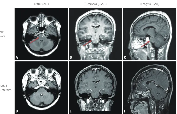
A 57-year-old female visited our hospital with complaints of gait disturbance, dysarthria, and diplopia with a 2-week history. She had previously been healthy with no history of medications. She presented with ataxic gait with tilting on both sides, but she had no oth-er symptoms or signs such as motor and sensory focal deficits of the limbs. Normal find-ings were obtained in all of the applied laboratory tests, including for autoimmune and paraneoplastic antibodies in her serum, for oligoclonal bands, IgG index, viral markers, and cytology in her cerebrospinal fluid, and for infection, vasculitis, and demyelinating CNS disease. The findings of nerve conduction tests (including the blink reflex) and brain MR angiography were also normal. She underwent brain MRI with gadolinium enhancement, which revealed patchy increased signal intensities in the pons, with strong contrast en-hancement (Fig. 1). She was treated with a high-dose steroid pulse therapy (methylpredni-sone at 500 mg for 5 days). Her symptoms improved significantly after this treatment, and she was discharged on prescribed oral prednisone. Follow-up MRI was performed after 4 months, which revealed complete resolution of the enhancing abnormalities.
With CLIPPERS, as the name already implies, both clinical and radiological features are rapidly improved by treatment with high-dose intravenous methylprednisone. However, diagnosing CLIPPERS is challenging, primarily due to the lack of a gold-standard diagnosis method, and secondarily because its pathogenesis is poorly understood.2 These reasons ORCID
Byung Joon Kim
https://orcid.org/0000-0001-8938-7667 Kang Min Park
130 https://doi.org/10.14253/acn.2020.22.2.129 http://www.e-acn.org
Annals of Clinical Neurophysiology Volume 22, Number 2, October 2020
mean that the most-important findings for distinguishing CLIPPERS from other diseases are its typical radiological findings including multiple, homogeneous, small (<3 mm in diameter), and curvilinear gadolinium-enhanced nodules in the pons and cerebellum.1 Clinicians suspicious of CLIPPERS
with the characteristic radiologic pattern could diagnose it based on the diagnostic criteria for CLIPPERS proposed by Tobin et al.3 Despite its rarity, this treatable condition still
needs to be considered.
Conflicts of Interest
The authors declare no conflicts of interest relevant to this article.
REFERENCES
1. Pittock SJ, Debruyne J, Krecke KN, Giannini C, van den Ameele J, De Herdt V, et al. Chronic lymphocytic inflammation with pon-tine perivascular enhancement responsive to steroids (CLIPPERS). Brain 2010;133:2626-2634.
2. Dudesek A, Rimmele F, Tesar S, Kolbaske S, Rommer PS, Benecke R, et al. CLIPPERS: chronic lymphocytic inflammation with pon-tine perivascular enhancement responsive to steroids. Review of an increasingly recognized entity within the spectrum of in-flammatory central nervous system disorders. Clin Exp Immunol 2014;175:385-396.
3. Tobin WO, Guo Y, Krecke KN, Parisi JE, Lucchinetti CF, Pittock SJ, et al. Diagnostic criteria for chronic lymphocytic inflammation with pontine perivascular enhancement responsive to steroids (CLIP-PERS). Brain 2017;140:2415-2425.
Fig. 1. FLAIR axial (A, D) and gadolinium-enhanced T1-weighted coronal (B, E) and sagittal (C, F) brain magnetic resonance imaging (MRI). Initial MRI revealed foci of gadolinium enhancement with a punctate and curvilinear pattern, predominantly in the pons (arrows). Follow-up MRI performed 4 months after steroid therapy revealed a marked reduction in the extent of gadolinium-enhanced lesions.
T2 flair Gd(+) Before steroids 4 months after steroids T1 coronal(+) Gd(+) T1 sagittal Gd(+) A D B E C F