Vol. 19, No. 2, December, 2006
상지 구획 증후군 이후 발생한 치명적인 뇌출혈
포천 중문의과대학교 분당차병원 신경외과학교실, 정형외과학교실*, 연세대학교 의과대학 신경외과학교실**
한인보∙정영선∙신동은*∙허
@륭∙정상섭∙안정용**
─ Abstract ─
A Fatal Intracerebral Hemorrhage Complicated by
Compartment Syndrome of the Upper Arm
In-Bo Han, M.D., Young-Sun Chung, M.D., Dong Eun Shin, M.D.*, Ryoong-Huh, M.D., Sang-Sup Chung, M.D., and Jung-Yong Ahn, M.D.**
Departments of Neurosurgery, Orthopedic Surgery*, Pochon CHA University College of Medicine, Sungnam, Korea Departments of Neurosurgery, Yonsei University College of Medicine**, Seoul, Korea
Compartment syndrome has a wide spectrum from muscle pain to a life- threatening condition, such as acute renal failure and disseminated intravascular coagulation (DIC). Intracerebral hemorrhage (ICH) due to com-partment syndrome has not been reported. We report a patient who presented with ICH leading to death. A 25-year-old female with no significant past history developed extensive compartment syndrome followed by rhab-domyolysis, acute renal failure, DIC, and ICH. Although the patient underwent a fasciotomy and hemodialysis and received aggressive resuscitation with massive transfusions of blood and intravenous fluids, she died. This case stresses the importance of early diagnosis and prompt treatment of compartment syndrome to prevent dev-astating complications. (J Korean Soc Traumatol 2006;19:178-182)
Key Words: Intracerebral hemorrhage, Compartment syndrome, Disseminated intravascular coagulation
� Address for Correspondence : Jung Yong Ahn, M.D.
Department of Neurosurgery, Yonsei University College of Medicine, 146-92 Dogok-dong, Gangam-gu, Seoul 135-720, Korea
Tel : 82-2-2019-3391, Fax : 82-2-3461-9929, E-mail : [email protected]
접수일: 2006년 8월 18일, 심사일: 2006년 10월 11일, 수정일: 2006년 10월 18일, 승인일: 2006년 11월 15일
Ⅰ. Introduction
Most compartment syndromes develop in the forearm or the leg.(1-3) There are numerous causes of the pressure elevation, and these include muscle trauma, tourniquet use, infiltra-tion of intravenous fluids, external compression
caused by narcotic overdose, or intraoperative positioning leading to muscle ischemia and subse-quent necrosis.(1-4) If this condition goes unrec-ognized, it can be devastating, and delays in the diagnosis and treatment may lead to life threat-ening complications such as severe metabolic dis-turbance, cardiac arrhythmia, cardiac arrest,
acute renal failure (ARF), and disseminated intravascular coagulation (DIC).(1-5) This report describes a case of a woman who developed who intracerebral hemorrhage (ICH) secondary to com-partment syndrome of the upper arm
Ⅱ. Case Report
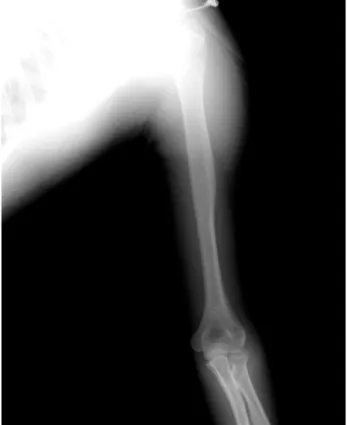
A 25-year-old woman was admitted to the orthopedic clinic for contusion of his left upper arm caused by blunt impact after fall. The patient was alert at the time of injury. Progressive swelling and pain developed in the left upper arm. Four days later, she was referred to our hospital due to sudden loss of conscious-ness. The patient didn’t exhibit fever. Physical examination revealed swollen and stiff lesions on the left side of her upper arm. Radiographs of the upper arm showed soft tissue swelling around left upper arm. But there was no definite frac-ture (Fig. 1). Chest radiograph and electrocardio-gram showed no abnormal features. Neurological examination showed coma with fully dilated and
fixed pupils. Brain computed tomography showed a large ICH of the right frontal lobe with severe swelling (Fig. 2). Doppler examination demon-strated normal arterial and venous signals, but showed increased muscular echo on the left proxi-mal upper arm. Edema formation was suspected within that compartment, but there was no hematoma. The pressure in the anterior compart-ment of the left upper arm was measured because compartment syndrome was suspected. The pres-sure was 48 mm Hg and emergent fasciotomy was performed.
Significant laboratory results included elevated serum blood urea nitrogen (138.4 mg/dL), creati-nine (7.1 mg/dL), myoglobin (>3000 ng/mL), potassium (5.7 mEq/L), aspartate ferase (AST) (1498 IU/L), alanine aminotrans-ferase (ALT) (1033 IU/L), uric acid (17.7 mg/dL), phosphorus (8.4 mg/dL), and creatine kinase (12372 U/L). Serum calcium was decreased (5.8 mg/dL). The patient’s urine looked dark, and its analysis was positive for myoglobin. Urine output was less than 300 ml/day. These lab find-ings were consistent with rhabdomyolysis and acute renal failure. Muscle biopsy from left upper arm done on 4th day of injury showed extensive rupture of skeletal muscle cells without inflam-mation. Regenerating fibers and degenerating fibers were also observed in most of fascicles
Fig. 1. Radiographs of the upper arm shows soft tissue swelling around left upper arm without fracture.
Fig. 2. Brain computed tomography scan shows huge intrac-erebral hemorrhage of the right frontal lobe with severe brain swelling.
(Fig. 3). The histological changes were suggestive of rhabdomyolysis. The levels of coagulation fac-tors were measured to rule out bleeding tendency disorder. Coagulation factor assay showed normal factor II, factor VII, factor VIII, factor IX, factor XII, and factor XIII. Factor V and factor XI decreased (18% and 33% respectively). The level of protein C5 (13.60 mg/dl), protein C3 (75.20 mg/dl), and C1 (13.4 mg/dl) was normal. Protein C4 (6.04 mg/dl) was decreased. Serum analysis was negative for factor VIII and factor Anti-IX. Other laboratory results were hemoglobin: 10.0 g/dL, hematocrit: 29.8%, white blood cell count: 22.7/cubic mm, neutrophil: 84%, platelets: 266,000/uL, erythrocyte sedimentation rate (ESR): 14 mm/hr, C-reactive protein (CRP): 19 mg/dL, prothrombin time (PT): more than 80.2 seconds, activated partial thromboplastin time (aPTT): more than 100 seconds, fibrinogen: less than 75 mg/dL, D-dimer: more than 8.0 ug/mL, fibrin degradation product (FDP): more than 80 ug/ml, and antithrombin III: 115.2%. DIC was suspected and replacement therapy with fresh frozen plasma (FFP) was started. Blood culture showed no bacteremia.
Since the ICH was very huge and the
evacua-tion of hematoma was not possible because of sus-pected uncontrollable bleeding and fatal patient’s condition, the initial treatment consisted of intracranial pressure control, replacement therapy with FFP, vigorous fluid replacement, and forced diuresis. Hemodialysis was also started from the day 1 of admission. But she died eventually on the day 24 of admission.
Ⅲ. Discussion
Compartment syndrome is a common condition characterized by an elevation of the interstitial pressure within the closed skeletal muscle com-partment leading to microvascular compromise.(1) Although the condition is well recognized in the leg and forearm, it is rare in the upper arm, as in our patient because upper arm contains a large number of flexible, broad, and rigid ligaments and tendons; thus, the brachial fascia yields more easily than in other areas.(1-3)
In the case presented here, the contusion of upper arm seems to cause muscle injury with sub-sequent edema and ischemia. Edema formation in a closed compartment will cause a rise in com-partmental pressure, further impairing the circu-lation and eventually leading to the development of a full-blown compartment syndrome with rhabdomyolysis. We could see an increased crea-tine kinase level and biochemical abnormalities such as elevated levels of AST, ALT, LDH, potassium, myoglobin and uric acid, accompanied by a low level of calcium. Among them, the ele-vation of creatine kinase level has been reported to be the most sensitive factor.(6)
Rrhabdomyolysis is an important cause of ARF. Around 33% of the episodes of rhabdomyolysis lead to ARF due to renal vasoconstriction, intra-luminal cast formation and direct myoglobin toxi-city. Rhabdomyolysis-induced ARF is mostly olig-uric and severe (ususally requiring dialysis) but reversible. So for patients strongly suspected of having rhabdomyolysis, aggressive hydration with normal saline should be initiated as soon as possi-ble to prevent the development of ARF. To assist in volume expansion and diuresis and to minimize Fig. 3. Photomiograph of the biceps-brachialis muscle biopsy
shows widespread muscle necrosis in each of the fasci-cles (H & E 400).
heme pigment deposition, mannitol is recommend-ed. Furosemide has also been used to assist in maintaining an adequate urine output. Urine alkalinization with sodium bicarbonate has been recommended to minimize the formation of myo-globin casts in renal tubule. Despite optimal treatment, some patients may develop renal fail-ure and require dialysis. However, complete recovery of renal function was frequently report-ed through hemodialysis.(6-9)
Muscle injury, regardless of etiology, can cause inflammatory response leading to endothelial cell damage followed by stimulation of clotting cas-cade. Highly activated and sustained inflamma-tion may give rise to DIC, which has been described in a wide variety of disorders such as infection, carcinoma, acute leukemia, and trauma including crush injury, burn, and rhabdomyoly-sis. DIC complicated by rhabdomyolysis may develop 12~72 hours after the acute insult. Patients who have severe DIC have a mortality rate of up to 85% according to underlying dis-eases. The massive activation of coagulation may result in depletion of platelets and coagulation factors, which may cause systemic hemorrhage. Hemorrhages from the skin and mucous mem-branes are the most common but intracranial hemorrhages are rare.(10-13) It has been reported that intracranial hemorrhages associated with DIC was complicated with solid tumors, leukemia, and aortic aneurysm and it usually occurred in the parenchymal or subdural space, and rarely in the subarachnoid space.(10,13) The patients present-ing with a sudden onset were fatal. The evacua-tion of hematoma was usually not possible due to suspected uncontrollable bleeding. However, some cases might have an indication for the evacuation of hematoma. It is important to check conditions carefully whether the patient has an indication for the operation. Treatment of patients with DIC depends on the etiology and severity of their con-dition, but generally involves maintaining the patient hematologically. So for patients suspected of having DIC, replacement of blood products such as fresh frozen plasma should be performed.
Since early diagnosis is very important,
com-partment pressure monitoring may be helpful. An intracompartmental pressure of 30 mmHg is often used as a basis for performing a fasciotomy. Some authors suggested that fasciotomy should be performed when the difference between mean arterial pressure and intracompartmental pressure is 40~50 mmHg.(14)
This case clearly shows that compartment syn-drome can lead to life threatening conditions such as ARF and DIC, possibly death. Although lethal complications can arise due to the severe nature of compartment syndrome itself, its late and sometimes incomplete initial treatment is a possi-ble causative factor. Early diagnosis and prompt treatment is crucial to prevent the devastating complications.
Ⅳ. Conclusion
Careful history and physical examination are the most important tools for diagnosis of compart-ment syndrome. Errors of omission in the diagno-sis of compartment syndrome may lead rapidly to life threatening complication, occasionally death. This case stresses the importance of early diagno-sis and prompt treatment of compartment syn-drome to prevent devastating complications.
REFERENCES
01) Diminick M, Shapiro G, Cornell C. Acute compart-ment syndrome of the triceps and deltoid. J Orthop Trauma 1999;13:225-7.
02) Palumbo RC, Abrams JS. Compartment syndrome of the upper arm. Orthopedics 1994;12:1144-7. 03) Yabuki S, Kikuchi S. Dorsal compartment
syn-drome of the upper arm: A case report. Clin Orthop Relat Res 1999;366:107-9.
04) Weinmann M. Compartment syndrome. Emerg Med Serv 2003;32:6.
05) Bacal D, Lampman RM, Hogikyan JV. Compartment syndrome of the arm and disseminat-ed intravascular coagulation. Am J Orthop 2001;30: 422-3.
06) Ana LH, Joseph V, Paul EM. Bench-to-bedside review: Rhabdomyolysis-an overview for clinicians. Crit Care 2005;9:158-69.
07) Daher ED, Silva Junior GB, Brunetta DM. Rhabdomyolysis and acute renal failure after
stren-uous exercise and alcohol abuse: case report and lit-erature review. Sao Paulo Med J 2005;123:33-7. 08) Poels PJ, Gabreel FJ. Rhabdomyolysis: a review of
the literature. Clin Neurol Neurosurg 1993;95:175-92.
09) Szewczyk D, Ovadia P, Abdullah F. Pressure induced rhabdomyolysis and acute renal failure. J Trauma 1998;44:384-8.
10) Kawakami Y, Ueki K, Chikama M. Intracranial hemorrhage associated with nontraumatic dissemi-nated intravascular coagulation-report of four cases. Neurol Med Chir (Tokyo) 1990;30:610-7.
11) Rodger LB. Disseminated intravascular coagulation. Current concepts of etiology, pathophysiology,
diag-nosis, and treatment. Hematol Oncol Clin N Am 2003;17:149-76.
12) Brick RL, Arun B. Disseminated intravascular coagulation. Clinical and pathophysiological mecha-nisms and manifestations. Haemostasis 1999;29:111-34.
13) Tasdemiroglu, Kaya. Neurologic complications of cancer: part 2: vascular, infectious, paraneoplastic, neuromuscular, and treatment-related complica-tions. Neurosurgery Quart 2004;14:133-53.
14) Kai M, David WL. Clinical spectrum of acute com-partment syndrome of the thigh and its relation to associated injuries. Clinical orthopedics and related research. 2004;425:223-9.