Copyright ⓒ 2012 by the Korean Society for Biochemistry and Molecular Biology
This is an open-access article distributed under the terms of the Creative Commons Attribution Non-Commercial License (http://creativecommons. org/licenses/ by-nc/3. 0) which permits unrestricted non-commercial use, distribution, and reproduction in any medium, provided the original work is properly cited.
Woo Jin Park
3and Pyung-Lim Han
1,2,4,5 1Department of Brain and Cognitive Sciences 2
Brain Disease Research Institute Ewha Womans University Seoul 120-750, Korea 3
Department of Life Science
Gwangju Institute of Science and Technology (GIST) Gwangju 500-712, Korea
4
Department of Chemistry and Nano Science Ewha Womans University
Seoul 120-750, Korea 5
Corresponding author: Tel, 82-2-3277-4130; Fax, 82-2-3277-3419; E-mail, [email protected] http://dx.doi.org/10.3858/emm.2012.44.12.082 Accepted 13 November 2012
Available Online 22 November 2012
Abbreviations: APP, amyloid precursor protein; NIa, nuclear inclusion a; PS1, presenilin 1; TuMV, Turnip mosaic virus
Abstract
The plant viral protease, NIa, has a strict substrate
spe-cificity for the consensus sequence of Val-Xaa-His-Gln,
with a scissoring property after Gln. We recently
re-ported that NIa efficiently cleaved the amyloid-β (Aβ)
peptide, which contains the sequence Val-His-His-Gln
in the vicinity of the cleavage site by α-secretase, and
that the expression of NIa using a lentiviral system in
the brain of AD mouse model reduced plaque
deposi-tion levels. In the present study, we investigated
wheth-er exogenous expression of NIa in the brain of AD
mouse model is beneficial to the improvement of
cog-nitive deficits. To address this question, Lenti-NIa was
intracerebrally injected into the brain of Tg-APPswe/
PS1dE9 (Tg-APP/PS1) mice at 7 months of age and
be-havioral tests were performed 15-30 days afterwards.
The results of the water maze test indicated that
hidden-platform and markedly enhanced navigation
near the maze-wall, and that such behavioral deficits
were significantly reversed in Tg-APP/PS1 mice
in-jected with Lenti-NIa. In the passive avoidance test,
Tg-APP/PS1 mice exhibited a severe deficit in their
contextual memory retention, which was reversed by
NIa expression. In the marble burying test, Tg-APP/PS1
mice buried marbles fewer than non-transgenic mice,
which was also significantly improved by NIa. After
be-havioral tests, it was verified that the Tg-APP/PS1 mice
with Lenti-NIa injection had reduced Aβ levels and
pla-que deposition when compared to Tg-APP/PS1 mice.
These results showed that the plant viral protease, NIa,
not only reduces Aβ pathology, but also improves
be-havioral deficits.
Keywords: Alzheimer disease; amyloid β-peptides;
disease models, animal; endopeptidases; maze
learning
Introduction
Alzheimer’s disease (AD) is a neurodegenerative disease distinguished by extracellular deposition of amyloid-β (Aβ) outside neurons and progressive cognitive dysfunction. Because Aβ is highly toxic to neural cells, accumulation of Aβ has been thought to cause the pathogenesis and cognitive impairment of AD (McLean et al., 1999; Larson and Lesne, 2001; Walsh et al., 2002). Aβ levels in the brain are regulated by the ratio between Aβ production and Aβ clearance (Tanzi and Bertram, 2005). Several endogenous proteases are known to play a role in clearance of Aβ in AD brains. Such endogenous proteases include neprilysin (NEP), insulin degrading enzyme (IDE), endothelin-converting enzyme (ECE), angiotensin-converting enzyme (ACE), plasmin/ tPA/uPA, cathepsin B, and MMP-2/MMP-9 (Wang et al., 2006; Haass and Selkoe, 2007; Miners et al., 2008), among which NEP and IDE have been most extensivelystudied. The pharmacological or genetic
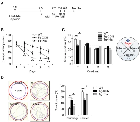
Figure 1. Spatial memory deficits displayed by Tg-APP/PS1 mice were improved by NIa expression. (A) Experimental design of Lenti-NIa injection and behavioral performance. Lenti-NIa was injected in Tg-APP/PS1 mice at 7 months of age and behavioral tests were started 15 days after the injection, for the two-week period, the sequence of which was the Morris water maze test (WM), passive avoidance test (PA), and marble burying test (MB). (B) Escape latency on a hidden platform of Tg-APP/PS1 mouse control (Tg-CON), Tg-APP/PS1 mice infused with Lenti-NIa (Tg+NIa), and their non-transgenic control (WT) in the Morris water maze test. Tg-APP/PS1 mice infused with Lenti-GFP are used as Tg-APP/PS1 mouse control (Tg-CON) throughout this work. Numbers of animals used: WT, 5 males and 5 females; Tg-CON, 5 males and 4 females; Tg+NIa, 5 males and 4 females. Two-way repeated measures ANOVA and Bonferroni post-hoc test: significant difference between animal groups [F(2,204)= 19.77, P < 0.0001], significant effect of time [F(4,204)= 32.94,
P < 0.0001] and significant animal group × time interaction [F(8,204)= 2.301, P < 0.05]. Data are presented as the means ± SEM. **denotes a differ-ence between WT and Tg-CON at P < 0.01 at the indicated time point. Separate two-way repeated measures ANOVA for the data groups between Tg-CON and Tg+NIa showed significant effects of treatment (NIa) [F(1,128)= 10.95, P < 0.01] and time [F(4,128)= 14.10, P < 0.0001], but no significant treatment × time interaction [F(4,128)= 0.8769, P = 0.4798]. Data are presented as the means ± SEM. (C) The percentage of time spent in the target quadrant in the spatial probe trial test. The dashed line represents the chance performance level (25%) at each quadrant. Tg-CON; Tg-APP/PS1 mouse control. Tg+NIa; Tg-APP/PS1 mice infused with Lenti-NIa. WT; non-transgenic control. Target (T), opposite (O), and adjacent quadrants (L, R) are de-picted on the right panel. Two-way ANOVA and Bonferroni post-hoc test: no difference between animal groups [F(2,96)= 0.0000, P = 1], but significant dif-ference between zones [F(3,96)= 14.92, P < 0.0001] and significant animal group × zone interaction [F(6,96)= 3.997, P < 0.01]. * and ** denote differ-ences between indicated groups in the T zone, at P < 0.05 and P < 0.01, respectively. Data are presented as the means ± SEM. (D) The percentage of time spent in the center vs. periphery zones in the hidden platform version of the Morris water maze examined on day 5 (right panel). The center and periphery zones are depicted (left panels). The periphery zone is defined as the area between wall and the circle apart by 10-cm from the wall. Representative spatial navigations on the water maze pool for 30-sec period are presented for WT, Tg-CON, and Tg+NIa mice. One-way ANOVA and
Newman-Keuls post-hoc test; significant difference between animal groups in the periphery zone [F(2,24)= 8.693, P < 0.01] and in the center zone [F(2,24) = 8.715, P < 0.01]. * and ** denote differences between indicated groups at P < 0.05 and P < 0.01, respectively. Data are presented as the means ± SEM.
inhibition of NEP in mouse models of AD increased Aβ accumulation and memory impairment (Mouri et al., 2006; Farris et al., 2007), while the over-expression of NEP was shown to reduce Aβ levels, plaque deposition, and plaque-related pathology (Marr et al., 2003). IDE immunoreactivity was shown to be significantly reduced in the brain of those diagnosed with AD (Miners et al., 2008), while mice
with a deficiency of the IDE gene exhibited an accumulation of Aβ in the brain (Farris et al., 2003). Therefore, the enzymatic degradation of Aβ has been considered as an attractive strategy for the mitigation of Aβ accumulation in the AD brains (Haass and Selkoe, 2007; Miners et al., 2008). However, NEP overexpression did not reduce oligomeric Aβ levels and Aβ oligomer-induced
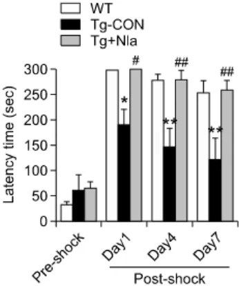
Figure 2. The contextual memory deficit of Tg-APP/PS1 mice was re-versed by NIa expression. The entry latency into the dark chamber for Tg-APP/PS1 mouse control (Tg-CON), Tg-APP/PS1 mice infused with Lenti-NIa (Tg+NIa), and their non-transgenic control (WT) in the passive avoidance test. The latency to enter into the dark chamber after shock (post-shock) is used as an indicator of learning and memory. Two-way re-peated measures ANOVA and Bonferroni post-hoc test for data groups at post-shock days: significant difference between animal groups [F(2,46)= 11.34, P < 0.001], time [F(2,46)= 10.08, P < 0.001], but no animal group × time interaction [F(4,46)= 0.3358, P = 0.8524]; * and ** denote differences between WT and Tg-CON at P < 0.05 and P < 0.01, re-spectively; # and ## denote difference between Tg-CON and Tg+NIa at P <0.05 and P < 0.01, respectively. Data are presented as the means ±SEM. Numbers of animals used: WT, 5 males and 5 females; Tg-CON, 4 males and 4 females; Tg+NIa, 4 males and 4 females. degrade many cellular proteins, these proteases are
likely to present many disadvantages through their overexpression in the brain. Therefore, a new strategy that permits the cleavage of both monomeric and oligomeric Aβ, but has a substrate specificity for Aβ, is necessarily sought.
The nuclear inclusion a protease of the Turnip mosaic virus, NIa, has a substrate specificity for the sequence of Val-Xaa-His-Gln with a cleaving property after Gln (Kang et al., 2001). We recently reported that Aβ peptides contain the sequence Val-His-His-Gln in the vicinity of the cleavage site by α-secretase, and that NIa degraded Aβ in vitro. Moreover, overexpression of NIa using a Lentiviral system reduced plaque levels in the brain of AD mouse model (Han et al., 2010). In the present study, we investigated whether NIa expression in the brain of AD mouse model is beneficial towards the improvement of cognitive deficits at the behavioral level in a mouse model of AD.
Results
To investigate whether the exogenous expression of NIa improved cognitive function, Lenti-NIa virus was stereotaxically injected into the lateral ventricles at both hemispheres in the brain of Tg-APP/PS1 mice which were 7 months old, and at this stage where plaque deposition in the brain has been determined to show at low levels in various brain regions (Kim et al., 2012). Tg-APP/PS1 mice infused with Lenti-GFP in the same manner were used as a control. Starting 15 days after the injection, a series of behavioral tests were performed for the following 15 days (Figure 1A).
Cognitive impairments displayed by Tg-APP/PS1
mice were improved by NIa expression
In the Morris water maze test, the performance of Tg-APP/PS1 mice (carrying Lenti-GFP) in finding the hidden platform was much reduced when compared to that of the non-transgenic control, indicating that the special recognition learning/memory in Tg-APP/PS1 mice was severely impaired. On the contrary, the behavioral performance of Tg-APP/PS1 mice
injected with Lenti-NIa was significantly improved when compared with that of control mice (Figure 1B). In a probe trial test, Tg-APP/PS1 mice showed reduced dwelling time in the target quadrant compared with the non-transgenic control. Whereas Tg-APP/PS1 mice with Lenti-NIa spent more time in the target quadrant compared with that of Tg-APP/PS1 mice (Figure 1C). These results suggest that the expression of NIa through the Lentivial system in the brain was beneficial for improving cognitive function of Tg-APP/PS1 mice. It was unexpectedly noticed that Tg-APP/PS1 mice spent more time in the periphery of the water maze pool than the non-transgenic control during the 90 sec of the test period. Whereas NIa expression reversed the stereotyped behavior in the periphery to the control level (Figure 1D). In the passive avoidance test, the post-shock latency for Tg-APP/PS1 mice to enter into the dark chamber was shorter than that displayed by the non-transgenic control (Figure 2). Moreover, this defective memory retention of Tg-APP/PS1 mice was decayed faster than the non-transgenic control in 7 days of repeated tests (Figure 2A). Whereas expression of NIa improved the impaired memory retention to the level of the non-transgenic control (Figure 2). These results suggest that the expression of NIa in the brain of Tg-APP/PS1 mice significantly improved context-dependent memory as well. In the marble burying test, Tg-APP/PS1 mice
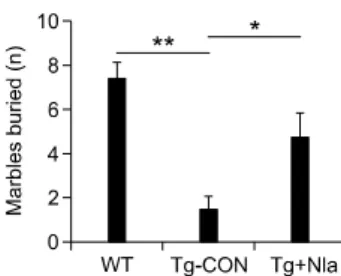
Figure 3. Behavioral changes in the marble burying test were restored by NIa expression. The numbers of glass marbles buried under wood chips by Tg-APP/PS1 mouse control (Tg-CON), Tg-APP/PS1 mice in-fused with Lenti-NIa (Tg+NIa), and their non-transgenic control (WT) for the 30-min period are presented. NIa expression significantly reverted marble burying behavior. One-way ANOVA and Newman-Keuls post-hoc test: [F(2,24)= 13.81, P = 0.0001]. * and ** denote difference at P < 0.05 and P < 0.01, respectively. Data are presented as the means ± SEM. Numbers of animals used: WT, 5 males and 5 females; Tg-CON,4 males and 4 females; Tg+NIa, 4 males and 4 females.
buried marbles fewer than the non-transgenic control, while expression of NIa significantly reversed the behavioral change displayed by Tg-APP/PS1 mice (Figure 3).
Suppression of Aβ pathology in the brain with NIa
expression
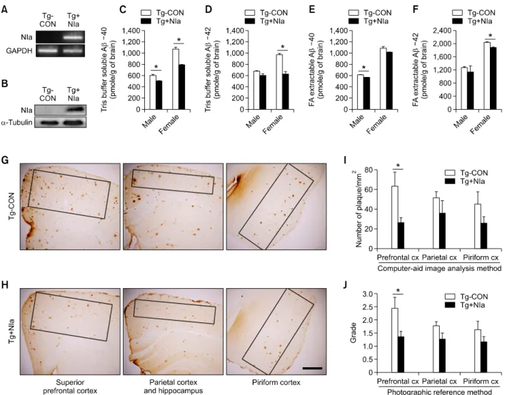
To verify that intracerebral injection of Lenti-NIa reduced Aβ pathology, the plaque pathology in the prefrontal cortex was examined. RT-PCR and Western blot analyses were used to confirm the expression of NIa in the brain of Tg-APP/PS1 mice infused with Lenti-NIa (Figures 4A and 4B). ELISA assay showed that the levels of Aβ (40) in the Tris-buffer soluble faction as well as the formic acid-extractable fraction of male were significantly reduced. The levels of Aβ (40) in the Tris-buffer soluble faction, but not in the formic acid-extractable faction, of female also was significantly reduced (Figures 4C and 4E). The levels of Aβ (42) in both the Tris-buffer soluble, and formic acid-extractable factions of female were significantly reduced, although the reduction of Aβ (42) in male was not statistically significant (Figures 4D and 4F).
Immunohistochemical analysis revealed that plaque deposition levels in the prefrontal cortex, parietal cortex, and piriform cortex tended to decrease in the brain of Tg-APP/PS1 mice infused with Lenti-NIa when compared to the levels in Tg-APP/PS1 mouse control (Figures 4G and 4H). Quantification of plaque deposition levels, using a computer-aid image analysis system, indicated that the number of plaques in the prefrontal cortex was significantly reduced although very subtle reductions were observed for the parietal cortex and piriform cortex (Figure 4I). Quantification of plaque deposition levels
using the photographic plaque deposition reference panel (Kim et al., 2012) also yielded similar results (Figure 4J).
Discussion
Beneficial effects of NIa expression on the cognitive
function of Tg-APP/PS1 mice
We recently demonstrated that the plant viral protease NIa effectively degraded Aβ in vitro, and that the expression of NIa in the brain of an AD model mice using a Lentiviral system reduced Aβ levels and plaque deposition (Han et al., 2010). Because NIa is a potent exogenous protease, it was questioned whether or not overexpression of NIa produces any detrimental effects on the brain, other than the reduction in the levels of Aβ. After the infusion of Lenti-NIa in the brain, the spatial memory task in the Morris water maze test (Figures 1B and 1C) and the contextual memory task in the passive avoidance test (Figure 2) were significantly improved, suggesting that overexpression of NIa has beneficial effects in alleviating cognitive dysfunction of the AD mouse model. As these beneficial or protective effects were produced by the expression of the Aβ-degrading protease NIa, the results of the present study support also that the cognitive deficits displayed by untreated Tg-APP/PS1 mice were due to the accumulation of Aβ in the brain. We believe that further studies on the effects of a more prolonged expression of NIa over a period of several months to a year, for example, are required in order to more fully elucidate the effects of Aβ regulation through NIa.
Anxiety is yet another of many problematic symptoms endured by patients with AD (Serra et al., 2010; Spalletta et al., 2010). Our results indicated that Tg-APP/PS1 mice had increased anxiety and this emotional change was also reversed by the expression of NIa (Figures 1D and 3), reducing the concern that overexpression of the foreign protease NIa in the brain could have detrimental effects on the emotional behavior.
Repression of Aβ pathology by NIa in the brain of
Tg-APP/PS1 mice
It was reported that Tg-APP/PS1 mice showed cognitive deficits at the age of 15 months, but not at 8 months in the Morris water maze test (Hooijmans et al., 2009). A further study reported cognitive deficits in Tg-APP/PS1 mice at the age of 10 months in the Morris water maze test (Timmer et al., 2010). The present study demonstrated that Tg-APP/PS1 mice before reaching to 8 months of age showed
Figure 4. Suppression of Aβ pathology by NIa expression in the brain of Tg-APP/PS1 mice. (A) RT-PCR showing the expression of NIa transcript in the
brain of Tg-APP/PS1 mice infused with Lenti-NIa (Tg+NIa).The expression of NIa was analyzed on the parietal cortex of Tg-APP/PS1 mice and their con-trol mice (Tg-CON). GAPDH is a concon-trol. (B) Western blots showing the expression of NIa protein in the brain of Tg-APP/PS1 mice infused with Lenti-NIa (Tg+NIa). The expression of NIa (molecular weight: 28 kD) in the prefrontal cortex was examined using anti-NIa. Tg-APP/PS1 mouse control (Tg-CON). α-tubulin was used as a loading control. (C-F) The amounts of Aβ (40) (C, E) and Aβ (42) (D, F) in Tris-buffer soluble (C, D) and formic acid-extractable (E, F) fractions in the brains of Tg-APP/PS1 mouse control (Tg-CON) and Tg-APP/PS1 mice infused with Lenti-NIa (Tg+NIa). (G, H) Photomicrographs showing anti-Aβ (Bam-10)-stained superior prefrontal cortex, parietal cortex and hippocampus, and piriform cortex of Tg-APP/PS1 mouse control (Tg-CON; G) and Tg-APP/PS1 mice infused with Lenti-NIa (Tg+NIa; H). (I, J) Quantification of the levels of plaques (mm2) in the prefrontal cortex, parietal cortex, and piriform cortex of Tg-APP/PS1 mouse control (Tg-CON) and Tg-APP/PS1 mice infused with Lenti-NIa. Plaque levels presented were counted using computer-aid image analysis program (I) and the 6-grade plaque photographic reference panels (J). All data were from female mice. Scale bar: 500 μm. Data are presented as the means ± SEM. *denotes difference between the indicated groups at P < 0.05 (Student t-test).
significant impairments in cognitive function in the Morris water maze test and the passive avoidance test (Figures 1 and 2). This discrepancy between previous result (Hooijmans et al., 2009) and the present study might be attributed to differing conditions in animal rooms or different handing skills in behavioral assays (Wahlsten et al., 2006). Given that Tg-APP/PS1 mice show visible plaque deposition from 6-6.5 months of age (Seo et al., 2011; Kim et al., 2012; unpublished observation), it is not surprising that Tg-APP/PS1 mice exhibited behavioral deficits prior to 8 months of age.
Considering that the cognitive deficits displayed by untreated Tg-APP/PS1 mice were attributed to the accumulation of Aβ in the brain, as discussed above, it would be interesting to specifically examine whether behavioral deficits in Tg-APP/PS1 mice could occur as early as 6.0-7.5 months of age, at the time when plaque deposition has begun to occur, presumably due to enhanced accumulation of Aβ. In the Morris water maze test, Tg-APP/PS1 mice spent approximately 50% of the time in navigation near the wall of the water maze, and this behavior was in sharp contrast to that of the non-transgenic
mice. This stereotyped behavior is apparently caused by Aβ pathology, as evidenced that the phenotype also was reversed by NIa expression (Figure 1D). We speculate that this stereotyped behavior is likely to reflect the increased anxiety of Tg-APP/PS1 mice, although we do not exclude the possibility of problems associated with their working memory. If the neurobiological feature of such behavior is defined more clearly, this stereotyped behavior might be used as an important behavioral indicator in analysis of transgenic mouse models of AD. The intracerebral injection of Lenti-NIa in Tg-APP/PS1 mice suppressed the Aβ levels and plaque deposition, the reduction of which was significant in the prefrontal cortex, but not so in the parietal and piriform cortices (Figure 4). However, a similar infusion of Lenti-NIa in Tg-APP/PS1 mice suppressed Aβ pathology within the same brain regions to a somewhat higher degree (Han et al., 2010). Although the present study did not provide further evidence, we speculate that the moderate and differential suppression of plaque pathology by NIa in different brain regions is, in part, due to the uneven penetration of Lenti-NIa in different brain regions. It is also possibly attributed to the fact that the final viral tire used in the present study was 0.8 ×10⁸ TU, which is slightly lower than the previous
tire of 1 × 10⁸ TU (Han et al., 2010). We speculate
that it is also related to the partial rescue of behavioral performance in the Morris water maze test, a behavioral test for a hippocampus-dependent spatial learning. Nonetheless, significant or complete rescue of behavioral performance in the Morris water maze test, passive avoidance test, and marble burying test indicated that a partial repression of Aβ levels was sufficient to provide a beneficial effect on behavior. In conjunction with this, it would be worth investigating whether local expression of NIa in specific brain regions produces specific or limited rescue of impaired brain function. Lenti-NIa expression in female Tg-APP/PS1 mice seemed to suppress Aβ contents more effectively than in male (Figures 4C-4F). We examined if there was any sexual difference in the learning and memory retention in relation to this differential effect in Aβ contents, but no evidence for the sexual difference was detected. There was also difference in Tris- and FA-extracted Aβ levels between male and female mice (Figures 4C-4F). Although the mechanisms underlying the differences remain unknown, it may be possible that this was in part due to differential expression of NIa or Aβ availability to given NIa levels in the brains with different genders. Considering that NEP and IDE do not effectively clear oligomeric Aβ (Haass and Selkoe, 2007; Meilandt et al., 2009) and have a broad substrate
specificity for cellular proteins, the results of the present study, together with the previous work (Han et al., 2010), suggest that because NIa cleaves both monomeric and oligomeric Aβ, and has a strict substrate specificity for Aβ, NIa is a good candidate to be used as a strategy for the improvement of Aβ pathology and the cognitive deficits of AD.
Methods
Transgenic AD model mice
Transgenic AD model mice, Tg-APPswe/PS1dE9 (Tg-APP/ PS1 for short), overexpressing human mutated APP and PS1 (APPswe/PS1dE9) were maintained in C57BL6 × C3H F1 hybrid mice as described previously (Jankowsky et
al., 2001; Seo et al., 2011). The mice were housed in pairs,
in normal plastic cages, with free access to food and water in a temperature- and humidity-controlled environment, un-der a 12 h light/dark cycle. They were fed a diet of lab chow and water ad libitum. All animals were handled in ac-cordance with the animal care guidelines of the Ewha Womans University School of Medicine.
Preparation of Lenti-NIa virus
The Lenti-NIa expression vector system was prepared as previously described (Han et al., 2010). In brief, the cDNA for NIa was inserted in the pCD513B-1 lenti viral ex-pression system (System Biosciences, Mountain View, CA). The resulting Lenti-NIa plasmid, together with the pPACK packing plasmid mix (System Biosciences), was transfected in HEK293T cells using a calcium (2.5 M CaCl2)-phosphate method. After 3 days, the supernatant containing Lentiviral particles was collected after filtration with a 0.45-μm filter (Millipore Corporation, Billerica, Massachusetts) and then concentrated by ultra-cen-trifugation as previously described (Han et al., 2010). The control Lenti-GFP viral vector was prepared similarly. The titers of Lenti-NIa and Lenti-GFP were estimated using a FACS analysis method, and the final tires obtained were 0.8 ×10⁸ TU and 0.9 × 10⁸ TU, respectively.
Intracerebroventricular injection of Lenti-NIa
Intracerebroventricular (i.c.v.) injection of Lenti-NIa was performed as previously described (Han et al., 2010). Tg-APP/PS1 mice at 7 months of age were randomized in-to the Lenti-NIa (n = 8; 5 males and 3 females), and Lenti-GFP (n = 9; 5 males and 4 females), and their non-transgenic control (n = 10; 5 males and 5 females) groups. Mice were anesthetized by intraperitoneal (i.p.) in-jection of 3.5:1 mixture of ketamine (50 mg/ml) and xyla-zine hydrochloride (23.3 mg/ml) at the dose of 1.0 μl/g body weight and placed on a stereotaxic apparatus (Stoelting Company, Wood Dale, IL) as described in a previous study (Kim et al., 2008; Han et al., 2010). The mice were intra-cerebroventricularly injected, on both sides with a total of 3 μl of Lenti-NIa (0.8 ×10⁸ TU in total) or Lenti-GFP with the same titer (0.8 ×10⁸ TU) at the speed of 1 μl/min
Immunohistochemistry and quantification of plaque
deposition levels
Immunohistochemical works were carried out as previously described (Han et al., 2010; Kim et al., 2012). Mice were sacrificed and perfused with 0.9% saline via a trans-car-diac method. One hemisphere of each animal was post-fixed in 4% paraformaldehyde in 0.1 M phosphate buf-fer (pH 7.4) overnight at 4oC. The other hemisphere of each animal was used for ELISA assay as described below. They were coronally cut into 40 μm-thick sections with a vibratome (Leica VT 1000S; Leica Instruments, Nussloch, Germany). Free-floating sections were blocked with 4% bovine serum albumin in PBS for 1 h, and were re-acted with monoclonal anti-β-amyloid protein antibody (1:2,000) (Bam10; # A5213, Sigma, St. Louis, MO) at 4oC overnight. The sections were washed with PBS, reacted with biotinylated secondary antibodies diluted 1:200 in PBS, and visualized using an ABC Elite kit (Vector Laboratories, Burlingame, CA).
Stained images were analyzed using an Olympus BX 51 microscope equipped with a DP71 camera and DP-manag-er and DP-ControllDP-manag-er software (Olympus Co., Tokyo, Japan). Plaque deposition levels in the prefrontal, parietal, and piriform cortices were quantified using the TOMORO ScopeEye 3.6 program (Techsan Community, Seoul, Korea). Plaque deposition levels were also quantified using the 6-grade plaque photographic reference panels (Kim et
al., 2012). The 6-grade plaque photographic reference
panels consist of a series of anti-Aβ (Bam-10)-stained brain sections images at the levels of the prefrontal cortex, parietal cortex, piriform cortex and hippocampus. The pho-tographic reference panels represent plaque deposition levels assigned with the numerical grades of 0, +1, +2, +3, +4, +5, and +6, where plaque deposition levels increase “sigmoidally” over grading scales. Such “sigmoidal” in-crease allows the referenced plaque grades to be used to for the semi-quantitative assessment of plaque levels. When necessary, increments of 0.5 were applied.
Assessment of Aβ levels
ELISA assays for Aβ42 and Aβ40 levels were described previously (Lee et al., 2009; Seo et al., 2011). Briefly, the prefrontal cortex of each animal was homogenized in Tris-buffered saline (20 mM Tris and 137 mM NaCl, [pH 7.6]) containing protease inhibitor mixtures (Complete Mini; Roche). Homogenates were centrifuged at 100,000 g for 1 h at 4oC, and the supernatant was used to measure the levels of Tris buffer-soluble forms of Aβ. The pellet was so-nicated in 70% formic acid and further centrifuged as above, and the resulting supernatant was used to measure the levels of the formic acid-extractable Aβ. The formic
frontal cortices from 5 Tg-APP/PS1 control mice and 5 Tg-APP/PS1 mice carrying Lenti-NIa were analyzed. Anti-NIa was produced in rabbits by repeated injections of NIa which was expressed in bacteria and purified. The tailed procedure for the production of anti-NIa will be de-scribed elsewhere. Anti-NIa antibody (1:1,000) and mono-clonal α-tubulin antibody (Calbiochem; 1:2,000) were used in Western blot analysis.
RT-PCR analysis
RT-PCR was carried out as described previously (Han et
al., 2010) using the following primer sets: NIa: 5'-ACG AAA
GAC GGC CAA TGC GGA-3' and 5'-ACC CGA CGG TTG CGA TGC TT-3'; GAPDH, 5'-TCC GTG TTC CTA CCC CCA ATG-3' and 5'-GGG AGT TGC TGT TGA AGT CGC-3'. The parietal cortices from five Tg-APP/PS1 control and five Tg-APP/PS1 mice carrying Lenti-NIa were used.
Behavioral assessments
Mice were brought to the testing room 30 min prior to the start of each behavioral test. Behavioral tests were per-formed with a computerized video-tracking system (SMART; Panlab S.I., Barcelona, Spain). At all times throughout the test, sound was masked with 60-65dB white noise.
Water maze test
The water maze test was performed as described pre-viously (Lee et al., 2004, 2006). Briefly, the water maze consisted of a 90-cm-diameter cylinder pool filled with opa-que milky water (22oC). A hidden platform (10 cm in diame-ter) was placed in a quadrant 2 cm below the opaque wa-ter surface. The pool was placed in the cenwa-ter of a room with environmental and artificial cues including a door, a chair, and two marked posters. The room was illuminated at 35-40 lux. In each daily testing, mice were allowed to swim for a maximum of 90 s. Whether succeeded to locate the platform or failed, the animals were permitted to stand on it for 30 s before the session was terminated. The ani-mals were tested twice a day, with a 6-h interval between trials. The latency of time and distance traveled prior to finding the platform for each trial were recorded for each trial. A spatial probe trial was conducted on day 6 after the 5-daytraining session, in the absence of the hidden platform. Animals were given a single 60-s probe trial to examine their spatial memory retention.
Passive avoidance test
previously (Lee et al., 2004, 2006). Briefly, the test appara-tus consisted of a brightly lit (1,700 Lux) white chamber, a darkened black chamber equipped with a shock-grid floor(each 15 ×15 ×15 cm), and a door between the two. On the first day of testing, each mouse was placed in the lighted chamber and left to freely explore the light and dark chambers for 5 min. On the second day, the mice were placed in the lighted chamber, after 30 sec, the middle door was opened, and the latency for the mouse to enter the dark chamber was recorded as pre-shock values. When a mouse entered the dark room, the door was closed and two successive electric foot-shocks (100 V, 0.3 mA, 2 sec for each shock and 5 sec for the interval) were delivered through the grid-floor. After this conditioning, mice were then returned to their home cages. One day (24 h), 4 days, and 7 days later, each mouse was replaced in the lighted chamber and the latency for the mouse to enter the dark chamber was measured with cut-off time of 300 sec, which was recorded as post-shock values.
Marble burying test
The marbles burying test was performed as described pre-viously (Deacon, 2006), but with a minor modification. In brief, empty cages were filled with bedding up to 5 cm from the cage floor, and 12 grass marbles were placed evenly throughout the cage. Mice were individually allowed to freely explore the cage with marbles for 30 min, and after-wards, the number of successfully buried marbles was counted. Marble “burying” was defined when less than 25% of a marble was visible.
Statistical analysis
Two-sample comparisons were carried out using the Student’s t-test, while multiple comparisons were made us-ing one-way or two-way ANOVA followed by post-hoc tests to compare selected pairs of data. GraphPad PRISM soft-ware 4.0 (GraphPad Softsoft-ware Inc., San Diego, CA was used to perform statistical analyses. All data are presented as the means± S.E.M., and statistical differences were ac-cepted at the 5% level.
Acknowledgements
This work was supported by grants (20110027540, 2012R1A2A1A03010177) from the National Research Foundation of Korea from the Ministry of Science and Technology, Republic of Korea.
References
Benilova I, Karran E, De Strooper B. The toxic Aβ oligomer and Alzheimer’s disease: an emperor in need of clothes. Nat Neurosci 2012;15:349-57
Chesneau V, Vekrellis K, Rosner MR, Selkoe DJ. Purified recombinant insulin-degrading enzyme degrades amyloid beta-protein but does not promote its oligomerization. Biochem J 2000;351:509-16
Dahlgren KN, Manelli AM, Stine WB Jr, Baker LK, Krafft GA, LaDu MJ. Oligomeric and fibrillar species of amyloid-beta peptides differentially affect neuronal viability. J Biol Chem 2002;277:32046-53
Deacon RM. Digging and marble burying in mice: simple methods for in vivo identification of biological impacts. Nat Protoc 2006;1:122-4
Farris W, Mansourian S, Chang Y, Lindsley L, Eckman EA, Frosch MP, Eckman CB, Tanzi RE, Selkoe DJ, Guenette S. Insulin-degrading enzyme regulates the levels of insulin, amyloid beta-protein, and the beta-amyloid precursor protein intracellular domain in vivo. Proc Natl Acad Sci USA 2003;100:4162-7
Farris W, Schütz SG, Cirrito JR, Shankar GM, Sun X, George A, Leissring MA, Walsh DM, Qiu WQ, Holtzman DM, Selkoe DJ. Loss of neprilysin function promotes amyloid plaque formation and causes cerebral amyloid angiopathy. Am J Pathol 2007;171:241-51
Han HE, Sellamuthu S, Shin BH, Lee YJ, Song S, Seo JS, Baek IS, Bae J, Kim H, Yoo YJ, Jung YK, Song WK, Han PL, Park WJ. The nuclear inclusion a (NIa) protease of turnip mosaic virus (TuMV) cleaves amyloid-β. PLoS One 2010;5: e15645
Haass C, Selkoe DJ. Soluble protein oligomers in neurodegeneration: lessons from the Alzheimer’s amyloid beta-peptide. Nat Rev Mol Cell Biol 2007;8:101-12 Hooijmans CR, Van der Zee CE, Dederen PJ, Brouwer KM, Reijmer YD, van Groen T, Broersen LM, Lütjohann D, Heerschap A, Kiliaan AJ. DHA and cholesterol containing diets influence Alzheimer-like pathology, cognition and cerebral vasculature in APPswe/PS1dE9 mice. Neurobiol Dis 2009;33:482-98
Jankowsky JL, Slunt HH, Ratovitski T, Jenkins NA, Copeland NG, Borchelt DR. Co-expression of multiple transgenes in mouse CNS: a comparison of strategies. Biomol Eng 2001;17:157-65
Kang H, Lee YJ, Goo JH, Park WJ. Determination of the substrate specificity of turnip mosaic virus NIa protease using a genetic method. J Gen Virol 2001;82:3115-7
Kim KS, Lee KW, Baek IS, Lim CM, Krishnan V, Lee JK, Nestler EJ, Han PL. Adenylyl cyclase-5 activity in the nucleus accumbens regulates anxiety-related behavior. J Neurochem 2008;107:105-15
Kim TK, Park SK, Lee JE, Lee KW, Seo JS, Im JY, Kim ST, Lee JY, Kim YH, Han PL. Analysis of differential plaque depositions in the brains of Tg2576 and Tg-APPswe/ PS1dE9 transgenic mouse models of Alzheimer disease. Exp Mol Med 2012;44:492-502
Larson ME, Lesne SE. Soluble Aβ oligomer production and toxicity. J Neurochem 2001;120 (Suppl 1):125-39
Lee KW, Im JY, Song JS, Lee SH, Lee H-J, Ha HY, Koh JY, Gwag BJ, Yang SD, Paik SG, Han PL. Progressive neuronal loss and behavioral impairments of transgenic C57BL/6 inbred mice expressing the carboxy terminus of amyloid precursor protein. Neurobiol Dis 2006;22:10-24
Gage FH, Verma IM, Masliah E. Neprilysin gene transfer reduces human amyloid pathology in transgenic mice. J Neurosci 2003;23:1992-6
McLean CA, Cherny RA, Fraser FW, Fuller SJ, Smith MJ, Beyreuther K, Bush AI, Masters CL. Soluble pool of Abeta amyloid as a determinant of severity of neurodegeneration in Alzheimer's disease. Ann Neurol 1999;46:860-6 Meilandt WJ, Cisse M, Ho K, Wu T, Esposito LA, Scearce-Levie K, Cheng IH, Yu GQ, Mucke L. Neprilysin overexpression inhibits plaque formation but fails to reduce pathogenic Abeta oligomers and associated cognitive deficits in human amyloid precursor protein transgenic mice. J Neurosci 2009; 29:1977-86
Miners JS, Baig S, Palmer J, Palmer LE, Kehoe PG, Love S. Abeta-degrading enzymes in Alzheimer's disease. Brain Pathol 2008;18:240-52
Mouri A, Zou LB, Iwata N, Saido TC, Wang D, Wang MW, Noda Y, Nabeshima T. Inhibition of neprilysin by thiorphan (i.c.v.) causes an accumulation of amyloid beta and impairment of learning and memory. Behav Brain Res 2006; 168:83-91
Ono K, Condron MM, Teplow DB. Structure-neurotoxicity relationships of amyloid beta-protein oligomers. Proc Natl Acad Sci USA 2009;106:14745-50
Qiu WQ, Walsh DM, Ye Z, Vekrellis K, Zhang J, Podlisny MB, Rosner MR, Safavi A, Hersh LB, Selkoe DJ. Insulin-degrading
symptoms of Alzheimer's disease directly associated with neurodegeneration? J Alzheimers Dis 2010;21:627-39 Spalletta G, Musicco M, Padovani A, Rozzini L, Perri R, Fadda L, Canonico V, Trequattrini A, Pettenati C, Caltagirone C, Palmer K. Neuropsychiatric symptoms and syndromes in a large cohort of newly diagnosed, untreated patients with Alzheimer disease. Am J Geriatr Psychiatry 2010;18:1026-35 Tanzi RE, Bertram L. Twenty years of the Alzheimer's disease amyloid hypothesis: a genetic perspective. Cell 2005;120:545-55
Timmer NM, van Dijk L, van der Zee CE, Kiliaan A, de Waal RM, Verbeek MM. Enoxaparin treatment administered at both early and late stages of amyloid β deposition improves cognition of APPswe/PS1dE9 mice with differential effects on brain Aβ levels. Neurobiol Dis 2010;40:340-7
Wahlsten D, Bachmanov A, Finn DA, Crabbe JC. Stability of inbred mouse strain differences in behavior and brain size between laboratories and across decades. Proc Natl Acad Sci USA 2006;103:16364-9
Walsh DM, Klyubin I, Fadeeva JV, Cullen WK, Anwyl R, Wolfe MS, Rowan MJ, Selkoe DJ. Naturally secreted oligomers of amyloid beta protein potently inhibit hippocampal long-term potentiation in vivo. Nature 2002;416:535-9
Wang DS, Dickson DW, Malter JS. Beta-amyloid degradation and Alzheimer's disease. J Biomed Biotechnol 2006;2006: 58406