저작자표시-비영리-변경금지 2.0 대한민국 이용자는 아래의 조건을 따르는 경우에 한하여 자유롭게 l 이 저작물을 복제, 배포, 전송, 전시, 공연 및 방송할 수 있습니다. 다음과 같은 조건을 따라야 합니다: l 귀하는, 이 저작물의 재이용이나 배포의 경우, 이 저작물에 적용된 이용허락조건 을 명확하게 나타내어야 합니다. l 저작권자로부터 별도의 허가를 받으면 이러한 조건들은 적용되지 않습니다. 저작권법에 따른 이용자의 권리는 위의 내용에 의하여 영향을 받지 않습니다. 이것은 이용허락규약(Legal Code)을 이해하기 쉽게 요약한 것입니다. Disclaimer 저작자표시. 귀하는 원저작자를 표시하여야 합니다. 비영리. 귀하는 이 저작물을 영리 목적으로 이용할 수 없습니다. 변경금지. 귀하는 이 저작물을 개작, 변형 또는 가공할 수 없습니다.
Graded prognostic assessment (GPA) based
therapeutic strategy in patients with lung
metastasis from hepatocellular carcinoma
Kangpyo Kim
Department of Medicine
The Graduate School, Yonsei University
[UCI]I804:11046-000000514270
[UCI]I804:11046-000000514270
[UCI]I804:11046-000000514270
[UCI]I804:11046-000000514270
[UCI]I804:11046-000000514270
Graded prognostic assessment (GPA) based
therapeutic strategy in patients with lung
metastasis from hepatocellular carcinoma
Directed by Professor Jinsil Seong
The Master's Thesis
submitted to the Department of Medicine,
the Graduate School of Yonsei University
in partial fulfillment of the requirements for the degree
of Master of Medical Science
Kangpyo Kim
This certifies that the Master's Thesis of Kangpyo
Kim is approved.
---
Thesis Supervisor : Jinsil Seong
---
Thesis Committee Member#1 : Do Young Kim
---
Thesis Committee Member#2 : Chang Geol Lee
The Graduate School
Yonsei University
ACKNOWLEDGEMENTS
First and foremost, I would like to express my sincere gratitude to my
thesis supervisor Prof. Jinsil Seong for the continuous support of my thesis
with her patience and knowledge. Her guidance helped me in all the time
of research and writing of this thesis. One simply could not wish for a
better supervisor. I also would like to express my gratitude to Prof. Chang
Geol Lee and Do Young Kim for giving me advices which were essential
for completing this article.
I am indepted to my family- My wife, Dr. Kyu Hee Jeong, who has been
by my side throughout my life, living every single minute of it, and to
darling Chan Yoon Kim for being such a good little baby.
<TABLE OF CONTENTS>
ABSTRACT ··· 1
I. INTRODUCTION ··· 3
II. MATERIALS AND METHODS ··· 5
1. Patient selection and endpoints ··· 5
2. Diagnosis of HCC and extrahepatic metastses ··· 4
3. Treatment policy and reponse evaluation for liver and
metastatic lesions ··· 6
4. Statistical analysis ··· 6
5. Development of GPA model ··· 7
III. RESULTS ··· 8
1. Patient characteristics and treatments ··· 8
2. Prognostic factors and GPA score based risk group stratification · 11
3. Survival analysis ··· 15
IV. DISCUSSION ··· 20
V. CONCLUSION ··· 24
REFERENCES ··· 25
LIST OF FIGURES
Figure 1. (A) schematic flow of patient stratification according to the
prognostic factors and GPA score and (B) overall survival curves for low,
intermediate and high risk group ··· 14
Figure 2. Stratified survival curves according to the risk group and local
intervention (A) low risk group (B) intermediate risk group (C) High risk
group ··· 16
LIST OF TABLES
Table 1. Patient characteristics and disease status at the time of lung
metastasis appearance ··· 9
Table 2. Factors associated with overall survival of patients with lung
metastasis : Univariate ad multivariate analysis··· 12
Table 3. Patient characteristics according to the application of local
1
ABSTRACT
Graded prognostic assessment (GPA) based therapeutic strategy in
patients with lung metastasis from hepatocellular carcinoma
Kangpyo Kim
Department of Medicine
The Graduate School, Yonsei University
(Directed by Professor Jinsil Seong)
Background: Although extrahepatic metastasis from hepatocellular carcinoma (HCC) represents poor prognosis, long term survivors are rarely observed. Especially for lung metastasis from HCC, which is the most common organ of extrahepatic metastasis, there are limited studies about the prognosis and optimal treatment strategy. This study aimed to develop graded prognostic assessment (GPA) to identify patient subset with better prognosis and apply proper treatment accordingly in the patients with lung metastasis from HCC.
Methods: Among 401 patients who were diagnosed with lung metastasis from HCC between 2011 and 2015 at our institution, 157 patients with lung only metastasis were selected. The characteristics of patients and treatment options for primary liver tumor and metastatic lesion were reviewed along with survival analysis. Overall survival was calculated from the time of lung metastasis development. Factors predicting prognosis were analyzed using multivariate cox regression analysis.
Results: In multivariate analysis, Child Pugh (CP) grade at the time of lung metastasis development, the number and time of lung metastasis development (synchronous or metachronous) were shown as significant prognostic factors; Patients with CP grade A showed better survival than those with grade B and C (2 yr survival, 28.1 % vs. 0%, p<0.001). In number of metastasis, 2 yr
2
survival was better in patients with 1~4 lung metastasis than in those with >4 (40.5 % vs.11.6 % p<0.001). Survival was better in metachronous metastasis than in synchronous ones (2 yr survival, 23.6 % vs. 7.3 % p<0.001). GPA was developed by weighing the magnitude of the regression coefficients of prognostic factors involving CP grade, number and time of metastasis. Patients were stratified by GPA score into low (0), intermediate (1), and high (2-4) risk and then the treatment outcome was analyzed. According to the risk group, 1 yr survival rates for low, intermediate, and high risk group were 80.6 %, 34.1 %, and 2.6 % and those of 2 yr were 52.0 %, 23.5 %, and 3.0 % respectively (p<0.001). In low risk group, 2 yr survival was 73.7 % with local treatment, well contrasted to 28.6 % by systemic treatment alone (p=0.004). Similarly in intermediate risk group, 2 yr survival was better with local treatment than with systemic treatment (40 % vs 18.4 %, p=0.027). In high risk group, only 4 patients received local treatment and they failed to achieve statistically better survival compared to those without local intervention. (2 yr survival 25.0 % vs 1.7 %, p=0.114)
Conclusion: We developed GPA using prognostic factors and stratified patients group into low, intermediate and high risk group by GPA. Local treatment involving radiotherapy and surgical resection improved survival rates in the low and intermediate risk group but not in the high risk group. GPA seems useful to identify the patient subgroups with different prognoses and apply treatments accordingly.
--- Key words : Hepatocellular carcinoma, lung metastasis, GPA model, local treatment
3
Graded prognostic assessment (GPA) based therapeutic strategy in
patients with lung metastasis from hepatocellular carcinoma
Kangpyo Kim
Department of Medicine
The Graduate School, Yonsei University
(Directed by Professor Jinsil Seong)
I. INTRODUCTION
The incidence of extrahepatic metastasis from HCC ranges from 13.5-42 %, and the most frequent site of distant spread is the lungs, accounting for 18-55 % in previously reported data1. Despite the considerable advances in treatment for HCC, frequent intrahepatic recurrence and distant spread of HCC present a devastating treatment result with median survival of less than 1 yr2-4. Sorafenib offered survival advantages with good tolerance by two randomized phase III trials, and it has been widely used as a new systemic treatment in advanced cases of HCC. However, the benefit of sorafenib remains unsatisfactory and no other effective systemic agents are available to date5,6. Without sufficient options of systemic agents, many institutions rarely apply local treatments for viable liver lesion or metastatic disease, as extrahepatic metastasis of HCC were once regarded as a terminal event6.
The concept of oligometastasis is being widely accepted in many kinds of solid tumors. According to the concept, we are anticipating better treatment results with aggressive local intervention for the primary tumor and limited metastatic lesions. Hepatic resection is considered as a standard treatment option for metastatic colorectal cancer and resulted in 10 yr survival rates of 20-26% with potency of cure7. In non-small cell lung cancer, randomized phase II study showed the promising result that local consolidate therapy (LCT) for oligometastatic sites induced the increase of the time to appearance of new lesions (median 11.9 months for LCT group vs 5.7 months for systemic
4
treatment maintenance or observation group8. There are also accumulating evidences that local treatment for extrahepatic metastasis sites from HCC can increase the expected survival for the patients with distant metastasis. In our institution, patients who received adrenalectomy for the single organ adrenal metastasis showed median survival of 21.4 months which is significantly longer than non surgical treatment group9. External beam radiotherapy (EBRT) for regional LN metastasis also resulted 9.4 months of median survival and it was significantly longer than non-EBRT group of 3.3 months10.
However, there are a limited number of studies that show the efficacy of local treatment for patients with lung only metastasis from HCC as lung metastasis mostly appears to be multifocal and unsuitable for local treatment3. Additionally, the prognostic factors for patients with lung metastasis remain unclear even though lung is the most common site of extrahepatic metastasis in HCC. In practice, we have found out some long term survivors whose diseases are characterized by limited disease burden and aggressively treated with local interventions for viable liver lesion and limited number of lung metastasis. In this study, we retrospectively analyzed a cohort of HCC with single organ lung metastasis to stratify the patients along prognosis and to find appropriate candidates for aggressive local intervention that could lead to better treatment results.
5 II. MATERIALS AND METHODS
1. Patient selection and endpoints
From 2011 to 2015, a total of 740 patients with HCC extrahepatic metastasis were diagnosed at Yonsei Cancer Center, among which 401 patients had lung metastasis. Finally, 157 patients with single organ lung metastasis were selected and analyzed. Both patients with synchronous and metachronous lung metastasis were included in the analysis. Patient characteristics, disease factors, and treatment modalities are evaluated. Regarding the disease and treatment modalities, number of metastasis, treatment strategies for viable liver tumor and metastatic lesions, and overall survival after extrahepatic metastasis development were reviewed.
The endpoint of this study was to find out the prognostic factors of the patients with lung metastasis and to finally make graded prognostic assessment (GPA), which can be useful to stratify the patients into prognostic subgroups. Additionally, we also investigated treatment related survival analysis according to the GPA score.
2. Diagnosis of HCC and extrahepatic metastases
Initial diagnosis of HCC was defined as pathologic confirmation of HCC or compatible radiological findings (hyperattenuation in the arterial phase and washout in the late phase in either computed tomography (CT) or magnetic resonance (MR) image) with the elevation of serum AFP level11. HCC staging was determined according to the BCLC staging system and modified UICC staging system12,13. Screening for extrahepatic metastases was not performed as a mandatory work up. When lung metastases were incidentally found in abdominal CT or chest X-ray image, chest CT was additionally obtained to evaluate intrathoracic lesions. Other examinations such as whole body bone scan or brain CT or MR studies were indicated when symptoms attributable to extrahepatic metastases appeared. Pathologic investigations for hepatic lesion or metastatic lung lesion were also undertaken when surgical resections were performed.
6
3. Treatment policy and response evaluation for liver and metastatic lesions
Patients who were in Eastern Cooperative Oncology Group (ECOG) performance status (PS) score 0, 1 or 2 and thought to be tolerable for the treatments were treated with surgical resection, cytotoxic chemotherapy, sorafenib, radiotherapy, radiofrequency ablation (RFA) or transarterial therapy. Patients with lung metastasis were treated with sorafenib or cytotoxic chemotherapy in general. Local treatment for viable liver tumors and metastatic lesions was performed when it was judged to be optimal treatment according to the patient’s condition and disease status. Patients suffering from symptoms induced by liver or lung lesions were also indicated for the palliative local therapy. Multidisciplinary tumor board determined the optimal treatment option in accordance with our instituion’s treatment policy for the patients with extrahepatic metastasis.
Imaging studies including abdominal CT or liver MR were obtained every 2~3 months to evaluate intrahepatic lesions. Based on CT and MR images, treatment responses were evaluated according to Response Evaluation Criteria in Solid Tumors (RECIST) guidelines14. For evaluation of complete remission (CR) in intrahepatic lesion, modified RECIST criteria for HCC was used15. In our study, patients with progression of either viable liver lesion or metastatic lung tumor were considered as criteria of progressive disease (PD).
4. Statistical analysis
A survival model for the paients with lung metastasis was built by using a Cox regression analysis. Overall survival was defined as the interval from the date of diagnosis of lung metastasis to the date of death from any cause or to the last visit. Patients who died before their first radiologic assessment after treatment were also considered as PD. Cumulative survival probability was calculated using Kaplan-Meier method. Patients lost to follow up were censored at the date of the last observation. Statistical analysis was performed with SPSS version 23 (SPSS, Chicago, IL, USA) and P values of less than 0.05 were considered significant.
7
5. Development of GPA model
GPA was developed by weighing the magnitude of the regression coefficients of significant prognostic factors. To obtain a prognostic score for the factors, partial score was assigned to each of significant prognostic factors derived from multivariate analysis. A partial score was derived by dividing the value of each significant regression coefficient by the smallest statistically significant regression coefficient and the score was rounded to the nearest integer. Partial score for each factor was summed all together to suggest GPA model and the patients were stratified into risk groups by using GPA scores16,17.
8 III. RESULTS
1. Patient characteristics and treatments
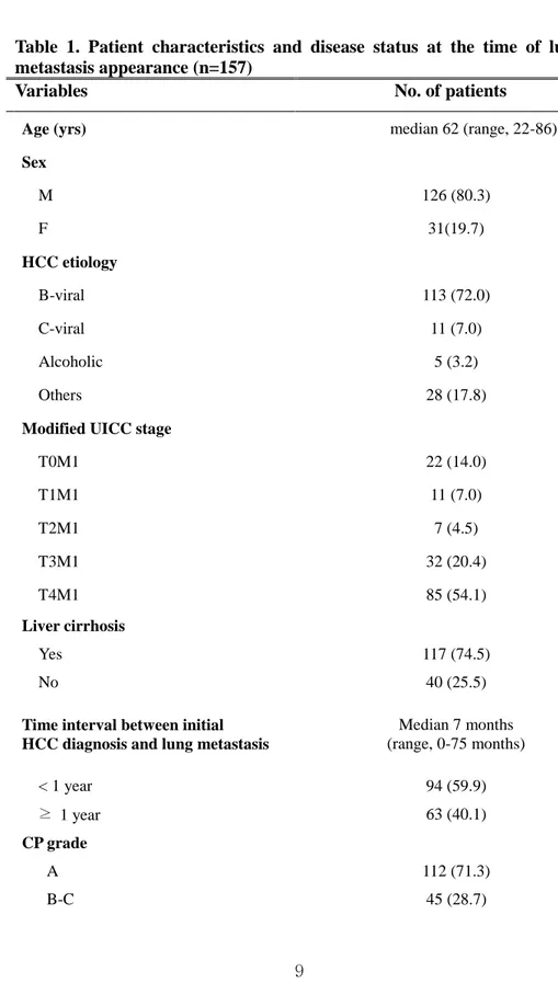
Table 1 indicates the characteristics of patients at the time of lung metastasis development. B-viral HCC predominated the incidence of HCC (72 %) and ratio of men to women was 4 : 1. About three quarters of patients had cirrhotic liver and showed liver function of CP grade A. Metastases detected within 4 weeks after the diagnosis of HCC were considered synchronous and forty-one patients (26.1 %) showed synchronous lung metastasis. Median time interval between initial HCC diagnosis and lung metastasis was 7 months. Viable liver tumor existed in more than 85 % of patients when lung metastasis firstly appeared. Modified UICC stage for viable liver lesion was evaluated and 117 patients were presenting T3-T4 disease. 43 patients (27.4 %) presented 1 to 4 pulmonary metastases and we named it as ‘limited number of metastases’ in our study. Patients were divided into two groups according to the tumor marker of AFP and PIVKA II respectively. There were 58 and 56 patients whose AFP and PIVKA II were less than 200 ng/ml and 200 mAU/ml, and the level of tumor marker was statistically significant in univariate survival analysis. Treatments for viable liver lesion and metastatic lung tumor were also shown on table 1. Twenty-six patients received local intervention for the metastatic lung lesion such as surgery or radiotherapy and 43 patients were treated with trans-arterial therapy or surgical resection as a local treatment method for viable liver tumors.
9
Table 1. Patient characteristics and disease status at the time of lung metastasis appearance (n=157)
Variables No. of patients
Age (yrs) median 62 (range, 22-86)
Sex M 126 (80.3) F 31(19.7) HCC etiology B-viral 113 (72.0) C-viral 11 (7.0) Alcoholic 5 (3.2) Others 28 (17.8)
Modified UICC stage
T0M1 22 (14.0) T1M1 11 (7.0) T2M1 7 (4.5) T3M1 32 (20.4) T4M1 85 (54.1) Liver cirrhosis Yes 117 (74.5) No 40 (25.5)
Time interval between initial HCC diagnosis and lung metastasis
Median 7 months (range, 0-75 months) < 1 year 94 (59.9) ≥ 1 year 63 (40.1) CP grade A 112 (71.3) B-C 45 (28.7)
10
Time of lung metastasis
Synchronous (<30 days) 41 (26.1)
Metachronous 116 (73.9)
Number of extrahepatic metastasis
1~4 43 (27.4)
≥5 114 (72.6)
Tumor marker, AFP
<200 (ng/ml) 58 (36.9)
≥200 (ng/ml) 99 (53.1)
Tumor marker, PIVKA-II
<200 (mAU/ml) 56 (35.7) ≥200 (mAU/ml) 101 (64.3)
Treatments for metastatic lesion
Surgery alone 9 (5.7)
Radiotherapy alone 7 (4.5) Surgery + sorafenib 2 (1.3) RT + sorafenib 7 (4.5) Surgery + RT + sorafenib 1 (0.6) Sorafenib or cytotoxic CTx only 96 (61.1)
Not treated 35 (22.3)
Treatments for liver lesion
Local intervention 43 (27.4) Systemic treatment only 101 (64.3)
No treatment 13 (8.3)
UICC; International Union Against Cancer, BCLC; Barcelona Clinic Liver Cancer, ECOG PS; Eastern Cooperative Oncology Group Performance Score, HCC; Hepatocellular Carcinoma, CP; Child Pugh, AFP; Alpha-feto protein, PIVKA-II; Protein induced by vitamine K absence of antagonist II
11
2. Prognostic factors and GPA score based risk group stratifications
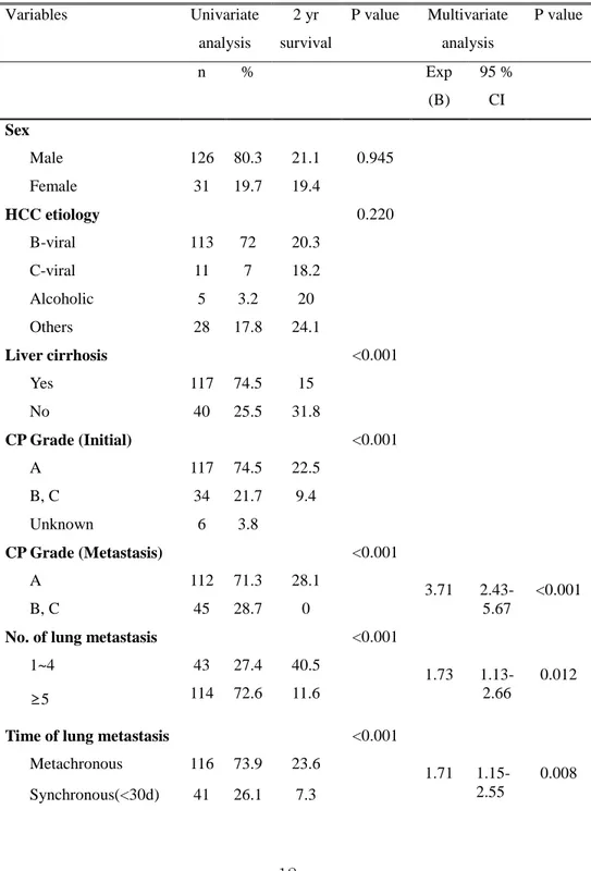
The result of univariate and multivariate analysis of prognostic factors is shown in table 2. In Cox regression analysis, CP grade, number of lung metastasis, and appearance time of lung metastasis (synchronous or metachronous) were shown to be significant for predicting the survival of patients with HCC lung metastasis. Patients with CP grade A showed better survival than those with grade B and C (2 yr survival, 28.1 % vs. 0%, p<0.001) and 2 yr survival for the patients with limited number of metastasis was better in those with multiple lung metastasis in the aspect of number of metastasis (40.5 % vs. 11.6 % p<0.001). Metachronous metastasis showed better survival than synchronous ones did (2 yr survival, 23.6 % vs. 7.3 % p<0.001).
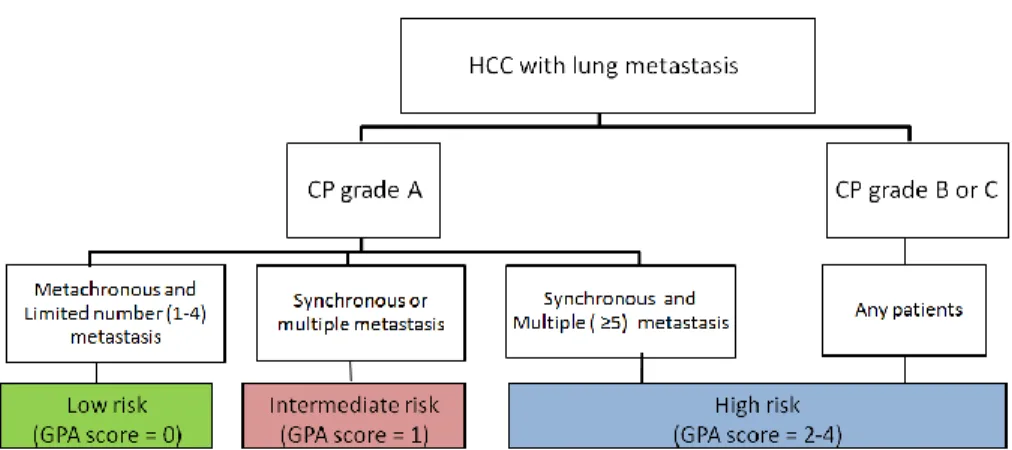
To make GPA score model, partial score for each significant prognostic factor was assigned according to the factor’s regression coefficients. Assigned partial scores for each factors were as follows; liver function at the time of lung metastasis development (CP grade A : 0, CP grade B or C : 2), number of lung metastasis (1~4 : 0, >4 : 1), appearance time of lung metastasis (metachronous : 0, synchronous : 1). As a result, 32, 61, 26, 20, and 18 patients were assigned the GPA scores of 0, 1, 2, 3, and 4 respectively. We divided patients into 3 groups according to the scores; score 0 for low risk, score 1 for intermediate risk, and score 2-4 for high risk. Schematic flow of patient stratification is shown on figure 1A. As patients group with GPA score 1 and 2 can be heterogeneous in disease status and patient characteristics, we investigated specific patient details regarding significant prognostic factors and disease status in GPA score 1 and 2 group. Three patients presented a limited number of metastasis and 58 patients showed metachronous metastasis in GPA score 1 group. Among 26 patients with GPA score 2, 7 patients had CP grade B or C, and other 19 patients showed synchronous multiple lung metastasis.
12
Table 2. Factors associated with overall survival of patients with lung metastasis: Univariate and multivariate analysis (n=157)
Variables Univariate analysis 2 yr survival P value Multivariate analysis P value n % Exp (B) 95 % CI Sex Male 126 80.3 21.1 0.945 Female 31 19.7 19.4 HCC etiology 0.220 B-viral 113 72 20.3 C-viral 11 7 18.2 Alcoholic 5 3.2 20 Others 28 17.8 24.1 Liver cirrhosis <0.001 Yes 117 74.5 15 No 40 25.5 31.8 CP Grade (Initial) <0.001 A 117 74.5 22.5 B, C 34 21.7 9.4 Unknown 6 3.8 CP Grade (Metastasis) <0.001 3.71 2.43-5.67 <0.001 A 112 71.3 28.1 B, C 45 28.7 0
No. of lung metastasis <0.001
1.73 1.13-2.66
0.012 1~4 43 27.4 40.5
≥5 114 72.6 11.6
Time of lung metastasis <0.001
1.71 1.15-2.55
0.008 Metachronous 116 73.9 23.6
13
Viable tumor at the time of metastasis development
<0.001
Yes 129 82.2 13 No 28 17.8 49.5
UICC stage of viable liver tumor at the time of metastasis
<0.001
T0-T1 32 20.4 44.8 T2-T4 125 79.6 13.6
Tumor marker, AFP <0.001 <200 (ng/ml) 58 36.9 32.5
≥200 (ng/ml) 99 63.1 13.9
Tumor marker, PIVKA II
<0.001
<200 (mAU/ml) 56 35.7 28 ≥200 (mAU/ml) 101 64.3 15.8
HCC; Hepatocellular Carcinoma, CP; Child Pugh, UICC; International Union Against Cancer, AFP; alpha-feto protein, PIVKA II; Protein induced by vitamine K absence of antagonist II
14 A.
B.
Figure 1. (A) Schematic flow of patient stratification according to prognostic factors and GPA score and (B) overall survival curves for low, intermediate and high risk group
P < 0.001
Low risk (n=32)
Intermediate risk (n=61) High risk (n=64)
15
3. Survival analysis
During the observation period, 147 patients died and HCC related death was 98.6 % (145 patients). The dominant cause of death was related to liver dysfunction in 109 patients (75.2 %), inducing hepatorenal syndrome, hepatic encephalopathy, or multiorgan failure. Variceal bleeding sometimes became a critical event for the patients even with endoscopic intervention. Lung metastasis and subsequent pneumonia or respiratory failure resulted in death in 26 patients’ deaths (17.7 %). Deaths of other 10 patients were caused by progression of other distant metastasis such as cerebral hemorrahges. With a median follow up of 24 months, 1 yr, 2 yr, and 3 yr cumulative survival rates after diagnosis of lung metastasis in our cohort was 36 %, 18.7 %, and 7 %. Comparison of Kaplan Meier survival curves according to the risk group is presented at figure 1B. 1 yr cumulative survival rates for low, intermediate, high risk group was 80.6 % vs 34.1 % vs 2.6 % and rate of 2 yr was 52.0 % vs 18.8 % vs 0 % respectively (p<0.001).
Kaplan-Meier survival curves were also drawn to evaluate the effect of local treatment for metastatic lesions. Low risk group and intermediate risk group patients survived significantly longer when they received local treatment for lung lesions. Low risk group patients showed 73.7 % of 2 yr survival rates when they received local intervention while patients without intervention showed 28.6 % of survival rates (Fig 2A. p=0.004). There was survival gain also in intermediate group patients with 2 yr survival rates of 50.0 % vs 13.7 % respectively when the metastatic lung lesions were treated with surgery or RT (Fig 2B, p=0.006). However, in high risk group patients, only 4 patients received local treatment for the metastatic lung lesion and they did not present better treatment result statistically even with local intervention. (Fig 2C. 2 yr survival 25.0 % vs 1.7 %, p=0.114).
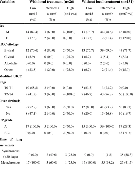
Obviously, local treatment such as metastaectomy or RT for the metastatic lesions were underwent more frequently in the patients with low and intermediate patients group. Thus, we reviewed further to find out whether any bias of disease status exists in the locally treated patients group compared to
16
others (Table 3). Among 22 patients of low and intermediate risk group, 19 patients (86.4 %) received local treatment for the metastatic lung lesion while only 3 patients were treated with local intervention in multiple number of metastasis (p<0.001). The majority of patients who received local therapy showed limited number of metastasis. However, time of lung metastasis, CP grade, tumor marker level, or presence of cirrhosis did not show significant statistical difference between two groups.
Local intervention for viable liver lesions was beneficial only for the low risk group patients. Among 32 patients of low risk group, 25 patients had viable liver tumor when lung metastasis developed and 16 patients received local intervention for viable liver tumor such as surgery, RT or RFA. The 2 yr survival rates of patients receiving local intervention was 72.7 % which was signifinalty better than the rate of 11.1 % for those without local treatment (<0.008). For intermediate and high risk group patients, there was no statistical survival benefit by applying local intervention for viable liver lesions. The 2 yr survival rate for intermediate group was 18.8 % with local treatment and 17.5 % without local intervention which was not statistically significant (p=0.518). Regardless of the administration of local intervention, the 2 yr survival rate for high risk group patients was 0 %.
17 A. B. P = 0.004
P = 0.027
P = 0.114
With local treatment (n=17) Without local treatment (n=15)With
local treatment
Without
local treatment
With
local treatment
Without
local treatment
P = 0.027 With local treatment (n=5) Without local treatment (n=56) P = 0.00418 C.
Figure 2. Stratified survival curves according to the risk group and local intervention. (A) Patients who received local treatment in low risk group showed median survival of 27 months which is significantly longer than those without local treatment. (27 months vs 16 months, p=0.004) (B) Patients in intermediate risk group also benefited from local treatment. (median survival 24 months vs 9 months, p=0.027) (C) High risk group patients with GPA score of 2 to 4 failed to benefit from local treatment for metastatic lesion. (median survival 5 months vs 3 months, p=0.114) P = 0.114 With local treatment (n=4) Without local treatment (n=60)
19
Table 3. Patient characteristics according to the application of local intervention and GPA based risk group.
Variables With local treatment (n=26) Without local treatment (n=131)
Low (n=17 (%)) Intermedia te (n=5 (%)) High (n=4 (%)) Low (n=15 (%)) Intermedia te (n=56 (%)) High (n=60 %)) Sex M 14 (82.4) 3 (60.0) 4 (100.0) 13 (76.7) 44 (78.6) 48 (80.0) F 3 (17.6) 2 (40.0) 0 (0.0) 2 (13.3) 12 (21.4) 12 (20.0) HCC etiology B-viral 12 (70.6) 4 (80.0) 2 (50.0) 13 (76.7) 39 (69.6) 43 (71.7) C-viral 1 (5.9) 0 (0.0) 1 (25.0) 1 (6.7) 3 (5.4) 5 (8.3) Alcoholic 0 (0.0) 0 (0.0) 0 (0.0) 0 (0.0) 2 (3.6) 3 (5.0) Others 4 (23.5) 1 (20.0) 1 (25.0) 1 (6.7) 12 (21.4) 9 (15.0) Modified UICC stage T0-T1 10 (58.8) 2 (40.0) 0 (0.0) 8 (53.3) 13 (23.2) 0 (0.0) T2-T4 7 (41.2) 3 (60.0) 4 (100.0) 7 (46.7) 43 (76.8) 60 (100.0) Liver cirrhosis Yes 9 (52.9) 3 (60.0) 2 (50.0) 12 (80.0) 41 (73.2) 50 (83.3) No 8 (47.1) 2 (40.0) 2 (50.0) 3 (20.0) 15 (26.8) 10 (16.7) CP grade A 17 (100.0) 5 (100.0) 2 (50.0) 15 (100.0) 56 (100.0) 17 (28.3) B-C 0 (0.0) 0 (0.0) 2 (50.0) 0 (0.0) 0 (0.0) 43 (71.7) Time of lung metastasis Synchronous (<30 days) 0 (0.0) 2 (40.0) 3 (75.0) 0 (0.0) 1 (1.8) 35 (58.3) Metachronous 17 (100.0) 3 (60.0) 1 (25.0) 15 (100.0) 55 (98.2) 25 (41.7)
20 Number of lung metastasis 1~4 17 (100.0) 2 (40.0) 0 (0.0) 15 (100.0) 1 (1.8) 8 (13.3) ≥5 0 (0.0) 3 (60.0) 4 (100.0) 0 (0.0) 55 (98.2) 52 (86.7) Tumor marker, AFP <200 (ng/ml) 10 (58.8) 3 (60.0) 0 (0.0) 7 (46.7) 29 (51.8) 5 (8.3) ≥200 (ng/ml) 7 (41.2) 2 (40.0) 4 (100.0) 8 (53.3) 27 (48.2) 55 (91.7) Tumor marker, PIVKA-II <200 (mAU/ml) 14 (82.4) 1 (20.0) 2 (50.0) 9 (60.0) 27 (48.2) 7 (11.7) ≥200 (mAU/ml) 3 (17.6) 4 (80.0) 2 (50.0) 6 (40.0) 29 (51.8) 53 (88.3)
HCC; Hepatocellular Carcinoma, UICC; International Union Against Cancer, CP; Child Pugh, AFP; Alpha-feto protein, PIVKA-II; Protein induced by vitamine K absence of antagonist II
21 IV. DISCUSSION
In our study, 1 yr survival for the patients with lung metastasis was 36 %, which is consistent with previously reported data2-4. Using 3 prognostic factors and GPA model, we divided patients into 3 groups according to the prognosis and low risk group patients showed promising treatment result with 80.6 % and 50.2 % of 1yr and 2 yr survival rates respectively. In low risk group and intermediate risk group, there was a survival benefit for the patients who were treated with local intervention for metastatic lung lesions.
In our cohorts of 157 patients, we found out that patients with a limited number of metachronous lung metastasis and good liver function showed longer survival than others, and these 3 prognostic factors coincide with other published data. One of the most frequently commented prognostic factors was the number of lung metastasis18-20. In 2010, thoracic and cardiovascular surgeons in our institution reported the prognosis of patients who underwent pulmonary metastatectomy from HCC18. Inclusion criteria for the pulmonary metastasectomy in the study were as follows: (1) primary tumor locally controlled or responding to treatment; (2) no extrathoracic metastasis; (3) the possibility of complete pulmonary metastasectomy, and (4) sufficient pulmonary function after pulmonary metastasectomy. 32 patients were evaluated and they showed better survival when the number of metastatic lesions were less than 3 (HR 3.858, p = 0.03). The metachronous metastasis resulted in good therapeutic outcomes in our study and some published reports insist that the better treatment result with metachronous pattern of spread originates from a slowly growing intrinsic tumor biology21,22. As a marker of tumor aggressiveness as well as tumor growth speed, disease free interval (DFI) > 1yr had been also reported as an associated factor with survival outcome after metastasectomy in HCC23,24. Along with reported data, it seems relatively evident that factors predicting tumor aggressiveness can affect patients’ survival even though the correlation between the time of metastasis and tumor aggressiveness is still not clear. Finally, as liver function is a well known prognostic factor of survival in HCC patients, we were able to make
22
GPA score risk model to select patients with promising prognosis even with extrahepatic metastasis.
As a result of GPA score based patient stratification, low risk and intermediate risk group patients benefited from local treatment for metastatic lung tumors. In low risk group patients, 1 yr and 2 yr overall survival rates were 80.6 % and 52.0 % respectively, and the rates surpass the previously known survival rates of the patients with extrahepatic metastasis. When local treatment was added to the low risk group patients, they survived even longer than those without local intervention. Intermediate risk group patients showed 1 yr and 2 yr survival rate of 34.1 % and 13.7 % and local intervention for lung metastasis has significantly increased treatment result with 1 yr and 2 yr survival of 83.3 % and 50.0% respectively. The result suggests that our GPA score based risk group model can select the patients with promising prognosis and find appropriate candidates for local intervention. In the aspect of treatment strategies, a dozen studies consistently insist that pulmonary metastasectomy in selected patients can result in better survival. Recently in 2016, Japanese retrospective multicenter trial reported 5 yr survival of patients who underwent pulmonary metastasectomy25. The inclusion criteria for the study were all of the patients who underwent surgical resection for pulmonary metastasis from HCC after complete resection for primary HCC. 93 patients were evaluated and 77 of them had single pulmonary metastasis. Estimated 5 yr overall survival was 41.4 % with 39.0 months of median survival time after metastasectomy. As most of the studies are dealing with a small number of patients with extrahepatic metastasis, there was a SEER database review of HCC extrahepatic metastasis patients to overcome the limitation of small number of the patients in most of studies. The study dealt with 4396 patients of stage IV HCC from 2010 to 2013 and local treatment to the primary tumor and surgery to the metastatic disease were associated with better overall survival and cancer specific survival26.
As far as we know, our study evaluated the largest number of patients in studying single organ lung metastasis of HCC, and so this is the most powerful strength of the study. Another advantage of our scoring system is that we don’t
23
need the specific details or extent of intrahepatic viable lesions. Frequently, repetitive transarterial treatment, RFA, or surgical resection of liver makes it hard to evaluate specific extent of recurrent viable liver lesions. Instead, our GPA model uses CP grade to evaluate the condition of liver to reflect the deterioration of liver function induced not only by viable intrahepatic HCC but also by chronic hepatitis. The additional strength of our result is that we directly compared the treatment results of patients according to the application of local intervention and systemic treatment. Most studies about local intervention for pulmonary metastasis from HCC were single arm retrospective studies for limited number of metastasis with well controlled primary liver lesions, and it was difficult to conclude whether local intervention for the metastatic lesions is more advantageous than sorafenib or systemic therapy alone group. Our result presented comparable survival results with other single arm studies treating the patients with aggressive local interventions and the patients survived significantly longer than the patients group with systemic treatment only.
Local treatment application for viable liver lesion was only beneficial in low risk group patients. The result corresponds to another study that aggressive control of viable liver lesion can induce better survival even with extrahepatic metastasis27. Furthermore, 13 patients without intrahepatic recurrence in our cohort showed 1 yr and 2 yr survival rates of 90.9 % and 54.5 % and the rates are comparable to our low risk group. Of these 13 patients, only 7 were in low risk group, 5 were in the intermediate risk group and 1 was in the high risk group. Further studies are required as our number of patients is limited. This study has some limitation from its retrospective nature. First, the
application of treatment strategies was not balanced according to the number of metastasis. Local treatment for the metastatic lesions was mostly undergone in patients with limited number of lung metastasis, which was only 43 patients in this study. The effect of local treatment needs to be studied further with larger number of patients. Second, our GPA score needs further external validation. We selected the patients from the year of 2011. Since 2011, sorafenib has been covered by national health insurance of South Korea and it
24
made us to reduce the heterogeneity in assessing the treatment strategy of metastatic lesions. Therefore we were not able to analyze sufficient number of patients and it made us difficult to internally validate our GPA score. External validation of our GPA score based risk group stratification is required.
V. CONCLUSION
In conclusion, we developed GPA model using prognostic factors of CP grade and time and number of lung metastasis, and the model stratified patients into low, intermediate and high risk group. Local treatment involving radiotherapy and surgical resection improved survival rates for patients in low and intermediate risk group but not in high risk group. GPA seems useful to identify the patient subgroup with different prognosis and to apply treatment accordingly.
25
REFERENCES
1. Nakamura M, Nagano H, Wada H, Noda T, Ota H, Damdinsuren B, et al. A case of hepatocellular carcinoma with multiple lung, spleen, and remnant liver metastasis successfully treated by combination chemotherapy with the novel oral DPD-inhibiting chemotherapeutic drug S-1 and interferon-α. Journal of gastroenterology 2006;41:1120-5.
2. Natsuizaka M, Omura T, Akaike T, Kuwata Y, Yamazaki K, Sato T, et al. Clinical features of hepatocellular carcinoma with extrahepatic metastases. Journal of gastroenterology and hepatology 2005;20:1781-7.
3. Uka K, Aikata H, Takaki S, Shirakawa H, Jeong SC, Yamashina K, et al. Clinical features and prognosis of patients with extrahepatic metastases from hepatocellular carcinoma. World journal of gastroenterology: WJG 2007;13:414.
4. Yang Y, Nagano H, Ota H, Morimoto O, Nakamura M, Wada H, et al. Patterns and clinicopathologic features of extrahepatic recurrence of
hepatocellular carcinoma after curative resection. Surgery 2007;141:196-202. 5. Llovet JM, Bruix J. Systematic review of randomized trials for
unresectable hepatocellular carcinoma: chemoembolization improves survival. Hepatology 2003;37:429-42.
6. Bruix J, Sherman M. Management of hepatocellular carcinoma. Hepatology 2005;42:1208-36.
7. Timmerman RD, Bizekis CS, Pass HI, Fong Y, Dupuy DE, Dawson LA, et al. Local surgical, ablative, and radiation treatment of metastases. CA: a cancer journal for clinicians 2009;59:145-70.
8. Gomez DR, Blumenschein GR, Lee JJ, Hernandez M, Ye R, Camidge DR, et al. Local consolidative therapy versus maintenance therapy or
observation for patients with oligometastatic non-small-cell lung cancer
without progression after first-line systemic therapy: a multicentre, randomised, controlled, phase 2 study. The Lancet Oncology 2016;17:1672-82.
26
9. Park JS, Yoon DS, Kim KS, Choi JS, Lee WJ, Chi HS, et al. What is the best treatment modality for adrenal metastasis from hepatocellular carcinoma? Journal of surgical oncology 2007;96:32-6.
10. Zeng Z-C, Tang Z-Y, Fan J, Qin L-X, Ye S-L, Zhou J, et al. Consideration of role of radiotherapy for lymph node metastases in patients with HCC: retrospective analysis for prognostic factors from 125 patients. International Journal of Radiation Oncology* Biology* Physics
2005;63:1067-76.
11. Park JW. Practice guideline for diagnosis and treatment of
hepatocellular carcinoma. The Korean journal of hepatology 2004;10:88-98. 12. de Lope CR, Tremosini S, Forner A, Reig M, Bruix J. Management of
HCC. Journal of hepatology 2012;56:S75-S87.
13. Edge SB, Compton CC. The American Joint Committee on Cancer: the 7th edition of the AJCC cancer staging manual and the future of TNM. Annals of surgical oncology 2010;17:1471-4.
14. Therasse P, Arbuck SG, Eisenhauer EA, Wanders J, Kaplan RS, Rubinstein L, et al. New guidelines to evaluate the response to treatment in solid tumors. Journal of the National Cancer Institute 2000;92:205-16. 15. Lencioni R, Llovet JM. Modified RECIST (mRECIST) assessment for
hepatocellular carcinoma. Seminars in liver disease: © Thieme Medical Publishers; 2010. p.052-60.
16. Chow E, Abdolell M, Panzarella T, Harris K, Bezjak A, Warde P, et al. Predictive model for survival in patients with advanced cancer. Journal of Clinical Oncology 2008;26:5863-9.
17. Chow E, Ding K, Parulekar WR, Wong RK, Van Der Linden YM, Roos D, et al. Predictive model for survival in patients having repeat radiation treatment for painful bone metastases. Radiotherapy and Oncology
2016;118:547-51.
27
resection for pulmonary metastasis from hepatocellular carcinoma: analysis of prognosis in relation to primary control. Journal of surgical oncology
2010;101:239-43.
19. Kawamura M, Nakajima J, Matsuguma H, Horio H, Miyoshi S, Nakagawa K, et al. Surgical outcomes for pulmonary metastases from hepatocellular carcinoma. European Journal of Cardio-Thoracic Surgery 2008;34:196-9.
20. Han KN, Kim YT, Yoon J-H, Suh K-S, Song JY, Kang CH, et al. Role of surgical resection for pulmonary metastasis of hepatocellular carcinoma. Lung Cancer 2010;70:295-300.
21. Kumar R, Price TJ, Beeke C, Jain K, Patel G, Padbury R, et al. Colorectal cancer survival: An analysis of patients with metastatic disease synchronous and metachronous with the primary tumor. Clinical colorectal cancer 2014;13:87-93.
22. Tanvetyanon T, Robinson LA, Schell MJ, Strong VE, Kapoor R, Coit DG, et al. Outcomes of Adrenalectomy for Isolated Synchronous Versus Metachronous Adrenal Metastases in Non–Small-Cell Lung Cancer: A Systematic Review and Pooled Analysis. Journal of Clinical Oncology 2008;26:1142-7.
23. Cho S, Ryu K-M, Hwang Y-J, Lee EB. Prognostic factors for pulmonary metastasectomy in the treatment of hepatocellular carcinoma. Journal of Thoracic Oncology 2010;5:1251-4.
24. Nakagawa T, Kamiyama T, Nakanishi K, Yokoo H, Kamachi H, Matsushita M, et al. Pulmonary resection for metastases from hepatocellular carcinoma: factors influencing prognosis. The Journal of thoracic and cardiovascular surgery 2006;131:1248-54.
25. Takahashi Y, Ikeda N, Nakajima J, Sawabata N, Chida M, Horio H, et al. Prognostic analysis of surgical resection for pulmonary metastasis from hepatocellular carcinoma. World journal of surgery 2016;40:2178-85.
28
26. Oweira H, Petrausch U, Helbling D, Schmidt J, Mehrabi A, Schöb O, et al. Prognostic value of site-specific extra-hepatic disease in hepatocellular carcinoma: a SEER database analysis. Expert Review of Gastroenterology & Hepatology 2017:1-7.
27. Yoo DJ, Kim KM, Jin YJ, Shim JH, Ko GY, Yoon HK, et al. Clinical outcome of 251 patients with extrahepatic metastasis at initial diagnosis of hepatocellular carcinoma: does transarterial chemoembolization improve survival in these patients? Journal of gastroenterology and hepatology 2011;26:145-54.
29
ABSTRACT(IN KOREAN)
간세포 암의 폐 전이 환자에서 단계별 예후 평가 모델 (GPA)
에 따른 치료 전략
<지도교수 성 진 실>
연세대학교 대학원 의학과
김 강 표
배경: 간세포 암의 간 외 전이는 굉장히 좋지 않은 예후를
보이고 있음에도 불구하고, 일부 환자에서 오래 생존하는
사람들이 발견된다. 특히 폐 전이는 가장 흔히 발견되는 간 외
전이 임에도 불구하고 그 예후와 최적화된 치료 방법과
관련된 연구가 부족한 상태이다. 이 연구의 목표는 간 세포
암의 폐 전이를 가진 환자에서 더 좋은 치료 예후를 보일 수
있는 단계별 예후 평가 모델을 개발하고, 이에 따른 적절한
치료를 적용하고자 한다.
방법: 2011년부터 2015년까지 연세 암 병원에서 간 세포암의
폐 전이로 진단받은 총 401 명의 환 자중 폐 단일 장기
전이를 가진 157 명의 환자를 분석하였다. 환자의 특징들과
함께, 간 종양과 전이된 폐 병변에 대한 치료법을 리뷰하고,
이에 따른 생존 분석을 실시하였다. 전체 생존기간은 폐
전이가 발생된 시점부터 계산하였으며, 예후를 예측하는
인자의 분석은 다 변수 Cox 회귀 분석을 이용하였다.
결과: Cox 회귀 분석 결과, CP grade 와 폐 전이 개수, 그리고
폐 전이의 시간이 통계적으로 유의미한 예후 인자로
30