Gastrointestinal complications after
concurrent chemoradiotherapy in locally
advanced pancreatic cancer
Kyong Joo Lee
Department of Medicine
Gastrointestinal complications after
concurrent chemoradiotherapy in locally
advanced pancreatic cancer
Directed by Professor Si Young Song
The Master's Thesis
submitted to the Department of Medicine
the Graduate School of Yonsei University
in partial fulfillment of the requirements for the degree of
Master of Medical Science
Kyong Joo Lee
This certifies that the Master's Thesis of
Kyong Joo Lee is approved.
---
Thesis Supervisor : Si Young Song
---
Thesis Committee Member#1: Woo Jung Lee
---
Thesis Committee Member#2: Jinsil Seong
The Graduate School
Yonsei University
ACKNOWLEDGEMENTS
This page is exclusively designed to note my gratitude and
respect for those who helped me to complete my thesis. I am
deeply indebted to my supervisor Prof. Dr. Si Young Song for
his kind help, guidance, support and encouragement throughout
my study. Sincere gratitude goes out to my reviewers, Prof. Dr.
Woo Jung Lee and Prof. Dr. Jinsil Seong who had the patience
and fortitude to read my thesis and provided constructive
criticism to help me defend it. Their guidance not only improved
my dissertation but also will benefit my future works. I also
sincerely thank my colleagues, Eun Suk Jung, Hyun Jung Lee,
Hui Won Jang, Young Eun Chon and Kyu Sik Jung for their
supports. Finally, this thesis would not have been possible
without my family.
<TABLE OF CONTENTS>
ABSTRACT ··· 1
I. INTRODUCTION ··· 3
II. MATERIALS AND METHODS ··· 5
1. Patients ··· 5
2. Treatment ··· 5
3. Gastrointestinal toxicities ··· 5
4. Statistical analysis ··· 6
III. RESULTS ··· 7
1. Patient Characteristics ··· 7
2. Gastrointestinal toxicities ··· 9
3. Gastrointestinal bleeding ··· 13
4. Risk factors for gastrointestinal complications ··· 16
5. Survival ··· 19
IV. DISCUSSION ··· 22
V. CONCLUSION ··· 24
REFERENCES ··· 25
LIST OF FIGURES
Figure 1A. Radiation gastritis ··· 11
Figure 1B. Gastric ulcer ··· 11
Figure 2. GI bleeding occurrence rate ··· 12
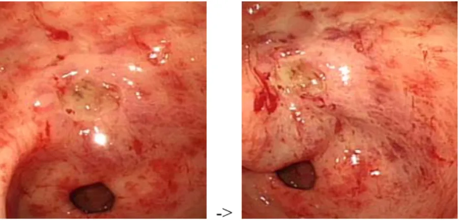
Figure 1C. Gastric ulcer with bleeding ··· 15
Figure 1D. Duodenal ulcer bleeding ··· 15
Figure 3. Overall survival ··· 20
Figure 4. Survival comparison between the GI bleeding
group and the non GI bleeding group ··· 21
LIST OF TABLES
Table 1. Baseline characteristics of all patients ··· 8
Table 2. Gastrointestinal toxicities after CRT according to the
NCI CTC 3.0 version ··· 10
Table 3. Gastrointestinal bleeding after CCRT ··· 14
Table 4. Risk factors of GI toxicities in all patients ··· 17
Table 5. Cox regression analysis for effect of GI bleeding
1
ABSTRACT
Gastrointestinal complications after concurrent chemoradiotherapy in
locally advanced pancreatic cancer
Kyong Joo Lee
Department of Medicine
The Graduate School, Yonsei University
(Directed by Professor Si Young Song)
Objectives
Locally advanced pancreatic cancer has a short survival of six to ten
months. Chemoradiotherapy (CRT) is considered a treatment of choice.
There is little information about the gastrointestinal toxicities of CRT in
pancreatic cancer. Clinical features of gastrointestinal toxicities in
patients with locally advanced pancreatic cancer underwent CRT and the
effect of gastrointestinal toxicities on survival were investigated.
Methods
Patients enrolled in this study had received concurrent CRT for
pathologically proven locally advanced pancreatic cancer. Their medical
records were retrospectively analyzed.
Results
One hundred fifty-six cases with locally advanced pancreatic cancer
between August 2005 and March 2009 were enrolled (Table 1). The
median age was 65 years and male patients 61.5%. The chemotherapy
included 5-FU-based regimen (30.8%), gemcitabine-based regimen
(59.6%), and 5-FU/gemcitabine-based regimen (9.6%). The delivered
radiotherapy modalities included 3D conformal radiotherapy (76.3%) and
intensity-modulated radiotherapy (23.7%). The median follow-up period
from the start of CRT was 13.2 months (2-52.2 months). Gastrointestinal
2
toxicities are summarized in Table 2; Abdominal pain or dyspepsia
developed in 30 patients and nausea/vomiting in 4 patients with grade 1-2
toxicity. There were two patients with anorexia with greater than grade 3
toxicity. Fifty-three patients had significant complications such as gastric
ulcer (n=26), duodenal ulcer (n=17), radiation gastritis (n=17), and
radiation duodenitis (n=5). Forty patients had upper gastrointestinal
bleeding, such as hematemesis and melena. Eight patients were dead due
to uncontrolled bleeding. The median onset time of gastrointestinal
complication was 5.2 months (0.8-50.8 months). Acute gastrointestinal
complications (less than 90 days) occurred in 13 patients (24.5%) and
late complications (more than 90 days) in 40 patients (75.5%). The
location of the tumor (body, P=0.033) and chemotherapy regimen
(5-FU+gemcitabine, P=0.015) were related with the risk factors of
gastrointestinal complications. The median overall survival was 13.1
months in the non-gastrointestinal complication group and 14.0 months
in the gastrointestinal complication group.
Conclusions
Gastrointestinal bleeding after CRT does not reduce survival of patients
with LAPC. However, gastrointestinal complications are common, and
bleeding is highly prevalent and may be fatal. Further investigation is
needed to reduce serious radiation-induced gastrointestinal
complications.
---
Key words : Gastrointestinal complications, chemoradiotherapy, locally
advanced pancreatic cancer, Gastrointestinal bleeding
3
Gastrointestinal complications after concurrent chemoradiotherapy in
locally advanced pancreatic cancer
Kyong Joo Lee
Department of Medicine
The Graduate School, Yonsei University
(Directed by Professor Si Young Song)
I. INTRODUCTION
Pancreatic cancer is the fourth leading cause of cancer-related death in the United States1. Surgical resection is the only curative treatment for the disease.
However, only 5-25% of the patients are candidates for pancreatectomy2-4.
Locally advanced pancreatic cancer is a disease with surgically unresectable but non-metastatic condition. The tumor is unresectable in the cases of extensive peripancreatic involvement, lymph node involvement, or major vasculatures involvement5. Locally advanced pancreatic cancer has a short median survival of
six to ten months. Although there is a controversy, the treatment to increase survival is known as concurrent chemoradiotherapy (CRT) as compared to radiotherapy alone and chemotherapy alone6-14. However, overall toxic effects of CRT is higher than those of chemotherapy alone15-19. These toxicities of CRT
can limit the maximum dose of radiotherapy and chemotherapy and may lead to unfavorable treatment results.
Generally, gastrointestinal toxicities of CRT include non-specific gastrointestinal symptoms, such as nausea, vomiting, and abdominal pain, as well as life-threatening gastrointestinal hemorrhages. Gastrointestinal toxicities of CRT on locally advanced pancreatic cancer are unique because the stomach and duodenum are included in the radiation field. The stomach and duodenum are readily approached by conventional endoscopy, and therapeutic endoscopy can manage gastrointestinal toxicities of CRT. However, to our knowledge, there are few data about gastrointestinal toxicities of CRT on locally advanced pancreatic cancer. In addition, its effect on survival has not been evaluated. The
4
information about clinical characteristics of gastrointestinal toxicities of CRT on locally advanced pancreatic cancer is necessary to help prevent adverse events and to develop methods to reduce the occurrence. In addition, a gastrointestinal endoscopist’s role will become important in patients receiving CRT on locally advanced pancreatic cancer.
5
II. MATERIALS AND METHODS Patients
Patients who had locally advanced pancreatic cancer and received concurrent chemoradiotherapy at Severance Hospital (Seoul, Korea) were selected. The inclusion criteria were pathologically-proven pancreatic adenocarcinoma, the ages of over 20 years, and Eastern Cooperative Oncology Group (ECOG) performance status of 0-2. The exclusion criteria included patients who received chemotherapy or surgery before CRT, and the dose of scheduled radiotherapy less than 4000 cGy. We also excluded patients who had not finished their scheduled radiation therapy. Treatment
Chemotherapy regimens were classified as follows: (1) gemcitabine group: gemcitabine (gemcitabine of 1000 mg/m2 was given on days 1, 8, and 15 of a
four-week regimen) or gemcitabine (same as above) plus cisplatin (cisplatin of 70 mg/m2 was given on day 1 of a four-week regimen) , (2) 5-fluorouracil (5-FU) group: 5-FU (5-FU of 1000 mg/m2 was given on days 1-3 of a four-week regimen)
or TS-1 (TS-1 of 60-80 mg for two weeks of a four-week regimen) or 5-FU plus etoposide plus cisplatin (5-FU of 1000 mg/m2 was given on days 1-3, etoposide of 100 mg/m2 was given on days 1-3, cisplatin of 70 mg/m2 was given on day 1 of a four-week regimen) , and (3) 5-FU plus gemcitabine group (5-FU of 1000 mg/m2 was given on days 1-3, and gemcitabine of 1000 mg/m2 was given on days 1, 8, and 15 of a four-week regimen).
The radiation therapy was classified into two groups either three-dimensional conformal radiotherapy (total dose: 4000-5400 cGy, one dose: 180-250 cGy, fraction: 28) or intensity modulated radiotherapy (total dose: 4200-6000 cGy, one dose: 200-293 cGy, fraction: 25).
Gastrointestinal toxicities
Gastrointestinal toxicities were classified according to the common-terminology criteria for the adverse events version 3.0.
6
Endoscopically, radiation-induced injuries were defined as telangiectasia, diffuse erythema of mucosa, shallow or deep ulcers, and scar formation20,21.
Statistical analysis
x2 was used to find the risk factors of gastrointestinal toxicities, and a
logistic regression was used for multivariate analysis. Cox-regression test was used to evaluate the risk of gastrointestinal bleeding for survival. The Kaplan-Meyer method and the log-rank test were used to compare survival. All analyses were performed using statistical software SPSS version 11 (SPSS, Chicago, IL, USA). P values less than 0.05 indicated statistical significance.
7
III. RESULTS Patient Characteristics
Between August 2005 and March 2009, 156 patients with locally advanced pancreatic cancer were eligible for analysis (Table 1). The median age at the time of the diagnosis of pancreatic cancer was 65 years, ranging from 35 to 90 years. The male patients were 61.5%. 43 patients had hypertension and 48 patients had diabetes mellitus with medications. The tumors were mostly located at the head (63.5%). The median size of the tumor was 3.0 cm with a range from 1.1 to 7.0 cm. The median level of CA19-9 was 384 U/mL with a range from 0.1 to 20000 U/mL. Three-dimensional conformal radiotherapy was delivered to 119 patients (76.3%) and intensity modulated radiotherapy was delivered to 37 patients (23.7%). The median delivered dose was 5040 cGy (4000-5400 cGy) for three-dimensional conformal radiotherapy and 5842 cGy (4200-6000 cGy) for intensity-modulated radiotherapy. The median follow-up period was 13.2 months with a range from 2 to 52.2 months.
8 Table 1. Baseline characteristics of all patients
CCRT: concurrent chemoradiotherapy
Variable No. of Patients
Total 156
Age (median) 65 years (39-90 years)
Sex (Male : Female) 96 (61.5%): 60 (38.5%)
Hypertension 43 (27.6%) Diabetes mellitus 48 (30.8%) Location of tumor Head 99 (63.5%) Body 44 (28.2%) Tail 13 (8.3%)
Size of tumor (median) 3.0 cm (1.1-7.0 cm)
CA 19-9 at diagnosis (median) 384 U/mL (0.1-20000 U/mL)
Chemotherapy
Gemcitabine based regimen 93 (59.6%)
5-FU based regimen 48 (30.8%)
5-FU + Gemcitabine regimen 15 (9.6%)
Radiation modality and dose
3D conformal radiotherapy 119 (76.3%)
Radiation dose (median) 5040 cGy (4000-5400 cGy)
Intensity modulated radiotherapy 37 (23.7%)
Radiation dose (median) 5842 cGy (4200-6000 cGy)
Surgery after CCRT
No 126 (80.8%)
Yes 30 (19.2%)
Time to GI complication occurred (median) 5.2 months (0.8-50.8 months)
≤90 days (acute) 13 (24.5%)
>90 days (late) 40 (75.5%)
9
Gastrointestinal toxicities
The overall incidence of gastrointestinal toxicities was 57.7% (Table 2). There were 30 patients with grade 1 or 2 abdominal pain or dyspepsia. Two patients had grade 3 anorexia. Nausea and vomiting developed in four patients, and these were well controlled with appropriate medications. Forty patients had bleeding: nine patients with hematemesis (22.5%), 21 patients with melena (52.5%), and 10 patients with hematochezia (25%). There were 18 patients (42.5%) with grade 3 or 4 gastrointestinal bleeding and eight (22.5%) with grade 5 (death) bleeding.
Endoscopy after CRT revealed mucositis (Fig 4A) in 27.2%, ulcer (Fig 4B) in 45%, and gastrointestinal bleeding in 65% greater than a grade 3 toxicity. The median gastrointestinal bleeding occurrence was 5.4 months (Fig 2).
10
Table 2. Gastrointestinal toxicities after CRT according to the NCI CTC 3.0 version
Variable No. of patients
Total Portion of
G3-G5
G1 G2 G3 G4 G5
Abdominal pain or Dyspepsia 30 (19.2%) 0 16 14 0 0 0
Anorexia 5 (3.2%) 2 (40%) 2 1 2 0 0
Nausea 3 (1.9%) 0 0 3 0 0 0
Vomiting 1 (0.6%) 0 0 1 0 0 0
Mucositis 22 (14.1%) 6 (27.2%) 15 1 6 0 0
Stomach 17 (10.8%) 5 (29.4%) 11 1 5 0 0
Small bowel (duodenum) 5 (3.2%) 1 (20%) 4 0 1 0 0
Ulcer 37 (23.7%) 15 (9.6%) 11 11 14 1 0
Stomach 22 (14.1%) 7 (31.8%) 7 8 7 0 0
Small bowel (duodenum) 15 (9.6%) 8 (5.1%) 4 3 7 1 0
Other
GI hemorrhage 40 (25.6%) 26 (65%) 0 14 17 1 8
Stomach 20 (12.8%) 9 (45%) 0 11 9 0 0
Duodenum 15 (9.6%) 12 (80%) 0 3 8 1 3
11
Figure 1A. Radiation gastritis. Mucosal erythema with telangiectasia and several superficial ulcerations on the antrum.
Figure 1B. Gastric ulcer. Oval shaped healing-staged ulceration with some surrounding regenerating epithelium on the antrum.
12
Figure 2. Cumulative incidence of GI bleeding in 40 patients. Median GI bleeding occurr ence was 5.4 months.
13
Gastrointestinal bleeding
Table 3 shows the characteristics of gastrointestinal bleeding. The median onset time was 5.4 months (range: 0.8-50.8 months). Forty patients presented themselves to the hospital for gastrointestinal bleeding. The median initial hemoglobin was 10.1 g/dL (range: 7.1-15.3 g/dL), which decreased to 7.1 g/dL (range: 3.5-10.8 g/dL) at bleeding. Endoscopy showed the cause of bleeding to be a gastric ulcer (Fig 4C) in 15 patients (37.5%), duodenal ulcer (Fig 4D) in 15 (37.5%), and radiation gastritis in five (15%). Endoscopy was not performed in five patients upon their guardians’ rejection. As the patients were in terminal status, the guardians did not want them to undergo any examinations. These five patients died without receiving an endoscopic evaluation and treatment. The remaining 35 patients received endoscopic treatment. Bleeding was successfully stopped by endoscopic treatment in 31 patients (77.5%), but not in four patients. Thus, embolization was performed in one of the four patients, and the bleeding was finally stopped, but three others died due to bleeding. The mortality of gastrointestinal bleeding was eight patients in total. The median time to gastrointestinal bleeding from CCRT was 5.4 months (range: 0.8-50.8 months) and the median overall survival was 13.5 months (range: 2.8-50.8 months).
14 Table 3. Gastrointestinal bleeding after CCRT (n=40)
No. of patients Hemoglobin (median) Initial 10.1 g/dL (7.1-15.3 g/dL) At bleeding 7.1 g/dL (3.5-10.8 g/dL) Origin of GI bleeding Gastric ulcer 15 (37.5%) Duodenal ulcer 15 (37.5%) Radiation gastritis 5 (15.0%) Unknown 5 (15.0%)
Extent of bleeding origin
Focal 27 (77.1%)
Diffuse 8 (22.9%)
Treatment
Endoscopic treatment 35 (87.5%)
Success 31 (77.5%)
Angiography and embolization 1 (2.5%)
No treatment 5 (12.5%)
Mortality of GI bleeding 8 (20%)
Time to GI bleeding from CCRT(median) 5.4 months (0.8-50.8 months)
Survival (median) with GI bleeding 13.5 months (2.8-50.8
15
Figure 1C. Gastric ulcer with bleeding. Oval shaped active ulceration with bleeding (stigmata of
recent bleeding) on the antrum. Bleeding was stopped by human plasma thrombin injection
following hypertonic saline-epinephrine injection successfully.
->
Figure 1D. Duodenal ulcer bleeding. Oval shaped active ulceration surrounded by edematous
mucosal crater at duodenal bulb. Bleeding was stopped by argon plasma coagulation following hemoclipping.
16
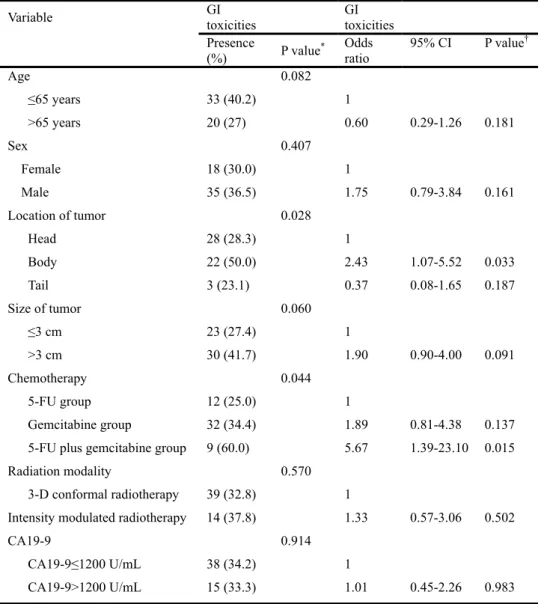
Risk factors for gastrointestinal complications
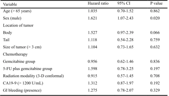
The association between clinical parameters and the risk of gastrointestinal complications are summarized in Table 4. In univariate analysis, the location of the tumor (body, P=0.028) and chemotherapy regimen (5-FU+gemcitabine, P=0.015) were related with the risk factors of gastrointestinal complications. In multivariate analysis, the location of the tumor (body, P=0.033) and chemotherapy regimen were significant for the risk factor of gastrointestinal complications. The hazard ratio was 1.27 for the effect of GI bleeding on survival in Cox regression, but it was not significant (P=0.329, Table 5). Male had higher hazard ratio than female and it was significant (Hazard ratio = 1.621, P=0.020). However, there were no difference in the number of hypertension, diabetes mellitus between male and female.
17
Table 4. Risk factors of GI toxicities in all patients (n=156)
* Chi-square test was used. † Logistic regression was used.
Variable GI toxicities GI toxicities
Presence (%) P value* Odds ratio 95% CI P value† Age 0.082 ≤65 years 33 (40.2) 1 >65 years 20 (27) 0.60 0.29-1.26 0.181 Sex 0.407 Female 18 (30.0) 1 Male 35 (36.5) 1.75 0.79-3.84 0.161 Location of tumor 0.028 Head 28 (28.3) 1 Body 22 (50.0) 2.43 1.07-5.52 0.033 Tail 3 (23.1) 0.37 0.08-1.65 0.187 Size of tumor 0.060 ≤3 cm 23 (27.4) 1 >3 cm 30 (41.7) 1.90 0.90-4.00 0.091 Chemotherapy 0.044 5-FU group 12 (25.0) 1 Gemcitabine group 32 (34.4) 1.89 0.81-4.38 0.137
5-FU plus gemcitabine group 9 (60.0) 5.67 1.39-23.10 0.015
Radiation modality 0.570
3-D conformal radiotherapy 39 (32.8) 1
Intensity modulated radiotherapy 14 (37.8) 1.33 0.57-3.06 0.502
CA19-9
CA19-9≤1200 U/mL
0.914
38 (34.2) 1
18
Table 5. Cox regression analysis for effect of GI bleeding on survival
Variable Hazard ratio 95% CI P value
Age (> 65 years) 1.035 0.70-1.52 0.862 Sex (male) 1.621 1.07-2.43 0.020 Location of tumor Body 1.527 0.97-2.39 0.066 Tail 1.118 0.54-2.28 0.759 Size of tumor (> 3 cm) 1.104 0.73-1.65 0.632 Chemotherapy Gemcitabine group 0.956 0.62-1.46 0.836
5-FU plus gemcitabine group 1.598 0.78-3.25 0.197
Radiation modality (3-D conformal) 0.915 0.57-1.45 0.708
CA19-9 (> 1200 U/mL) 1.312 0.87-1.97 0.192
19
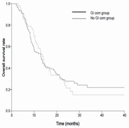
Survival
In a total of 156 patients, 117 patients (75%) died at the time of final analysis. Figure 3 shows the Kaplan-Meier curves for overall survival. The median overall survival was 13.1 months (range: 11.3-14.9 months) in LAPC from the start of CRT. The median overall survival was 13.1 months (range: 9.9-16.3 months) in the non-gastrointestinal complication group and 14.0 months (range: 12.3-15.7 months) in the gastrointestinal complication group. Although overall survival was longer in the GI complication group, this difference was not significant (P=0.755, Fig.4)
20 Figure 3. Overall survival
21
Figure 4. Survival comparison between the GI complication group and the non GI complication group. Overall survival was 13.1 months in the non GI complication group and 14.0 months in the GI complication group.
22
IV. DISCUSSION
It was shown in this study that gastrointestinal bleeding after CRT does not reduce survival of patients with locally advanced pancreatic cancer. The median overall survival was similar with other studies12,15,22,23. However, the prevalence of
CRT-induced gastrointestinal bleeding is considerable and serious.
CRT was first introduced in the GITSG trial10. The survival benefit was
found to be higher in the group with 5-FU + radiation than in the group with radiotherapy alone. There was no difference in survival benefit between the groups that received different doses of RT (4000 and 6000 cGy). After that, many studies reported the benefit of CRT, and CRT became one of the treatment options for pancreatic cancer11,16-18. Based on the results of several studies, the RT dose of 50-60 Gy (182 Gy/day) is generally used24,25. A study reported that toxicity was
higher in the LAPC group where the radiation dose increased up to 55 Gy than in the LAPC group with the dose up to 50 Gy, but that the compliance was similar between the groups, and the treatment performance in the former was better than that in the latter26. In studies comparing CRT and chemotherapy, however, more
cases of toxicity were found in the CRT group; thus, care must be taken with regard to the use of CRT6,27. Indeed, during clinical practice, we experienced many
patients with CRT-induced gastrointestinal bleeding, which led to this study. Due to the low awareness of bleeding, the frequency of endoscopy was quite low. Of the total of 156 patients, 20 received endoscopy before CRT and 78 after CRT. Very few patients who had bleeding received endoscopy before the onset of bleeding. Had endoscopy also been performed in the other patients, the chances of finding complications such as radiation gastritis would have been higher. The locations of the tumor and chemotherapy regimen were related with the risk factors for gastrointestinal complications. As the body of the pancreas is closely located to both the stomach and duodenum, they are affected by radiotherapy. The combination of 5-FU and gemcitabine increased the toxicity of gastrointestinal complications. Also poor prognosis is expected in male with gastrointestinal bleeding. There were no difference in the number of hypertension and diabetes
23
mellitus between male and female. Other risk factors need to be found in male group.
This study has several limitations. First, the results were obtained by retrospectively reviewing the medical charts. Second, of CRT, the chemotherapy-induced adverse effects could not be excluded. In this study, the number of patients who received gemcitabine was higher than the number of those who received 5-FU. Several studies reported that gemicitabine was shown to be more toxic than 5-FU. In addition, it was difficult to ascertain the cause of bleeding after surgery as well as the change in the chemotherapy regimen after
CRT28,29. Third, the low number of patients who received endoscopy before CRT
made it impossible to determine if the ulcer existed even before CRT. Fourth, the patients who received 3D conformal radiotherapy and intensity modulated radiotherapy were analyzed together, and 37 of them received intensity modulated radiotherapy. To the best of our knowledge, there has been no study that compared 3D conformal radiotherapy and intensity modulated radiotherapy. Thus, a study is required to investigate if there is any difference between the two therapies.
There is no consensus on the best time to perform endoscopy after radiotherapy. After CRT, however, gastrointestinal complications are likely to develop at anytime. Therefore, it is recommended that endoscopy be performed as was done in this study. If abnormal findings are found in endoscopy before CRT, preemptive treatment is necessary. Moreover, endoscopy as a baseline study is recommended for the comparison with the endoscopic results. Usually CRT is followed by chemotherapy or surgery about one month later, endoscopy is recommended before such therapies as gastrointestinal ulcer or bleeding can occur even before 90 days after CRT. As ulcerative bleeding is well responsive to PPI, its early detection and treatment may prevent adverse events. Although the best frequency of endoscopy may be debatable, yearly or more frequent endoscopy, particularly in patients with a history of bleeding, is recommended considering the possibility of delayed ulcer and bleeding.
24
V. CONCLUSION
Gastrointestinal bleeding after CRT is highly prevalent and may be fatal. This study shows that patients with LAPC are likely to develop a gastric ulcer, duodenal ulcer, or radiation gastritis after CRT, and a large number of them developed bleeding. Some patients in this study died or stopped receiving treatment because of bleeding. Extensive studies are required to compare the benefits and risks in terms of survival rate and complications between CRT and chemotherapy. In addition, studies are required to uncover the tests or treatments that can reduce CRT-induced gastrointestinal complications.
25
REFERENCES
1. Jemal A, Siegel R, Ward E, Hao Y, Xu J, Thun MJ. Cancer statistics, 2009. CA Cancer J Clin 2009;59:225-49.
2. Pellegrini CA, Heck CF, Raper S, Way LW. An analysis of the reduced morbidity and mortality rates after pancreaticoduodenectomy. Arch Surg 1989;124:778-81.
3. Winek T, Hamre D, Mozell E, Vetto RM. Prognostic factors for survival after pancreaticoduodenectomy for malignant disease. Am J Surg 1990;159:454-6.
4. Yeo CJ, Cameron JL, Lillemoe KD, Sitzmann JV, Hruban RH, Goodman SN, et al. Pancreaticoduodenectomy for cancer of the head of the pancreas. 201 patients. Ann Surg 1995;221:721-31; discussion 31-3.
5. Willett CG, Czito BG, Bendell JC, Ryan DP. Locally advanced pancreatic cancer. J Clin Oncol 2005;23:4538-44.
6. Treatment of locally unresectable carcinoma of the pancreas: comparison of combined-modality therapy (chemotherapy plus radiotherapy) to chemotherapy alone. Gastrointestinal Tumor Study Group. J Natl Cancer Inst 1988;80:751-5.
7. de Lange SM, van Groeningen CJ, Meijer OW, Cuesta MA, Langendijk JA, van Riel JM, et al. Gemcitabine-radiotherapy in patients with locally advanced pancreatic cancer. Eur J Cancer 2002;38:1212-7.
8. Klaassen DJ, MacIntyre JM, Catton GE, Engstrom PF, Moertel CG. Treatment of locally unresectable cancer of the stomach and pancreas: a randomized comparison of 5-fluorouracil alone with radiation plus concurrent and maintenance 5-fluorouracil--an Eastern Cooperative Oncology Group study. J Clin Oncol 1985;3:373-8.
9. Magnino A, Gatti M, Massucco P, Sperti E, Faggiuolo R, Regge D, et al. Phase II trial of primary radiation therapy and concurrent chemotherapy for patients with locally advanced pancreatic cancer. Oncology 2005;68:493-9.
10. Moertel CG, Frytak S, Hahn RG, O'Connell MJ, Reitemeier RJ, Rubin J, et al. Therapy of locally unresectable pancreatic carcinoma: a randomized comparison of high dose (6000 rads) radiation alone, moderate dose radiation (4000 rads + 5-fluorouracil), and high dose radiation + 5-fluorouracil: The Gastrointestinal Tumor Study Group. Cancer 1981;48:1705-10.
26
Phase II study of radiotherapy combined with gemcitabine for locally advanced pancreatic cancer. Br J Cancer 2004;91:673-7. 12. Shinchi H, Takao S, Noma H, Matsuo Y, Mataki Y, Mori S, et al.
Length and quality of survival after external-beam radiotherapy with concurrent continuous 5-fluorouracil infusion for locally unresectable pancreatic cancer. Int J Radiat Oncol Biol Phys 2002;53:146-50.
13. Sultana A, Tudur Smith C, Cunningham D, Starling N, Tait D, Neoptolemos JP, et al. Systematic review, including meta-analyses, on the management of locally advanced pancreatic cancer using radiation/combined modality therapy. Br J Cancer 2007;96:1183-90.
14. Yip D, Karapetis C, Strickland A, Steer CB, Goldstein D. Chemotherapy and radiotherapy for inoperable advanced pancreatic cancer. Cochrane Database Syst Rev 2006;3:CD002093. 15. Chauffert B, Mornex F, Bonnetain F, Rougier P, Mariette C, Bouche O, et al. Phase III trial comparing intensive induction chemoradiotherapy (60 Gy, infusional 5-FU and intermittent cisplatin) followed by maintenance gemcitabine with gemcitabine alone for locally advanced unresectable pancreatic cancer. Definitive results of the 2000-01 FFCD/SFRO study. Ann Oncol 2008;19:1592-9.
16. Huguet F, Andre T, Hammel P, Artru P, Balosso J, Selle F, et al. Impact of chemoradiotherapy after disease control with chemotherapy in locally advanced pancreatic adenocarcinoma in GERCOR phase II and III studies. J Clin Oncol 2007;25:326-31. 17. Morganti AG, Massaccesi M, La Torre G, Caravatta L, Piscopo A,
Tambaro R, et al. A systematic review of resectability and survival after concurrent chemoradiation in primarily unresectable pancreatic cancer. Ann Surg Oncol 2010;17:194-205.
18. Nakachi K, Furuse J, Kinoshita T, Kawashima M, Ishii H, Ikeda M, et al. A phase II study of induction chemotherapy with gemcitabine plus S-1 followed by chemoradiotherapy for locally advanced pancreatic cancer. Cancer Chemother Pharmacol 2009.
19. Sawaki A, Hoki N, Ito S, Matsumoto K, Mizuno N, Hara K, et al. Clinical impact of radiotherapy for locally advanced pancreatic cancer. J Gastroenterol 2009;44:1209-14.
20. DeCosse JJ, Rhodes RS, Wentz WB, Reagan JW, Dworken HJ, Holden WD. The natural history and management of radiation induced injury of the gastrointestinal tract. Ann Surg 1969;170:369-84.
27
Radiol Ther Phys Biol 1966;4:289-97.
22. Chung HW, Bang SM, Park SW, Chung JB, Kang JK, Kim JW, et al. A prospective randomized study of gemcitabine with doxifluridine versus paclitaxel with doxifluridine in concurrent chemoradiotherapy for locally advanced pancreatic cancer. Int J Radiat Oncol Biol Phys 2004;60:1494-501.
23. Li CP, Chao Y, Chi KH, Chan WK, Teng HC, Lee RC, et al. Concurrent chemoradiotherapy treatment of locally advanced pancreatic cancer: gemcitabine versus 5-fluorouracil, a randomized controlled study. Int J Radiat Oncol Biol Phys 2003;57:98-104.
24. Boz G, De Paoli A, Innocente R, Rossi C, Tosolini G, Pederzoli P, et al. Radiotherapy and continuous infusion 5-fluorouracil in patients with nonresectable pancreatic carcinoma. Int J Radiat Oncol Biol Phys 2001;51:736-40.
25. Mehta VK, Poen JC, Ford JM, Oberhelman HA, Vierra MA, Bastidas AJ, et al. Protracted venous infusion 5-fluorouracil with concomitant radiotherapy compared with bolus 5-fluorouracil for unresectable pancreatic cancer. Am J Clin Oncol 2001;24:155-9. 26. Henry AM, Ryder WD, Moore C, Sherlock DJ, Geh JI, Dunn P, et al.
Chemoradiotherapy for locally advanced pancreatic cancer: a radiotherapy dose escalation and organ motion study. Clin Oncol (R Coll Radiol) 2008;20:541-7.
27. Krishnan S, Rana V, Janjan NA, Varadhachary GR, Abbruzzese JL, Das P, et al. Induction chemotherapy selects patients with locally advanced, unresectable pancreatic cancer for optimal benefit from consolidative chemoradiation therapy. Cancer 2007;110:47-55. 28. Crane CH, Abbruzzese JL, Evans DB, Wolff RA, Ballo MT, Delclos
M, et al. Is the therapeutic index better with gemcitabine-based chemoradiation than with 5-fluorouracil-based chemoradiation in locally advanced pancreatic cancer? Int J Radiat Oncol Biol Phys 2002;52:1293-302.
29. Huguet F, Girard N, Guerche CS, Hennequin C, Mornex F, Azria D. Chemoradiotherapy in the management of locally advanced pancreatic carcinoma: a qualitative systematic review. J Clin Oncol 2009;27:2269-77.
28
ABSTRACT(In Korean)
진행성
췌장암에서 동시 항암화학 방사선 치료후
발생
가능한 소화기계 합병증
<지도교수 송시영>
연세대학교 대학원 의학과
이 경 주
국소 진행성 췌장암의 생존률은 6개월에서 10개월 사이로
알려져 있다. 최근 항암화학 방사선 치료가 여러 연구를 통해
치료의 선택이 될 수 있다. 하지만 항암화학 방사선 치료의
소화기계 합병증에 대해서는 많은 연구가 되어 있지 않다. 이번
연구에서는 항암화학 방사선 치료가 어떤 소화기계 합병증을
일으키고
생존에는
어떤
영향을
미치는지
알아보았다.
세브란스병원에서 2005년 8월부터 2009년 3월까지 국소 진행성
췌장암에서 항암화학 방사선 요법을 받은 환자 156명을
대상으로 조사하였다. 중간 나이는 65세였고 남자는 61.5%였다.
항암제는 5-FU base (30.8%), Gemcitabine base (59.6%) 그리고
5-FU/Gemcitabine base (9.6%) 였다. 방사선치료는 3D 입체 조형
방사선 치료 (76.3%) 였고 강도 변조 방사선 치료가 (23.7%)
였다.
항암화학
방사선
치료일로부터 13.2 개월간
추적관찰하였다. 소화기합병증으로는 다음과 같이 나타났다.
Grade 1 또는 2 독성을 가지는 복통과 속쓰림 환자가 30명에서
나타났고
오심 또는 구토는 4명의 환자에서 나타났다. Grade 3
이상의
독성을 가지는 식욕 부진이 2명의 환자에서 나타났다.
53명의 환자에서 궤양 또는 방사선으로 유도된 위염 또는
십이지장염이
발견 되었다. 위궤양이 26명, 십이지장 궤양이
17명, 방사선위염이 17명그리고 방사선십이지장염이 5명이였다.
이중
40명의 환자에서는 토혈이나 흑색변으로 상부위장관
29