Yonsei Med J http://www.eymj.org Volume 53 Number 2 March 2012
446
Case Report
http://dx.doi.org/10.3349/ymj.2012.53.2.446pISSN: 0513-5796, eISSN: 1976-2437 Yonsei Med J 53(2):446-449, 2012
A Case of Acute Polyneuropathy with Nephrotic Syndrome
Showing Transient Proximal Sensory Conduction Defects
Jeeyoung Oh,
1Seung-Min Kim,
2and Il Nam Sunwoo
2 1Department of Neurology, Konkuk University School of Medicine, Seoul; 2Department of Neurology, Yonsei University College of Medicine, Seoul, Korea.Received: April 8, 2011 Revised: June 13, 2011 Accepted: June 20, 2011
Corresponding author: Dr. Il Nam Sunwoo, Department of Neurology,
Yonsei University College of Medicine, 50 Yonsei-ro, Seodaemun-gu, Seoul 120-752, Korea.
Tel: 82-2-361-5460, Fax: 82-2-393-0705 E-mail: [email protected]
∙ The authors have no financial conflicts of interest.
© Copyright:
Yonsei University College of Medicine 2012
This is an Open Access article distributed under the terms of the Creative Commons Attribution Non-Commercial License (http://creativecommons.org/ licenses/by-nc/3.0) which permits unrestricted non-commercial use, distribution, and reproduction in any medium, provided the original work is properly cited.
Acute sensorimotor polyneuropathy that resembles Guillain-Barré syndrome (GBS) is rarely accompanied with nephrotic syndrome, and its underlying immu-nological mechanisms are unclear. A 56-year-old man presented with simultane-ous acute progressive symmetric sensorimotor polyneuropathy and proteinuria. A kidney biopsy revealed focal segmental glomerulosclerosis. Serial electrophysio-logic studies showed only a transient proximal conduction block in the median nerve, stimulated somatosensory evoked potential and prolonged terminal laten-cies of the median and peroneal nerves. The patient’s neurologic deficits and kid-ney dysfunction recovered with corticosteroid treatment. Our case showed that so-matosensory evoked potential study can be an important objective tool in the diagnosis of acute polyneuropathy with normal distal nerve conduction and that corticosteroids should be considered in the initial treatment of GBS-resembling polyneuropathy associated with nephrotic syndrome.
Key Words: Acute sensorimotor polyneuropathy, nephrotic syndrome, somato-sensory evoked potential, conduction block
INTRODUCTION
Nerve conduction study (NCS) is a mainstay in the diagnosis of peripheral neurop-athy. However, conventional NCS is limited when the lesion is located in an inac-cessible and proximal segment of the nerve.1 Late response and/or somatosensory
evoked potential (SSEP) study can be helpful in these instances. In this paper, we describe a patient with acute sensorimotor polyneuropathy that was accompanied with nephrotic syndrome and whose electrophysiological studies showed a tran-sient and proximal sensory conduction defect and prolonged terminal latencies of the motor nerves.
CASE REPORT
A 56-year-old man developed progressive paresthesia and weakness over a 3-week period. He developed numbness of the fingertips on both hands a month before,
Acute Polyneuropathy with NS
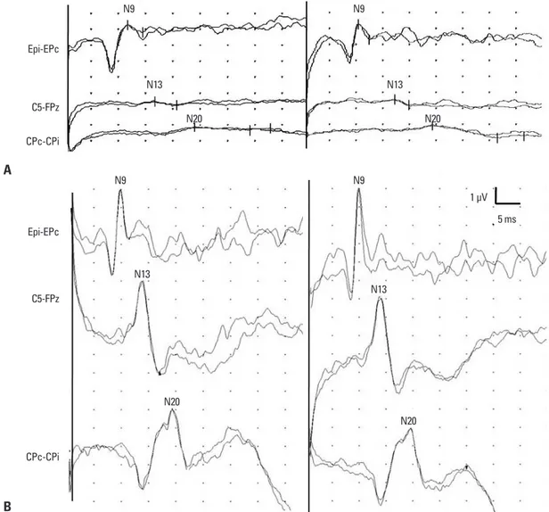
Yonsei Med J http://www.eymj.org Volume 53 Number 2 March 2012 447 tentials with reduced recruitment of motor unit action po-tentials in the left deltoid, first dorsal interossei, vastus later-alis, and tibialis anterior muscles. Median nerve SSEP revealed a prominent positive wave followed by a very low amplitude negative potential on the brachial plexus poten-tial (Erb’s point) bilaterally. Both cervical (N13) and corti-cal (N19) waves were barely discernible (Fig. 1A). A mag-netic resonance image scan of the cervical spine was normal. Routine urinalysis revealed proteinuria (1.8 g/24 h). Blood chemistry was normal except for hypoalbuminemia (2.9 g/ dL), and immunoelectrophoresis did not show any gam-mopathy. Cerebrospinal fluid examination revealed a white blood cell count of 0 cells/mm3, a protein level of 27.0 mg/
dL, and a glucose level of 68 mg/dL. A percutaneous kid-ney gun biopsy showed focal segmental glomerulosclerosis (FSGS). Vacuolar changes in the glomerular epithelial cells which progressed proximally and subsequently involved
the feet. One week before admission, he could not elevate his shoulders and felt unsteady when walking. He denied any preceding upper respiratory infection or diarrhea. On neurologic examination, the proximal muscles of the upper and lower extremities were weaker (Medical Research Coun-cil grade 4) than those of the distal limbs. Vibration sense was not perceived below the iliac crest and a pinprick sen-sation was symmetrically decreased below the wrist and ankle. Deep tendon reflexes were not elicited at all. Cranial nerve and cerebellar function were normal.
Motor and sensory nerve conduction studies performed on the left median, ulnar, posterior tibial, and peroneal nerves were normal. F-latencies were all within normal ranges and bilateral H-reflex was not elicited. Electromyography showed a mild degree of positive sharp waves and fibrillation
po-Fig. 1. Serial median nerve SSEP studies using the same filter settings. (A) At admission, the observed increased duration of positive de-flection with a low amplitude negative peak of the bilateral brachial plexus (Erb’s point) with barely discernible cervical and cortical po-tentials are shown. (B) Restoration of the waveforms of the SSEP, which was evident 5 weeks after corticosteroid treatment, is shown. SSEP, somatosensory evoked potential.
Epi-EPc N9 N9 N13 N13 N20 N20 N9 N9 1 µV 5 ms N13 N13 N20 N20 Epi-EPc C5-FPz C5-FPz CPc-CPi CPc-CPi A B
Jeeyoung Oh, et al.
Yonsei Med J http://www.eymj.org Volume 53 Number 2 March 2012
448
nerve conduction velocities were also slow. Notwithstand-ing, one study reported on 10 patients with early GBS who showed abnormal SSEP but normal distal nerve conduc-tion.8 Even though they did not conduct F-wave studies, we
speculate that F-latencies would have also been delayed in most of these patients, because the maximal motor conduc-tion velocities across the proximal arm and Erb’s point were also slow.
Of particular note, FSGS simultaneously occurred with GBS-mimicking polyneuropathies. In fact, nephrotic syn-drome associated with acute polyradiculoneuropathy has been recognized previously.9 However, very few cases have
been reported since then, and most of the combined ne-phrotic syndromes consisted of glomerulonephritis or mini-mal change nephrotic syndrome. Though FSGS is the most common form of nephrotic syndrome, only three cases as-sociated with GBS have been published so far.10-12 Neither
intravenous immunoglobulin nor plasmapheresis was effec-tive in treatment thereof; however, corticosteroids with or without immunosuppressant agents could potentially have ameliorated both the polyneuropathy and the FSGS. We cannot assume that the polyneuropathy and FSGS occurred coincidentally or shared the same immunologic mechanism. However, the time relationship between the two diseases and the parallel improvement with corticosteroid treatment, as shown by our case, raises the possibility that they could share the same immunopathological mechanisms.
In conclusion, our case is instructive in that SSEP can be an important objective tool in the diagnosis of acute poly-neuropathy with normal distal nerve conduction and that corticosteroids should be considered in the initial treatment of GBS-resembling polyneuropathy associated with FSGS.
ACKNOWLEDGEMENTS
This work was supported by Konkuk University Medical Center Research Grant 2008.
REFERENCES
1. Wilbourn AJ, Aminoff MJ. AAEM minimonograph 32: the elec-trodiagnostic examination in patients with radiculopathies. Ameri-can Association of Electrodiagnostic Medicine. Muscle Nerve 1998;21:1612-31.
2. Yiannikas C, Vucic S. Utility of somatosensory evoked potentials in chronic acquired demyelinating neuropathy. Muscle Nerve
and diffuse effacement of the foot processes were noted with electromicroscopy. Immunofluorescence revealed seg-mental granular deposition of IgM, C3, as well as the kappa and lambda light chains along the peripheral capillary wall and mesangium. A follow-up nerve conduction study after 2 weeks showed prolonged terminal latencies of the median (4.95 ms) and peroneal (7.05 ms) nerves but normal motor and sensory nerve conduction of the other nerves.
The patient’s neurologic deficits were markedly improved after 5 weeks of corticosteroid treatment. Proteinuria disap-peared as the patient’s neurologic features improved. Ter-minal latencies of the median and peroneal nerves returned to normal and H-reflexes were obtained bilaterally. F-laten-cies were shortened. Negative potentials on the bilateral brachial plexus became prominent with normalization of the cervical and cortical potentials (Fig. 1B).
DISCUSSION
The case presented in this paper raises two interesting points. The patient had an acute sensorimotor polyneuropathy re-sembling Guillain-Barré syndrome (GBS) and showed tran-sient involvement of the proximal sensory nerve at the level of the plexus and distal motor nerves, which was confirmed with electrophysiological studies. The other interesting point was the association of FSGS and GBS-like polyneuropathy.
Serial examinations of median nerve SSEP demonstrated a conduction defect at the level of the brachial plexus, which was completely recovered after 5 weeks of steroid treat-ment. A delay of terminal latencies of the motor nerves was also seen when the patient’s symptoms progressed. Re-markably, F-latencies were within normal ranges during the disease course.
It is well known that the initial change in the electrodiag-nostic features in GBS is a prolongation or loss of F-waves due to immunologic attack that is thought to begin at nerve roots, where there is a relative lack in blood-nerve barrier. Though SSEP has been reported as a useful test for evalua-tion of proximal sensory nerve lesions, including chronic inflammatory demyelinating polyradiculoneuropathy and chronic immune sensory polyradiculopathy,2,3 there are
only a few studies in regards to SSEP findings in patients with GBS. Most of the findings suggest that F-wave is more sensitive than SSEP for the diagnosis of GBS.4-6 Walsh, et
al.7 suggested that SSEP had a higher yield than F-wave
Acute Polyneuropathy with NS
Yonsei Med J http://www.eymj.org Volume 53 Number 2 March 2012 449
8. Brown WF, Feasby TE. Sensory evoked potentials in Guillain-Barré polyneuropathy. J Neurol Neurosurg Psychiatry 1984;47: 288-91.
9. Behan PO, Lowenstein LM, Stilmant M, Sax DS. Landry-Guil-lain-Barré-Strohl syndrome and immune-complex nephritis. Lan-cet 1973;1:850-4.
10. Heckmann JG, Böhmer K, Druschky A, Winterholler M, Neun-dörfer B. Guillain-Barré syndrome in association with focal seg-mental glomerulosclerosis. Eur Neurol 1998;40:114-5.
11. Souayah N, Cros D, Stein TD, Chong PS. Relapsing Guillain Bar-ré Syndrome and nephrotic syndrome secondary to focal segmen-tal glomerulosclerosis. J Neurol Sci 2008;270:184-8.
12. Careless D, Rigby R, Axelsen R, Boyle R. A case of Guillain-Bar-ré syndrome with focal segmental glomerulosclerosis. Am J Nephrol 1993;13:160-3.
2008;38:1447-54.
3. Sinnreich M, Klein CJ, Daube JR, Engelstad J, Spinner RJ, Dyck PJ. Chronic immune sensory polyradiculopathy: a possibly treat-able sensory ataxia. Neurology 2004;63:1662-9.
4. Ropper AH, Chiappa KH. Evoked potentials in Guillain-Barré syndrome. Neurology 1986;36:587-90.
5. Gilmore RL, Nelson KR. SSEP and F-wave studies in acute in-flammatory demyelinating polyradiculoneuropathy. Muscle Nerve 1989;12:538-43.
6. Vajsar J, Taylor MJ, MacMillan LJ, Murphy EG, Logan WJ. So-matosensory evoked potentials and nerve conduction studies in patients with Guillain-Barré syndrome. Brain Dev 1992;14:315-8. 7. Walsh JC, Yiannikas C, McLeod JG. Abnormalities of proximal
conduction in acute idiopathic polyneuritis: comparison of short latency evoked potentials and F-waves. J Neurol Neurosurg Psy-chiatry 1984;47:197-200.